- Physical Examination
- Surgical Examination
- Ophthalmology
- Clinical Skills
- Orthopedics
- Surgery Videos
- Laparoscopy
- Pediatrics
- Funny Videos
- Cardiothoracic Surgery
- Nursing Videos
- Plastic Surgery
- Otorhinolaryngology
- Histology and Histopathology
- Neurosurgery
- Dermatology
- Pediatric Surgery
- Urology
- Dentistry
- Oncology and Cancers
- Anatomy Videos
- Health and Fitness
- Radiology
- Anaesthesia
- Physical Therapy
- Pharmacology
- Interventional Radiology
- Cardiology
- Endocrinology
- Gynecology
- Emergency Medicine
- Psychiatry and Psychology
- Childbirth Videos
- General Medical Videos
- Nephrology
- Physiology
- Diet and Food Health
- Diabetes Mellitus
- Neurology
- Women Health
- Osteoporosis
- Gastroenterology
- Pulmonology
- Hematology
- Rheumatology
- Toxicology
- Nuclear Medicine
- Infectious Diseases
- Vascular Disease
- Reproductive Health
- Burns and Wound Healing
- Other
Neurosurgery

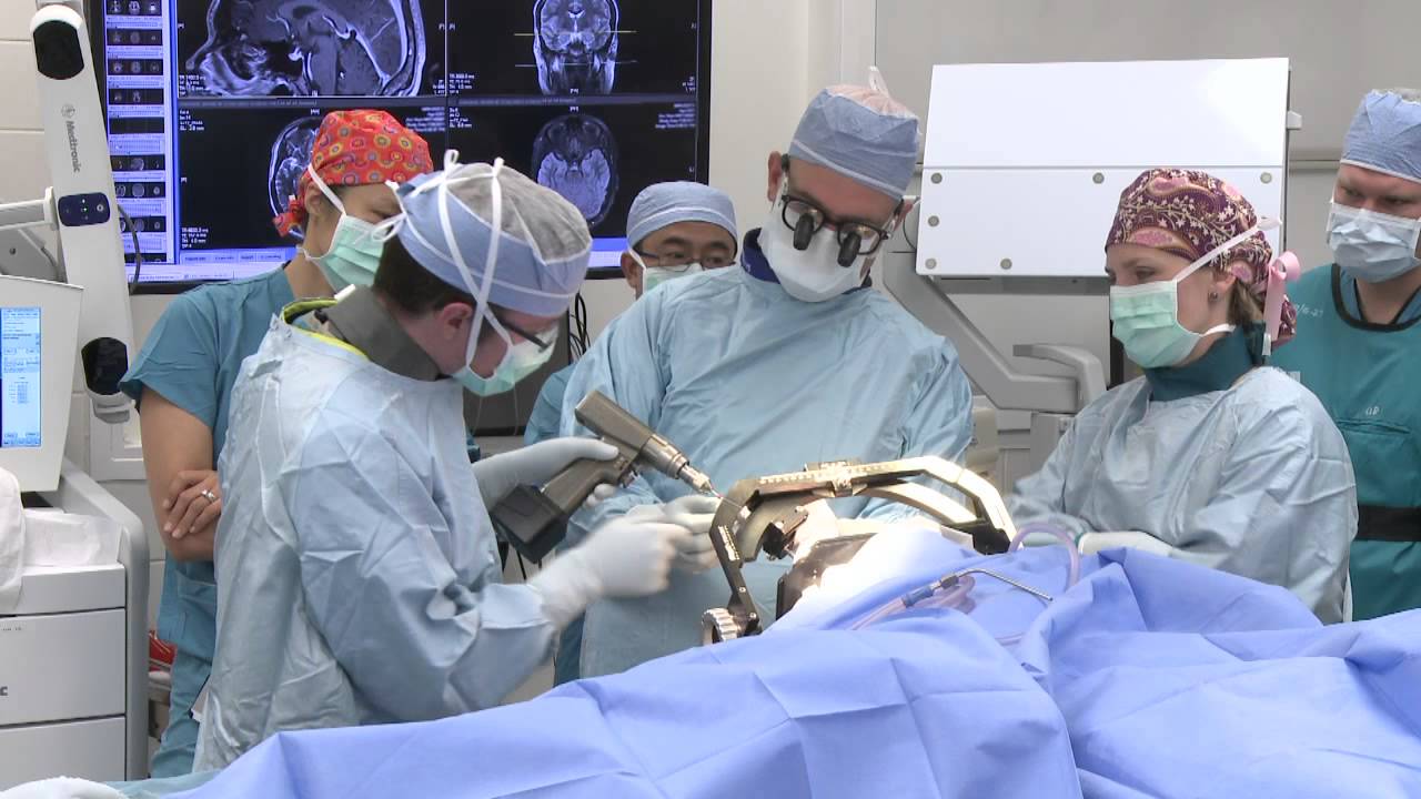
Patient Greg Grindley communicates with host Bryant Gumbel and his wife for the first time while undergoing deep brain stimulation surgery at University Hospital's Case Medical Center in Cleveland, Ohio.
➡ Subscribe: http://bit.ly/NatGeoSubscribe
About National Geographic:
National Geographic is the world's premium destination for science, exploration, and adventure. Through their world-class scientists, photographers, journalists, and filmmakers, Nat Geo gets you closer to the stories that matter and past the edge of what's possible.
Get More National Geographic:
Official Site: http://bit.ly/NatGeoOfficialSite
Facebook: http://bit.ly/FBNatGeo
Twitter: http://bit.ly/NatGeoTwitter
Instagram: http://bit.ly/NatGeoInsta
Greg's First In-Surgery Conversation | Brain Surgery Live
https://youtu.be/zvqV_2zncNU
National Geographic
https://www.youtube.com/natgeo

Kendall Lee, M.D., describes deep brain stimulation surgery, and how it is is typically done with patients who remain awake, so neurological functions can be measured and maintained. For more information on deep brain stimulation, visit http://mayocl.in/2A09T80.
Dr. Jeffrey Ojemann, director of epilepsy surgery at Seattle Children's Hospital, explains a cutting-edge treatment for epilepsy: minimally invasive MRI-guided laser ablation surgery. Laser ablation surgery is much safer and more precise than other treatments, with fewer side effects.
A special thanks to patient Keoni Giauque.
For more information, visit: http://www.seattlechildrens.or....g/clinics-programs/n
"One Last Look" music rights via RoyaltyFreeMusic.com

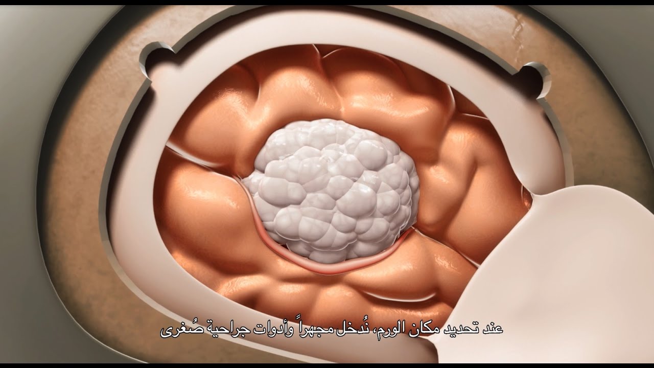
This minimally invasive technique allows surgeons to remove skull base tumors as large as softballs through the nose, with less trauma to the brain and critical nerves than with a traditional craniotomy.
To learn more, please visit https://www.upmc.com/

Brain port surgery is a minimally invasive surgical technique performed through a specially designed tube about the size of a dime. Using neuronavigation GPS-like guidance, the brain port is inserted into the brain with millimeter accuracy and is used as a channel to guide the surgeon and his/her instruments to various regions of the brain. Colloid cysts, metastatic tumors, and a variety of tumors within the ventricles are often candidates for this approach.

If left untreated, these “brain blisters” can lead to stroke. Get unprecedented access inside the angiosuite to see how Babak Jahromi, MD, PhD, treats a cerebral aneurysm without ever opening the skull. #InsideTheOR

Alexandra J. Golby, MD, Director, Image-guided Neurosurgery at Brigham and Women’s Hospital, discusses technological advancements to improve the precision of surgery to remove brain tumors.
It’s estimated that each year nearly 80,000 people are diagnosed with primary brain tumors and 100,000 with metastatic brain tumors. Nearly everybody is at risk for developing a brain tumor. Brain tumors can affect people from childhood to the last years of their lives. Men are slightly more affected than women and the causes of most brain tumors are not known.
There are a number of unique challenges in treating brain tumors. One challenge is that primary tumors can have indistinct margins that are difficult to see. Another challenge is that the tissue around a brain tumor is uniquely important and may impact things like language, visual and motor function.
The AMIGO Suite, opened in 2011 at Brigham and Women’s Hospital, is the Advanced Multimodality Image Guided Operating Suite. It's an NIH-funded national center which was developed with the goal of translating technological advances into improvements in surgical and interventional care for patients. In the AMIGO Suite, there is an intraoperative MRI scanner which can be brought in and out of the operating room during surgery to help surgeons visualize a patient’s tumor better.
Image-guided surgery uses the information obtained from advanced imaging and translates that into the planning and execution of surgery by acquiring high resolution and specialty structural images of the brain and also functional images of the brain. These images can be registered to one another and then to the patient's head during surgery. This allows surgeons to pinpoint the location of the tumor as well as the areas that we would like to preserve, areas that serve critical brain functions are located.
One of the big challenges, even with image-guided surgery, is that as we perform the surgery, the configuration of the brain is changing, and we call that brain shift. And it's due to changes in the brain itself and also as we remove tissue, things are constantly shifting and moving. When we're talking about doing brain tumor surgery, a few millimeters of movement can be a big difference. How to measure and track brain shift is an important area of research and a number of technologies are being studied to understand how to measure brain shift during surgery.
The development of various intraoperative imaging technologies allows surgeons to provide the most accurate surgical treatment for each individual patient.
Learn more about precision brain surgery at Brigham and Women’s Hospital:
https://www.brighamandwomens.o....rg/neurosurgery/brai

It’s called gamma knife surgery, but there’s no cutting involved.
It’s been used at Mayo Clinic for 30 years as an alternative to open brain surgery.
The patient’s head is held still during the procedure with a headframe, which also serves as a map for the radiation. Using 3D imaging — typically an MRI — as a guide, the gamma knife is targeted directly at the tumor.
And with no hospital stay and minimal side effects, it’s a procedure that is efficient and can be lifesaving.
More health and medical news on the Mayo Clinic News Network. https://newsnetwork.mayoclinic.org/
Journalists: Clean and nat sound versions of this pkg available for download at https://newsnetwork.mayoclinic.org/
Register (free) at https://newsnetwork.mayoclinic.org/request-account/

Neurosurgeon Sujit Prabhu, M.D., discusses what happens after surgery and how a patient recovers.
Learn more: http://www.mdanderson.org/educ....ation-and-research/d
Request an appointment at MD Anderson by calling 1-877-632-6789 or online: https://my.mdanderson.org/requestappointment

Surgeons in London removed a woman's brain tumor during a very unusual procedure. CBS News' Tina Kraus reports, the patient's love of music helped guide the surgery.
Subscribe to the CBS News Channel HERE: http://youtube.com/cbsnews
Watch CBSN live HERE: http://cbsn.ws/1PlLpZ7
Follow CBS News on Instagram HERE: https://www.instagram.com/cbsnews/
Like CBS News on Facebook HERE: http://facebook.com/cbsnews
Follow CBS News on Twitter HERE: http://twitter.com/cbsnews
Get the latest news and best in original reporting from CBS News delivered to your inbox. Subscribe to newsletters HERE: http://cbsn.ws/1RqHw7T
Get your news on the go! Download CBS News mobile apps HERE: http://cbsn.ws/1Xb1WC8
Get new episodes of shows you love across devices the next day, stream CBSN and local news live, and watch full seasons of CBS fan favorites like Star Trek Discovery anytime, anywhere with CBS All Access. Try it free! http://bit.ly/1OQA29B
---
CBSN is the first digital streaming news network that will allow Internet-connected consumers to watch live, anchored news coverage on their connected TV and other devices. At launch, the network is available 24/7 and makes all of the resources of CBS News available directly on digital platforms with live, anchored coverage 15 hours each weekday. CBSN. Always On.
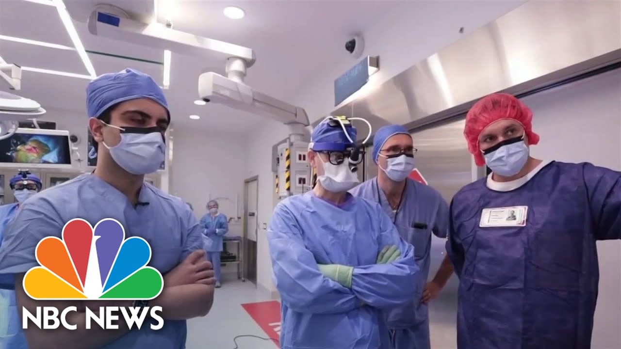
Dr. Akshay Syal takes us to NYU Langone Health where new A.I. technology is diagnosing brain tumors in record time, which opens the doors to possible new life-saving treatments.
» Subscribe to NBC News: http://nbcnews.to/SubscribeToNBC
» Watch more NBC video: http://bit.ly/MoreNBCNews
NBC News Digital is a collection of innovative and powerful news brands that deliver compelling, diverse and engaging news stories. NBC News Digital features NBCNews.com, MSNBC.com, TODAY.com, Nightly News, Meet the Press, Dateline, and the existing apps and digital extensions of these respective properties. We deliver the best in breaking news, live video coverage, original journalism and segments from your favorite NBC News Shows.
Connect with NBC News Online!
NBC News App: https://smart.link/5d0cd9df61b80
Breaking News Alerts: https://link.nbcnews.com/join/....5cj/breaking-news-si
Visit NBCNews.Com: http://nbcnews.to/ReadNBC
Find NBC News on Facebook: http://nbcnews.to/LikeNBC
Follow NBC News on Twitter: http://nbcnews.to/FollowNBC
Get more of NBC News delivered to your inbox: nbcnews.com/newsletters
#NBCNews #AI #Cancer

This innovative minimally invasive technique can remove large tumors located deep in the brain
To learn more, please visit http://brainsurgery.upmc.com
A craniotomy may be performed to treat brain tumors, blood clots, aneurysms, skull fractures, foreign objects, swelling of the brain, stroke or infection.

http://www.nucleushealth.com/ - This 3D medical animation depicts two operations, called craniotomy and craniectomy, in which the skull is opened to access the brain. The normal anatomy of the skull and tissues surrounding the brain are shown, including arteries and veins. The animation lists the common reasons for these procedures, and briefly introduces intracranial pressure.
Video ID: ANH13109
Transcript:
Your doctor may recommend a craniotomy or a craniectomy procedure to treat a number of different brain diseases, injuries, or conditions.
Your skull is made of bone and serves as a hard, protective covering for your brain. Just inside your skull, three layers of tissue, called meninges, surround your brain. The thick, outermost layer is the dura mater. The middle tissue layer is the arachnoid mater and the innermost layer is the pia mater. Between the arachnoid mater and the pia mater is the subarachnoid space, which contains blood vessels and a clear fluid called cerebrospinal fluid. Blood vessels, called bridging veins, connect the surface of your brain with the dura mater. Other blood vessels, called cerebral arteries, bring blood to your brain.
Inside your skull, normal brain function requires a delicate balance of pressure between the blood in your blood vessels, the cerebrospinal fluid that surrounds your brain, and your brain tissue. This is called normal intracranial pressure. Increased intracranial pressure may result from: brain tumors, head injuries, problems with your blood vessels, or infections in your brain or spinal cord. These conditions put pressure on your brain and may cause it to swell or change shape inside your skull, which can lead to serious brain injury.
Your doctor may recommend a craniotomy to remove: abnormal brain tissue, such as a brain tumor, a sample of tissue by biopsy, a blood clot, called a hematoma, excess cerebrospinal fluid, or pus from an infection, called an abscess.
A craniotomy may also be done to: relieve brain swelling,
stop bleeding, called a hemorrhage, repair abnormal blood vessels, repair skull fractures, or repair damaged meninges.
Finally, a craniotomy may also be done to: treat brain conditions, such as epilepsy, deliver medication to your brain, or implant a medical device, such as a deep brain stimulator.
The most common reason for a craniotomy is to remove a brain tumor.
#Craniotomy #Craniectomy #BrainSurgery
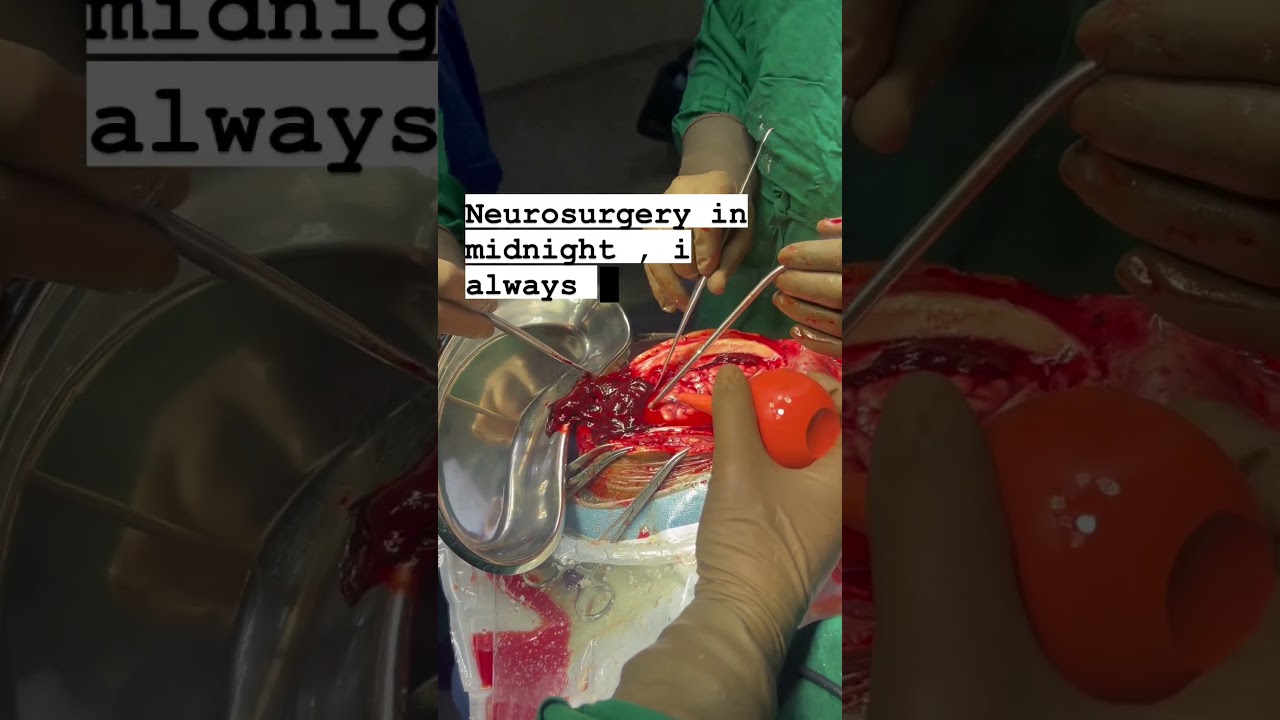
In this video, Dr Dhaval Patel, the best brain & spine surgeon in Surat South Gujarat, is performing Brain Hemorrhage Surgery. The Brain Hemorrhage Surgery was successfully done by the best neurosurgeon Dr Dhaval Patel in the midnight in Surat, South Gujarat.
Dr Dhaval Patel is the best and experienced brain & spine surgeon in Adajan, Vesu, Parvat Patiya, Surat, South Gujarat. Dr Dhaval is the expert of treatments and surgery for brain problems and spine problems.
.
Brain Hemorrhage Surgery, Best Brain & Spine Surgeon, Neurosurgeon, Brain Tumor Surgery, Brain Treatment Expert, Brain Expert, Brain & Spine Surgery, Neurosurgery in Surat, South Gujarat, Ahmedabad, Rajkot, Anand, Porbandar, patan, kutch, jamnagar, bhavnagar, junagadh, mehsana, nadiad, amreli, morbi, gandhinagar, verval, palanpur,godhra, gandhidham, botad, jetpur, kundal, kalol, disha, gondal, himatnagar, bhuj, modasa, lonavala, mandavi, kheda, khambhaliya, khambhat, dwarka, chhota udaipur, ambaji, dhoraji, idar, vallabhipur, una, dhandhuka, bhachau, mundra.
Dr. Dhaval Patel is an excellent neurosurgeon in Surat, South Gujarat. He is a Brain and Spine Surgeon; he is a reputable Neurosurgeon in Surat, South Gujarat. He has been practicing for the past five years. Till now, he has done 2500+ minor and major surgeries.
NEUROSURGEON DR. DHAVAL PATEL
Specialist in Brain & Spine Surgery
M.S.DNB (Neurosurgery - New Delhi)
Consultant Neurosurgeon
Surat Neuro Clinic Majura Gate, Ring Road, Surat.
Unity Hospital Parvat Patiya, Surat
United Green Hospital Adajan, Surat.
For more info. : +91-9687866766
#brainhemorrhage #brainsurgery #brainhemorrhagesurgery #brainstroke #heartdisease #brainconditions #brainsurgery #drdhavalpatel #spine #spinesurgery #unitedgreenhospital #surat_neuro_clinic #unity_hospital #drdhavalpatel #hormones #health #neuro #neurologiest #brain #surgery #recovery #patientreview #neurosurgeon #minimally_invasive #surgery #neurosurgery #stroke #heartattack #i3corporation

“Neurosurgery necessitates a very high level of detail involving complex procedures. I’m a very intense person inside the hospital and I feel like neurosurgery matched that level of intensity.”
It’s that intensity that made Dr. Jonathan Pindrik want to become a neurosurgeon. But it’s his certainty and skill inside the operating room that make him one of the best pediatric neurosurgeons in the country.
Dr. Pindrik is a neurosurgeon at Nationwide Children’s Hospital. While he performs multiple complex brain and spinal procedures each week, he also specializes in surgical intervention for children with epilepsy. Dr. Pindrik serves as co-director of the Epilepsy Surgery Program at Nationwide Children’s Epilepsy Center. It’s level-four accreditation means we offer the highest level of epilepsy care including advanced epilepsy surgery.
Connect with a specialist: http://bit.ly/2qdhDj7
Our team of neurosurgeons: http://bit.ly/2qcvxSl
Nationwide Children's Epilepsy Center: http://bit.ly/2qcGtj1
Learn more about Nationwide Children’s Level 4 Epilepsy Center: http://bit.ly/2qcGtj1
Meet our Chief of Neurosurgery: https://bit.ly/2GJSuYm

The moment doctors at University Hospital's Case Medical Center activate the electrode they implanted in patient Greg Grindley’s brain, the tremor in his right hand stops immediately.
➡ Subscribe: http://bit.ly/NatGeoSubscribe
About National Geographic:
National Geographic is the world's premium destination for science, exploration, and adventure. Through their world-class scientists, photographers, journalists, and filmmakers, Nat Geo gets you closer to the stories that matter and past the edge of what's possible.
Get More National Geographic:
Official Site: http://bit.ly/NatGeoOfficialSite
Facebook: http://bit.ly/FBNatGeo
Twitter: http://bit.ly/NatGeoTwitter
Instagram: http://bit.ly/NatGeoInsta
Tremor Relief at Last | Brain Surgery Live
https://youtu.be/iX-QKDnUbhg
National Geographic
https://www.youtube.com/natgeo

A patient at a British hospital played Mahler and Gershwin on the violin while surgeons removed a tumor from her brain, so doctors could preserve her ability to play music.
She left the hospital 3 days later and hopes to return to the symphony soon. https://abcn.ws/2SGY9mp
SUBSCRIBE to ABC NEWS: https://www.youtube.com/ABCNews/
Watch More on http://abcnews.go.com/
LIKE ABC News on FACEBOOK
https://www.facebook.com/abcnews
FOLLOW ABC News on TWITTER:
https://twitter.com/abc
GOOD MORNING AMERICA'S HOMEPAGE:
https://www.goodmorningamerica.com/

This 3D animation of brain surgery, shows how a ventriculostomy is performed, which is a neurosurgical procedure of creating a hole within a cerebral ventricle for drainage. It is most commonly performed on those with hydrocephalus, an abnormal buildup of fluid in the ventricles (cavities) deep within the brain. It's done by surgically penetrating the skull, dura mater, and brain such that the ventricular system ventricle of the brain is accessed.
When catheter drainage is temporary, it is commonly referred to as an external ventricular drain (EVD). When catheter drainage is permanent, it is usually referred to as a shunt.
There are many catheter-based ventricular shunts that are named for where they terminate, for example, a ventriculi-peritoneal shunt terminates in the peritoneal cavity, a ventriculoarterial shunt terminates within the atrium of the heart, etc. The most common entry point on the skull is called Kocher's point. An EVD ventriculostomy is done primarily to monitor the intracranial pressure as well as to drain cerebrospinal fluid (CSF), primarily, or blood to relieve pressure from the central nervous system (CNS).
For more information about custom medical animation, please visit https://www.amerra.com/.
Watch additional medical animations:
Craniectomy brain surgery - 3D animation: https://youtu.be/1RkseDeYS9g
Accessing an implantable port training - 3D animation: https://youtu.be/xSTpxjyv4O4
Open Suctioning with a Tracheostomy Tube - 3D animation: https://youtu.be/wamB7jpWCiQ
Suctioning the endotracheal tube - medical animation: https://youtu.be/pN6-EYoeh3g
Functional endoscopic sinus surgery (FESS) - 3D animation: https://youtu.be/qKTRyowwaLA
How to insert a nasogastric tube for NG intubation - 3d animation: https://youtu.be/Abf3Gd6AaZQ
Oral airway insertion - oropharyngeal airway technique - 3D animation: https://youtu.be/caxUdNwjt34
Nasotracheal suctioning (NTS) - 3D animation: https://youtu.be/979jWMsF62c
Learn about hemorrhoids with #3d #animation: https://youtu.be/R6NqlMpsiiY
LASIK eye surgery - 3D animation: https://youtu.be/Bb8bnjnEM00
CPR cardiopulmonary resuscitation - 3D animation: https://youtu.be/G87knTZnhks
What are warts (HPV)? - 3D animation: https://youtu.be/guJ1J7rRs1w
How Macular Degeneration Affects Your Vision - 3D animation: https://youtu.be/ozZQIZ_52YY
NeoGraft hair transplant procedure – animation: https://youtu.be/C-eTdH2UPXI

Thanks to Ben, Addenbrooke's and neuroscientist Yaara Erez from the University of Cambridge
