- Physical Examination
- Surgical Examination
- Ophthalmology
- Clinical Skills
- Orthopedics
- Surgery Videos
- Laparoscopy
- Pediatrics
- Funny Videos
- Cardiothoracic Surgery
- Nursing Videos
- Plastic Surgery
- Otorhinolaryngology
- Histology and Histopathology
- Neurosurgery
- Dermatology
- Pediatric Surgery
- Urology
- Dentistry
- Oncology and Cancers
- Anatomy Videos
- Health and Fitness
- Radiology
- Anaesthesia
- Physical Therapy
- Pharmacology
- Interventional Radiology
- Cardiology
- Endocrinology
- Gynecology
- Emergency Medicine
- Psychiatry and Psychology
- Childbirth Videos
- General Medical Videos
- Nephrology
- Physiology
- Diet and Food Health
- Diabetes Mellitus
- Neurology
- Women Health
- Osteoporosis
- Gastroenterology
- Pulmonology
- Hematology
- Rheumatology
- Toxicology
- Nuclear Medicine
- Infectious Diseases
- Vascular Disease
- Reproductive Health
- Burns and Wound Healing
- Other
Interventional Radiology
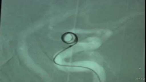
Endovascular Coiling of Unruptured Ophthalmic Artery Aneurysm
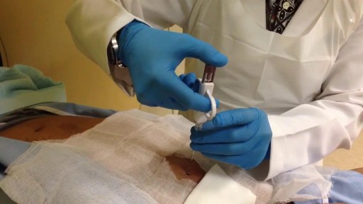
Computed tomography (CT)-guided transthoracic needle biopsy is a well-established, minimally invasive diagnostic tool for pulmonary lesions. Few large studies have been conducted on the diagnostic performance and adequacy for molecular testing of transthoracic core needle biopsy (TCNB) for small pulmonary lesions.
A computed tomography (CT) scan uses a special X-ray machine to take detailed pictures of the body’s organs and tissues. In a biopsy, a small piece of tissue is removed from your body. This tissue sample is then examined in the lab. A needle biopsy is the safest and easiest way to remove this tissue safely from the body. To do a needle biopsy, the radiologist will insert a needle through your skin and into your tissue. A syringe or an automated needle may be used to take the tissue sample.
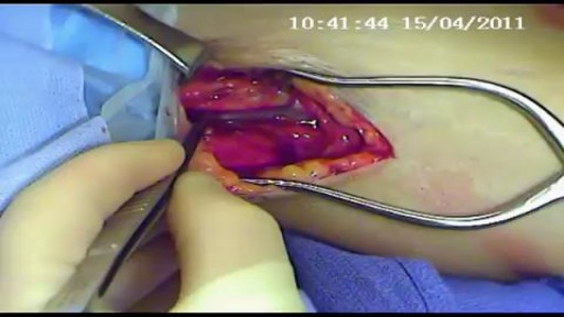
An AV fistula is a connection, made by a vascular surgeon, of an artery to a vein.Vascular surgeons specialize in blood vessel surgery. The surgeon usually places an AV fistula in the forearm or upper arm. An AV fistula causes extra pressure and extra blood to flow into the vein, making it grow large and strong.
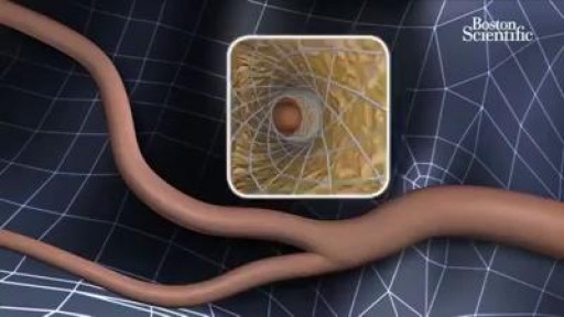
It involves placing a small, expandable tube called a stent in the narrowed artery. This procedure is also called carotid angioplasty and stenting. There are two carotid arteries-one on each side of the neck-that supply blood to the brain. These arteries can be narrowed and damaged by fatty deposits called plaque.
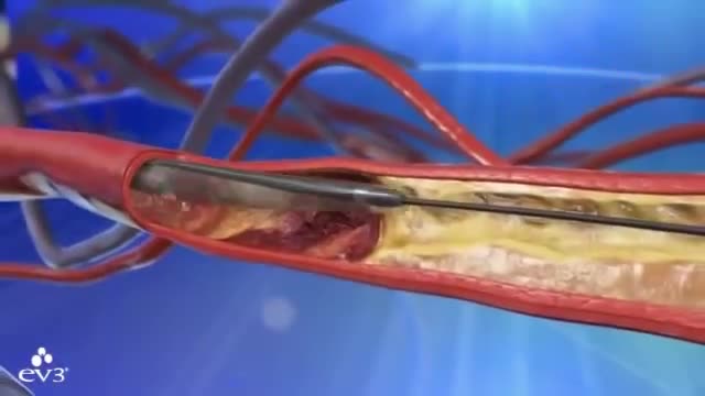
Preventing heart attacks and stroke can involve extensive surgery to remove plaque from your arteries, but as FOX17's Nick Paranjape shows us, there's a new procedure in Middle Tennessee that is less invasive and substantially cuts down on your recovery time. At 76, Jimmy Wilkie of Hendersonville exercises on his treadmill 3-4 times a week. Recently, he started having pain in his left leg. It was so bad, he couldn't even walk. Turned out, Mr. Wilkie had a blocked artery in his leg. In years past, this would've required major bypass surgery. Not anymore!"The Turbohawk Catheter has really opened a new door for us," says Dr. Dan Wunder.Dr. Wunder, an Interventional Radiologist at Premier Radiology in Madison, is talking about the Turbohawk. It's a device which is inserted into the blocked artery, and inside the Turbohawk are 4 tiny blades."It can cut the plaque and with that shape of the disc it cuts with it pushes it forward into the catheter," says Dr. Wunder.The one-hour procedure doesn't just push the plaque to the sides where it can re-grow, but instead grabs it and removes it!"We pull it back out and it fills up," says Dr. Wunder. "Empty it out, go back down and we can cut some more out."Before and after images really say it all."They used a roto rooter as he called it," says Wilkie.A roto rooter, Turbohawk, call it what you want, but Wilkie says all he knows is the procedure worked right away!"There wasn't any pain at all in my leg," says Wilkie.It's rare, but the outpatient procedure can have complications like plaque getting pushed down in the leg. Dr. Wunder says the main symptoms of a blockage in your legs is having severe pain or cramping when you're walking or exercising.
