- Physical Examination
- Surgical Examination
- Ophthalmology
- Clinical Skills
- Orthopedics
- Surgery Videos
- Laparoscopy
- Pediatrics
- Funny Videos
- Cardiothoracic Surgery
- Nursing Videos
- Plastic Surgery
- Otorhinolaryngology
- Histology and Histopathology
- Neurosurgery
- Dermatology
- Pediatric Surgery
- Urology
- Dentistry
- Oncology and Cancers
- Anatomy Videos
- Health and Fitness
- Radiology
- Anaesthesia
- Physical Therapy
- Pharmacology
- Interventional Radiology
- Cardiology
- Endocrinology
- Gynecology
- Emergency Medicine
- Psychiatry and Psychology
- Childbirth Videos
- General Medical Videos
- Nephrology
- Physiology
- Diet and Food Health
- Diabetes Mellitus
- Neurology
- Women Health
- Osteoporosis
- Gastroenterology
- Pulmonology
- Hematology
- Rheumatology
- Toxicology
- Nuclear Medicine
- Infectious Diseases
- Vascular Disease
- Reproductive Health
- Burns and Wound Healing
- Other
Latest videos
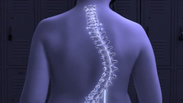
Pediatric orthopedic surgeons at Columbia are using a new device with magnetic technology that avoids the need for multiple spine-lengthening surgeries to correct early-onset scoliosis, a severe curvature of the spine in young children. In April 2014, Michael Vitale, MD, the Ana Lucia Professor of Pediatric Orthopedic Surgery at CUMC and 1995 graduate of P&S, performed the first procedure in the New York area, using the device to treat a 5-year-old boy. When braces and casts cannot control scoliosis in young children, surgeons turn to growing rods, which help correct the curve while allowing the spine to grow. When spinal maturity is near, the rods are removed and a spinal fusion can be performed. But during years of treatment with growing rods, patients must undergo surgery every six months to lengthen the rods to keep up with the patients’ growth. A patient may undergo eight to 10 procedures, which are costly and result in lost time for parents at work and children at school. The new device—MAGEC (MAGnetic Expansion Control) rods—contains a mechanism inside the growing rods that allows surgeons to lengthen the rods with a handheld external magnet, without surgery.
Inserting the Enlite Sensor with insulin pump
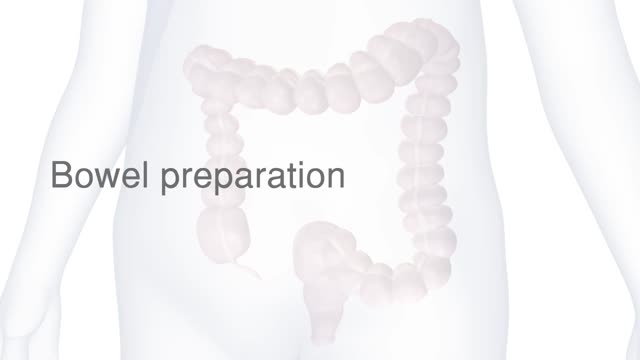
The camera sends images to an external monitor so the doctor can study the inside of your colon. The doctor can also insert instruments through the channel to take tissue samples (biopsies) or remove polyps or other areas of abnormal tissue. A colonoscopy typically takes about 20 minutes to an hour.
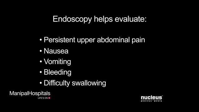
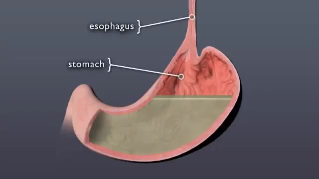
This animated video is an informative video that provides information regarding Upper Gastro-intestinal Endoscopy. An upper GI endoscopy procedure allows your doctor to view the mucus lining of the upper portion of your gastro-intestinal tract. This includes your oesophagus, stomach and duodenum. Upper endoscopy is used to evaluate symptoms of persistent upper abdominal pain, nausea, vomiting, bleeding, or difficulty in swallowing. The procedure is performed using an endoscope which is a long thin flexible tube a light and a tiny video camera attached to the end. The camera transmits the image to a monitor. Uncomplicated upper endoscopy takes 10-20 minutes, your doctor will gently insert the endoscope through your mouth and then slowly and carefully move it down your oesophagus until it reaches your stomach. An endoscopy for stomach may also be necessary in some cases.
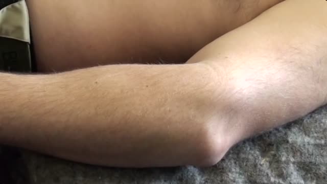
One technique to relocate a dislocated elbow with anatomy diagrammed out.
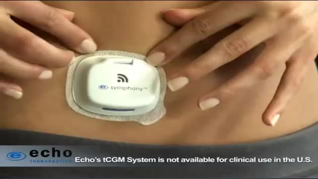
Echo Therapeutics Symphony tCGM Continuous Glucose Monitor
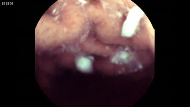
There’s a strange, mysterious world inside us, an alien-looking environment that turns the food we eat into nutrients that keep us alive. Michael Mosley swallows a camera to take a closer look.
Discover what happens to pill when it swallowed
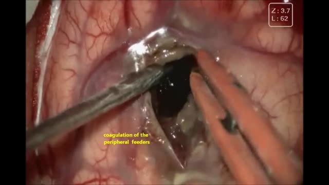
This 13 yrs young girl has had left temporo parietal cavernous angioma ,she came with acute bleed with raised ICT ,aphasia ,right hemiparesis ,leision was excised microsurgically with excellent out come
Risks & Benefits of Epilepsy Surgery | Epilepsy
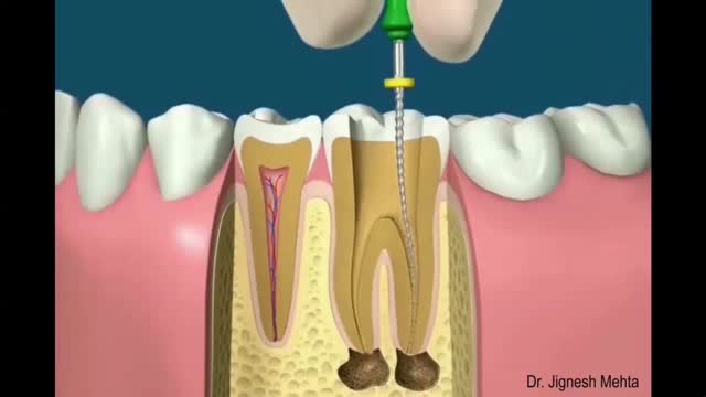
“Endo” is the Greek word for “inside” and “odont” is Greek for “tooth.” Endodontic treatment treats the inside of the tooth. Root canal treatment is one type of endodontic treatment. To understand endodontic treatment, it helps to know something about the anatomy of the tooth. Inside the tooth, under the white enamel and a hard layer called the dentin, is a soft tissue called the pulp. The pulp contains blood vessels, nerves, and connective tissue and creates the surrounding hard tissues of the tooth during development.
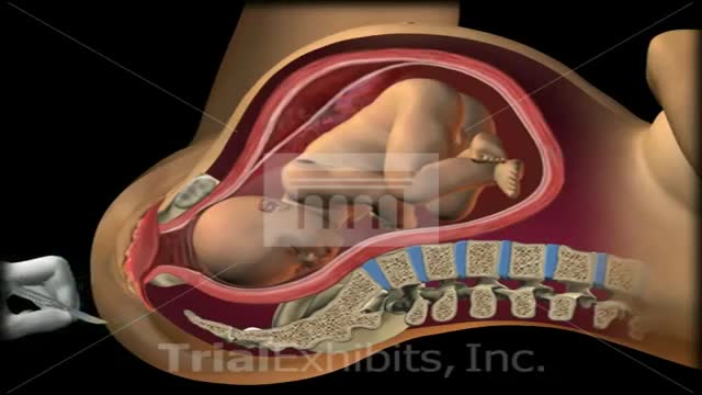
This medical 3D animation exhibit shows the left brachial plexus during birth and shoulder dystocia. Anatomy: symphysis pubis, uterus, sacrum, coccyx and fetus. "McRoberts Position". An episiotomy is cut. Brachial Plexus stretch injury. Retraction of head (turtle sign). Suprapubic pressure, gentle traction. To view our medical library of exhibits,
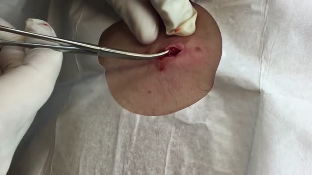
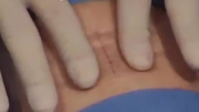
This cyst had been slowly growing for decades and created some redundant skin on the surface. A decision was made to make a slightly bigger incision in order to remove this tissue as well. As a result of this deeper process, 2 deep dermal sutures were added before the superficial interrupted sutures were put in place.
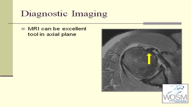
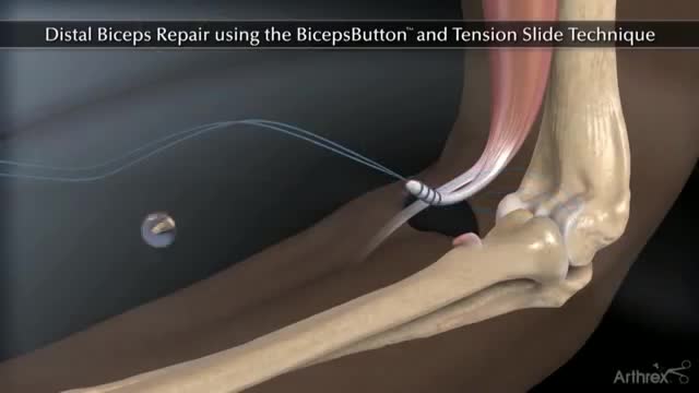
Biceps tendonitis, also called bicipital tendonitis, is inflammation in the main tendon that attaches the top of the biceps muscle to the shoulder. The most common cause is overuse from certain types of work or sports activities.
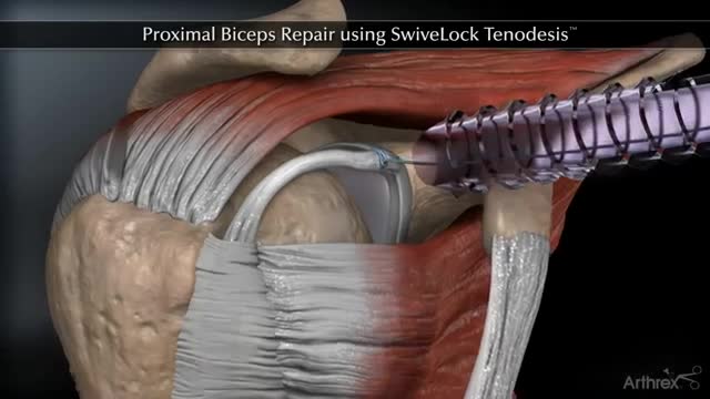
Proximal Biceps Repair using SwiveLock Tenodesis
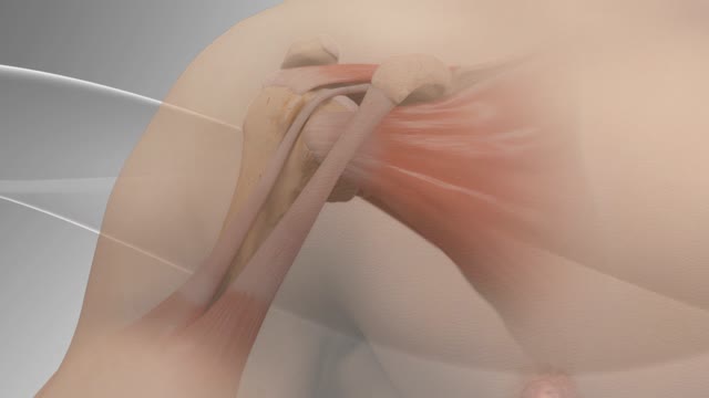
Biceps tenodesis surgery is performed when the biceps tendon is damaged, or the rotator cuff tendon or cartilage ring in the shoulder is torn. The biceps tendon is a strong rope‐like structure connecting the upper end of the biceps muscle to the bones in the shoulder. In biceps tenodesis surgery, the biceps tendon is separated from the shoulder and reattached to the humerus, or the upper arm bone.
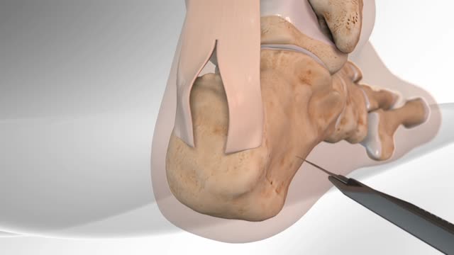
The Arthrex® Achilles SpeedBridge™ repair is a surgical technique system that combines fully threaded SwiveLock® anchors with FiberTape® suture. The surgeon may use the Achilles SpeedBridge to reattach the Achilles tendon to the heel bone after repairing the damaged portion of the Achilles tendon. The Achilles tendon connects the two large muscles at the back of the calf to the heel. Insertional Achilles tendinitis is a painful and disabling condition where the tendon attaches to the heel bone causing redness, pain and swelling. Patients who do not respond to the initial treatment may require surgical treatment.
An example of a technique I use in my surgical practice
The pituitary is a small gland found inside the skull just below the brain and above the nasal passages, which are above the fleshy back part of the roof of the mouth (known as the soft palate). The pituitary sits in a tiny bony space called the sella turcica. The nerves that connect the eyes to the brain, called the optic nerves, pass close by it.
Wound-closure technologies are becoming less painful and more efficient at closing wounds
