- Physical Examination
- Surgical Examination
- Ophthalmology
- Clinical Skills
- Orthopedics
- Surgery Videos
- Laparoscopy
- Pediatrics
- Funny Videos
- Cardiothoracic Surgery
- Nursing Videos
- Plastic Surgery
- Otorhinolaryngology
- Histology and Histopathology
- Neurosurgery
- Dermatology
- Pediatric Surgery
- Urology
- Dentistry
- Oncology and Cancers
- Anatomy Videos
- Health and Fitness
- Radiology
- Anaesthesia
- Physical Therapy
- Pharmacology
- Interventional Radiology
- Cardiology
- Endocrinology
- Gynecology
- Emergency Medicine
- Psychiatry and Psychology
- Childbirth Videos
- General Medical Videos
- Nephrology
- Physiology
- Diet and Food Health
- Diabetes Mellitus
- Neurology
- Women Health
- Osteoporosis
- Gastroenterology
- Pulmonology
- Hematology
- Rheumatology
- Toxicology
- Nuclear Medicine
- Infectious Diseases
- Vascular Disease
- Reproductive Health
- Burns and Wound Healing
- Other
Latest videos
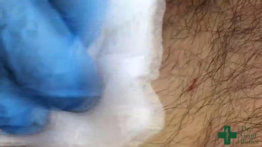
Watch that Huge Skin Cyst Removal Surgery

Watch that video to know What is Trypophobia? Do You Have it ?

Watch that video of Unreal Mutations and Medical Condition

Watch that Terrible Skin Jiggers Removal Video

Watch that video of a Black Salve Left an Inch Hole In Man's Hole

Watch that video of Disgusting! Parasites, zits, insects in people’s ears & more

Watch that video of people should have gone to the dentist sooner
Watch that video of an Ingrown hair turns into 140 Lbs tumor in man’s stomach
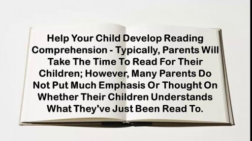
How To Help Your Child Learn To Read, Help My Child Learn To Read, Best Way To Teach Reading---- http://children-learning-reading.good-info.co -- how to help your child learn to read - Help My Child Learn to Read The ability to read is vital for success. It helps your child succeed in school, helps them build self-confidence, and helps to motivate your child. Being able to read will help your child learn more about the world, understand directions on signs and posters, allow them to find reading as an entertainment, and help them gather information. Learning to read is very different from learning to speak, and it does not happen all at once. There is a steady progression in the development of reading ability over time. The best time for children to start learning to read is at a very young age - even before they enter pre-school. Once a child is able to speak, they can begin developing basic reading skills. Very young children have a natural curiosity to learn about everything, and they are naturally intrigued by the printed texts they see, and are eager to learn about the sounds made by those letters. You will likely notice that your young child likes to look at books and thoroughly enjoys being read to. They will even pretend to behave like a reader by holding books and pretend to read them. As parents, you're the most important first step in your children's journey into the wonderful world of reading. It is up to you to create the most supportive environment that turns your child on to reading - such as reading aloud to them often during the day and before bedtime, and placing age appropriate books for children around the house, so that the child will have access to plenty of books. Reading often to your child will help develop their interest in books and stories, and soon they will want to read stories on their own. >>Teach your child to read and enable your child to become a fast and fluent reader! Click here to help your child learn to read http://children-learning-reading.good-info.co
Teaching Phonics To Children, How To Teach Phonics And Reading, How To Read Better, Teach Kid Read--- http://children-learning-reading.good-info.co --- Teaching phonics to children - How to Teach Phonics and Reading, Teaching children to read by teaching phonics activities is a lot like doing math, where you have to know what the numbers are, how to count, and you need to learn to add and subtract before learning to multiply and divide. Teaching phonics to children is no different where you follow a step by step approach by first teaching the child the alphabet letters and phonics sounds, and then teaching them the combination of different letters to create different words, and using words to form sentences. It is a very logical and sequential buildup of phonics knowledge and reading ability. Before a child can learn to read, he or she must first learn the alphabet letters, and know the sounds represented by the letters. It's usually easier to teach some consonants and short vowels first before moving on to more complicated things such as consonant digraphs (2 consonants formed to produce one sound, such as "ch" or "ph") and long vowels. As you can see, teaching children to read by the phonics method helps them develop phonemic awareness, and it is also a very logical and straight forward approach. Start off by teaching your child the phonics sounds. You can choose to teach your child in alphabetic order going from A to Z, or you can teach several commonly used consonant sounds and vowels, and go from there. For example, you may start teaching your child /a/, /c/, and /t/ (slashes denote sound of the letters). Once your child has learn to quickly recognize these letters and properly sound out their sounds, you can then teach them to blend /c/, /a/, /t/ to make the words "cat", or "tac", or "at". As you introduce more letters and phonics sounds in your lesson plans, you can generate more words, and slowly introduce short, simple sentences to your reading lessons. Depending on the age of your child, I would suggest keeping the phonics lessons relatively short - around 5 to 10 minutes. Sometimes, just 3 to 5 minutes for a short lesson is plenty, and you can easily teach these short phonics lessons 2 or 3 times each day for a total of 10 to 15 minutes. >> Teach your child to read today using our step-by-step, proven method for teaching young children to read http://children-learning-reading.good-info.co
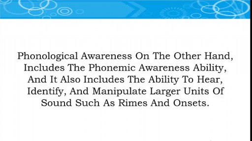
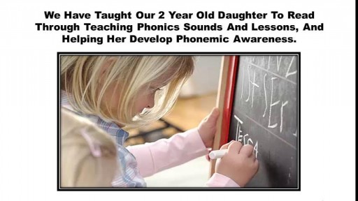
What Is Phonemic Awareness, Reading Program For Kids, Phonics For Children, Teach Your Baby To Read---- http://children-learning-reading.good-info.co----- What is Phonemic Awareness, Phonemic Awareness is defined as the ability to identify, hear, and work with the smallest units of sound known as phonemes. It is NOT the same as phonological awareness, instead, it is a sub-category of phonological awareness. For example, phonemic awareness is narrow, and deals only with phonemes and manipulating the individual sounds of words - such as /c/, /a/, and /t/ are the individual sounds that make up to form the word "cat". Phonological awareness on the other hand, includes the phonemic awareness ability, and it also includes the ability to hear, identify, and manipulate larger units of sound such as rimes and onsets. Phonemic awareness can be taught very early on, and will play a critical role in helping children learn to read and spell. While it's not set in stone on when a child can learn to read, however, I do believe that a child that can speak is a child that can learn to read. Children as young as two years old can learn to read by developing phonemic awareness, and they can learn to read fluently. Please see a video of a 2 year old (2yr11months) reading below. Below are several of the most common phonemic awareness skills that are often practiced with students and young children: Phonemic identity - being able to recognize common sounds in different words such as /p/ is the common sound for "pat", "pick", and "play". Phonemic isolation - being able to recognize the individual sounds of words such as /c/ is the beginning sound of "cat" and /t/ is the ending sound of "cat". Phoneme substitution - being able to change one word to another by substituting one phoneme. For example changing the /t/ in "cat" to /p/ now makes "cap". Word Segmenting - the parent says the word "lap", and the child says the individual sounds: /l/, /a/, and /p/. Oral blending - the parent says the individual sounds such as /r/, /e/, and /d/, and the child forms the word from the sounds to say "red". Studies have found that phonemic awareness is the best predictor of reading success in young children. Research has also found that children with a high level of phonemic awareness progress with high reading and spelling achievements; however, some children with low phonemic awareness experience difficulties in learning to read and spell. Therefore, it is important for parents to help their young children develop good phonemic awareness. Being able to oral blend and segment words helps children to read and spell. According to the National Reading Panel, oral blending helps children develop reading skills where printed letters are turned into sounds which combine to form words. Additionally, word segmenting helps children breakdown words into their individual sounds (phonemes), and helps children learn to spell unfamiliar words. As a young child begins to develop and master phonemic awareness skills, they will discover an entirely new world in print and reading. You will open up their world to a whole new dimension of fun and silliness. They will be able to read books that they enjoy, develop a better understanding of the world around them through printed materials, and have a whole lot of fun by making up new nonsense words through phonemic substitutions. For example, we taught our daughter to read at a young age - when she was a little over 2 and a half years old. Before she turned three, she would run around the house saying all types of silly words using phonemic substitution. One of her favorite was substituting the letter sound /d/ in "daddy" with the letter sound /n/. So, she would run around me in circles and repeatedly say "nanny, nanny, come do this" or "nanny, nanny, come play with me" etc... Of course, she only did this when she wanted to be silly and to make me laugh, at other times, she would of course properly refer to me as "daddy", and not "nanny". She is well aware of the differences between these words and is fully capable of using phonemic substitution to change any of the letters in the words to make other words. Give your child a head start, and.. pave the way for a bright, successful future..Click here to learn how to easily and quickly teach your child to read. http://children-learning-reading.good-info.co

Watch that video of Sperm Formation and Pathway Ejaculation

Watch that Huge Stomach Tumor Removal Medical Surgery

Watch How Snake Poison Could Turn Human Blood Into Jelly

Watch that video of a Snake bite causes girl’s leg to rot away with necrosis

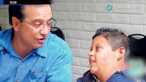
Watch that video of an Indian Boy Was Born With 232 Teeth Got Them Removed
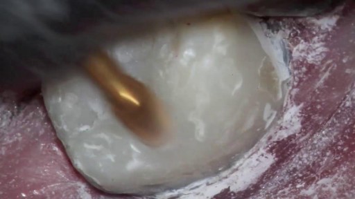
Watch that video of Filling Monster Tooth Cavity
Watch that Male Catheter Insertion Procedure

Watch that Female Recto-vaginal Exam Video

Watch that Cutting Inside Human Fat Body video
