

Colonoscopy

A video describing the procedure of colonoscopy or flexible fibre-optic examination of the colon.
Video Transcript
A narrated endoscopy-suite film explaining colonoscopy from preparation to discharge. After showing a normal examination, it tours findings the scope can identify — diverticular disease, Crohn's disease, ischemic colitis, polyps removed by snare, and cancers of the colon and rectum — then describes reaching the cecum, the careful second look on withdrawal, and recovery.
Read the full transcript
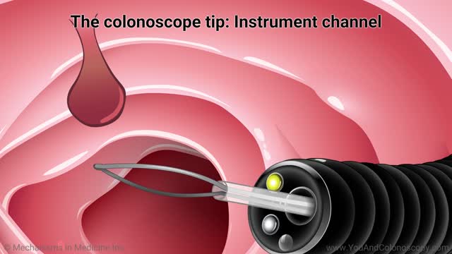
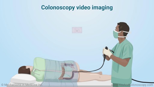
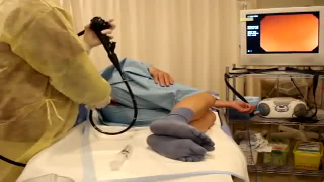
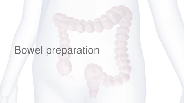
A colonoscopy is a flexible fibro optic scope examination of the large bowel, which is synonymous with the colon and rectum. A bowel preparation is administered beforehand so that a good view is obtained at the time of the procedure. The patient is brought into the endoscopy suite, placed in the left lateral position and conscious sedation administered. For practical purposes this is a light anaesthetic. The scope is inserted through the anus and then up through the rectum and into the colon. It is attached to a camera which relays an excellent view to the monitor as seen here.
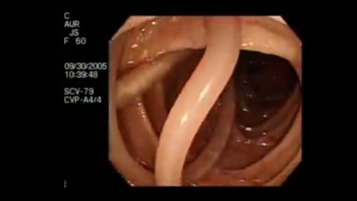
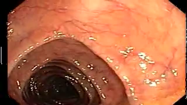
This is the view that can be seen from the monitor and in this particular case the lining of the colon appears entirely normal. It now follows a collection of pathologies that can be identified at colonoscopy.
Diabeticular disease is a condition that is characterized by numerous outpouchings of the bowel wall. Crohn's disease is a chronic inflammatory condition affecting the full thickness of the bowel wall. Ischemic colitis is inflammation and ulceration of the lining due to an inadequate blood supply. On the left hand side is a polyp which is a benign tumor attached to the lining of the bowel by a stalk. This is treated by excision and this is achieved by placing a snare diatheomy over the polyp and cutting through the stalk. The polyp is seen lying free on the left hand side which is then retrieved and a normal looking stalk on the right.
This polyp which is broad based and no more than a centimeter in size is treated in exactly the same way. Cancer of the colon otherwise known as a malignant tumor has a varied appearance. In this particular case there is a thick rolled edge and ulceration. This shows a mass that is almost totally obliterating the lumen of the bowel which is on the right hand side. And finally the scope has been turned through 180 degrees to demonstrate an obvious cancer arising within the lower part of the rectum.
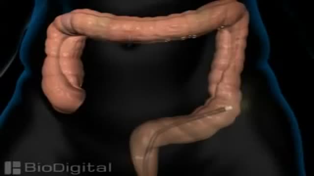
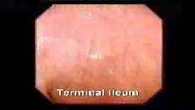
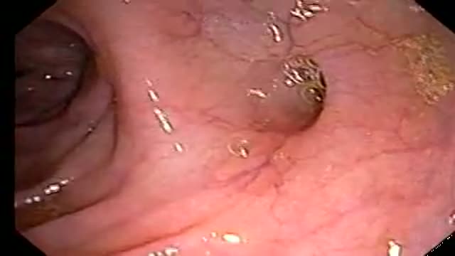
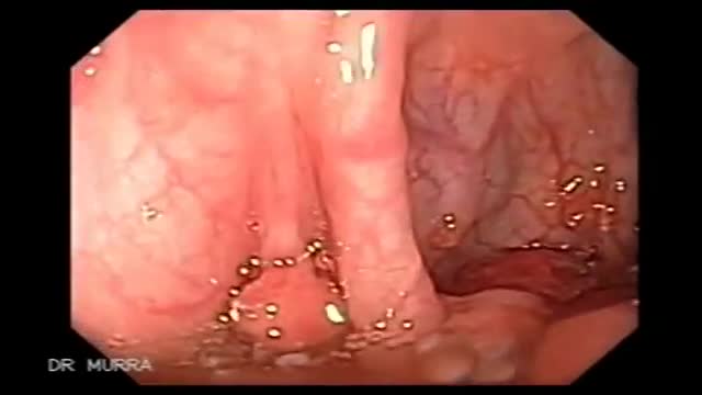
Colonoscopy involves traversing the whole length of the rectum and colon from the anus to the last part of the small bowel where it joins the colon otherwise known as the cecum. This structure can be seen here with the idasequal valve on the right hand side. It is possible to visualize the last part of the small bowel by entering through the idasequal valve if small bowel pathology is suspected. Confirmation that the cecum has been reached can be achieved in a number of ways. In this particular instance it is identification of the opening of the appendix as seen in the center of the picture here.
Upon withdrawal of the scope a second careful examination is made of the lining of the colon and rectum to ensure that no pathology has been missed on the way in. In this particular case the lining appears entirely normal. Once the scope has been removed the patient is transferred to the recovery room and after approximately 30 to 60 minutes they are ready to be discharged home. Patients who have had a colonoscopy under sedation should not drive a car or operate machinery for the remainder of the day. The following day they are completely back to normal.
Transcript auto-generated from the video’s audio for accessibility and study. Educational content only — not medical advice.
Video Overview
From our Surgical Examination collection: Pre-op assessments, wound and hernia exams, and clinical surgical skills demonstrated step by step by surgeons. Browse all Surgical Examination videos →
SORT BY-
Top Reacties
-
Laatste Reacties