






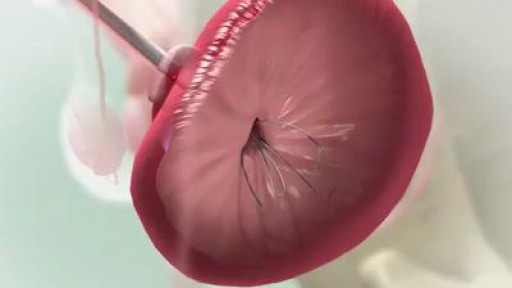
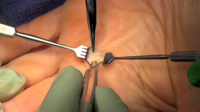
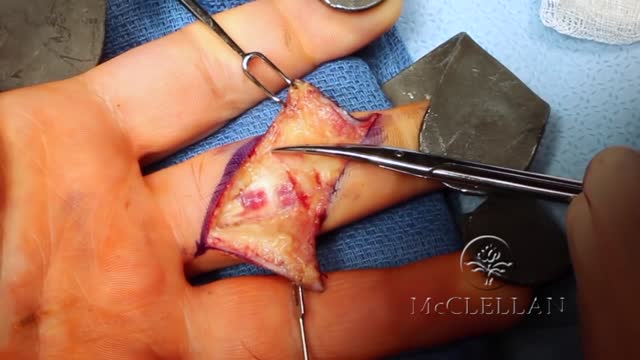
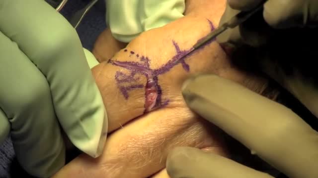


Giant Cell (Benign) Tumor of the Finger

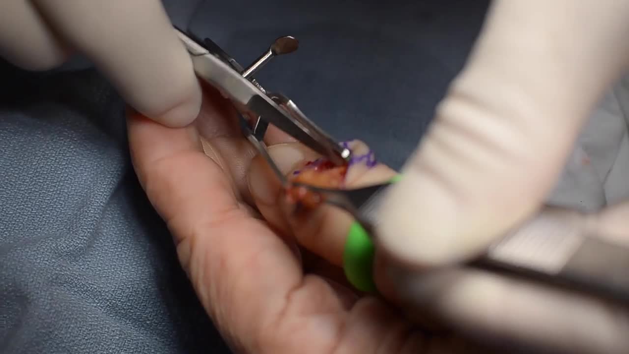
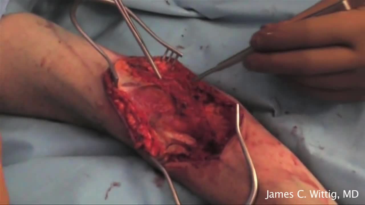
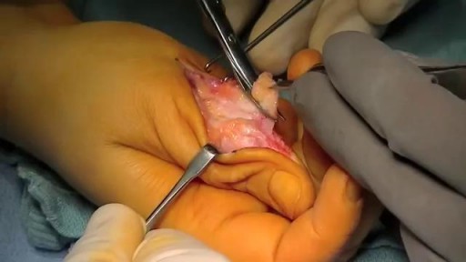
Giant cell tumors of the tendon sheath are common lesions and are the second most frequent tumors in the hand, after synovial cysts. They are diagnosed by means of clinical examination and complementary examinations (simple radiography and magnetic resonance). Erosion and invasion of the phalangeal bone affected may be seen on radiological examination. Magnetic resonance may show a “fluorescent or radiant effect” may be observed, caused by the high quantity of hemosiderin inside the tumor. Surgical treatment is the commonest practice, and complete excision is important for avoiding recurrence of the tumor, especially when bone invasion is observed on imaging examinations, which is generally related to greater tumor recurrence. In this paper, a case of a giant cell tumor of the tendon sheath in the middle phalanx of the third finger of a 45-year-old female patient is presented. This was successfully treated by means of surgery using a double access approach (dorsal and volar)
Video Overview
This video presents “Giant Cell (Benign) Tumor of the Finger” — Giant cell tumors of the tendon sheath are common lesions and are the second most frequent tumors in the hand, after synovial cysts. They are diagnosed by means of clinical examination and complementary examinations (simple radiography and. Whether you are preparing for a procedure, studying the technique, or learning how a condition is assessed, the recording shows the process as it is performed in practice.
From our Orthopedics collection: Orthopedic surgery videos: fracture fixation, joint replacement, arthroscopy and trauma procedures performed by orthopedic surgeons. Browse all Orthopedics videos →
Educational content only — not a substitute for professional medical advice.
SORT BY-
Najbolji komentari
-
Najnoviji komentari