- Physical Examination
- Surgical Examination
- Ophthalmology
- Clinical Skills
- Orthopedics
- Surgery Videos
- Laparoscopy
- Pediatrics
- Funny Videos
- Cardiothoracic Surgery
- Nursing Videos
- Plastic Surgery
- Otorhinolaryngology
- Histology and Histopathology
- Neurosurgery
- Dermatology
- Pediatric Surgery
- Urology
- Dentistry
- Oncology and Cancers
- Anatomy Videos
- Health and Fitness
- Radiology
- Anaesthesia
- Physical Therapy
- Pharmacology
- Interventional Radiology
- Cardiology
- Endocrinology
- Gynecology
- Emergency Medicine
- Psychiatry and Psychology
- Childbirth Videos
- General Medical Videos
- Nephrology
- Physiology
- Diet and Food Health
- Diabetes Mellitus
- Neurology
- Women Health
- Osteoporosis
- Gastroenterology
- Pulmonology
- Hematology
- Rheumatology
- Toxicology
- Nuclear Medicine
- Infectious Diseases
- Vascular Disease
- Reproductive Health
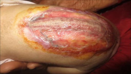
- Burns and Wound Healing
- Other
Latest videos
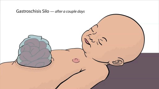
Gastroschisis is a birth defect of the abdominal (belly) wall. The baby’s intestines stick outside of the baby’s body, through a hole beside the belly button. The hole can be small or large and sometimes other organs, such as the stomach and liver, can also stick outside of the baby’s body. Gastroschisis occurs early during pregnancy when the muscles that make up the baby’s abdominal wall do not form correctly. A hole occurs which allows the intestines and other organs to extend outside of the body, usually to the right side of belly button. Because the intestines are not covered in a protective sac and are exposed to the amniotic fluid, the bowel can become irritated, causing it to shorten, twist, or swell.
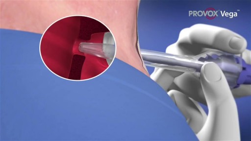
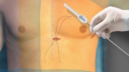
A voice prosthesis (plural prostheses) is an artificial device, usually made of silicone that is used to help laryngectomized patients to speak. During a total laryngectomy, the entire voice box (larynx) is removed and the windpipe (trachea) and food pipe (esophagus) are separated from each other.

Rare condition disorder known as Diprosopus, also known as craniofacial duplication. Diprosopus is a congenital defect also known as craniofacial duplication. The exact description of diprosopus refers to a fetus with a single trunk, normal limbs, and facial features that are duplicated to a certain degree. A less severe instance is when the fetus has a duplicated nose and the eyes are spaced far apart. In the most extreme instances, the entire face is duplicated, hence the name diprosopus, which is Greek for two-faced. Fetuses with diprosopus often also lack brains (anencephaly), have neural tube defects, or heart malformations. In some cases, if the brain is formed, it may have duplicated structures. Most infants with diprosopus are stillborn and there are fewer than fifty cases documented since 1864.
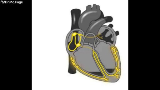
The human heart explained in 1 minute Video by Dr. Mo

According to a Danish study , frequent sex may help prevent pre-eclampsia. Researchers believe it's because of a protein found in sperm that can regulate the body's immune system. Yet because the cause of preeclampsia is unknown, it's important to keep your prenatal visits and talk to your doctor about your risk.
If you're pregnant or might become pregnant, it's critically important to get enough folic acid, the synthetic form of vitamin B9, also known as folate. Folic acid helps prevent neural tube defects (NTDs) – serious birth defects of the spinal cord (such as spina bifida) and the brain (such as anencephaly).

Is Air Travel During Pregnancy Safe? Traveling by air is considered safe for women while they are pregnant; however, the following ideas might make your trip safer and more comfortable. Most airlines allow pregnant women to travel through their eighth month.
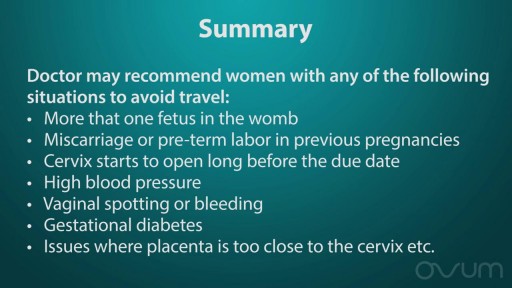
Airline travel. When you're pregnant, the safest time to travel is during your second trimester (18 to 24 weeks), when your risks for miscarriage and preterm labor are lowest. During your third trimester, it's best to stay within 300 miles of home, in case of sudden changes that need medical attention.
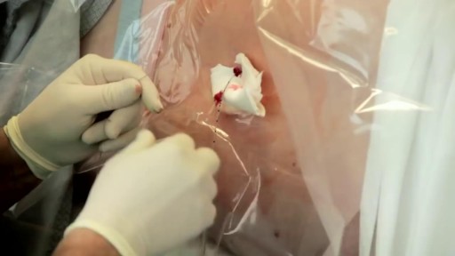
S-ICD leaves the heart and vasculature untouched. It may be implanted using only anatomical landmarks, thereby eliminating the need for fluoroscopy during implant and therefore reducing radiation exposure for both patients and physicians and eliminating the need for lead apron during implant.
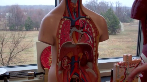
The digestive system is a group of organs working together to convert food into energy and basic nutrients to feed the entire body. Food passes through a long tube inside the body known as the alimentary canal or the gastrointestinal tract (GI tract).

The purpose of the organs of the male reproductive system is to perform the following functions: To produce, maintain, and transport sperm (the male reproductive cells) and protective fluid (semen) To discharge sperm within the female reproductive tract during sex To produce and secrete male sex hormones responsible for maintaining the male reproductive system

Start out with a visit to a doctor called a urologist. He'll give you a physical exam and ask you questions about your lifestyle and medical history, such as: Surgeries you've had Medications you take Your exercise habits Whether you smoke or take recreational drugs He may also have a frank discussion with you about your sex life, including any problems you've had or whether you have or ever had any STDs (sexually transmitted diseases). You'll probably be asked to give a sample of semen for analysis.
Most of the time, you won't know the exact day you got pregnant. Your doctor will count the start of your pregnancy from the first day of your last menstrual period. That's about 2 weeks ahead of when conception happens.
The drugs known as targeted therapy help stop cancer from growing and spreading. They work by targeting specific genes or proteins. These genes and proteins are found in cancer cells or in cells related to cancer growth, like blood vessel cells. Doctors often use targeted therapy with chemotherapy and other treatments.
If you’re considering an epidural to help manage the pain of childbirth, you’re not alone. More than 60 percent of women delivering at hospitals elect for an epidural during labor. And with good reason: An epidural is considered one of the safest methods of pain control, with just one in 3,000 pregnancies experiencing serious complications. It’s also good for you, since you’ll remain awake and alert during the birth, as well as for your baby, since the drugs will barely reach your bloodstream (so they can’t get into hers).

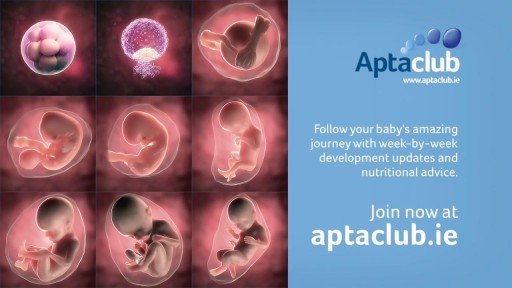
9 Months In The Womb: A Remarkable Look At Fetal Development Through Ultrasound
A heart attack is a frightening experience. If you have had a heart attack, or are close with someone who has, you are not alone: tens of thousands of Americans survive. As you work toward recovery, please use the following questions and answers to better understand what has happened to you and how you can help your heart heal so you can live a healthier, longer life.

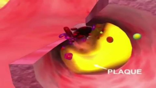
Atherosclerosis is a narrowing of the arteries caused by a buildup of plaque. It’s also called arteriosclerosis or hardening of the arteries. Arteries are the blood vessels that carry oxygen and nutrients from your heart to the rest of your body. As you get older, fat and cholesterol can collect in your arteries and form plaque. The buildup of plaque makes it difficult for blood to flow through your arteries. This buildup may occur in any artery in your body and can result in a shortage of blood and oxygen in various tissues of your body. Pieces of plaque can also break off, causing a blood clot. Atherosclerosis can lead to heart attack, stroke, or heart failure if left untreated.
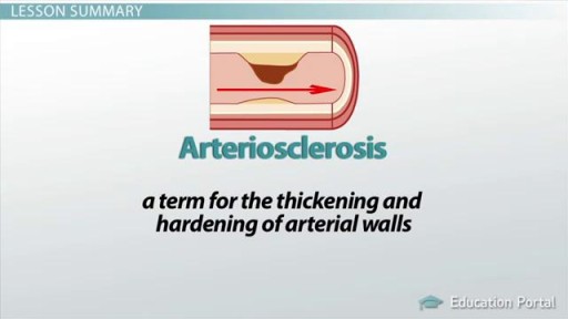
Arteriosclerosis occurs when the blood vessels that carry oxygen and nutrients from your heart to the rest of your body (arteries) become thick and stiff — sometimes restricting blood flow to your organs and tissues. Healthy arteries are flexible and elastic, but over time, the walls in your arteries can harden, a condition commonly called hardening of the arteries. Atherosclerosis is a specific type of arteriosclerosis, but the terms are sometimes used interchangeably. Atherosclerosis refers to the buildup of fats, cholesterol and other substances in and on your artery walls (plaques), which can restrict blood flow. These plaques can burst, triggering a blood clot. Although atherosclerosis is often considered a heart problem, it can affect arteries anywhere in your body. Atherosclerosis may be preventable and is treatable.
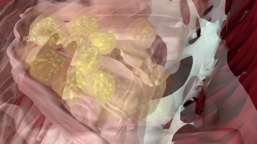
Atherosclerosis is a process in which blood, fats such as cholesterol, and other substances build up on your artery walls. Eventually, deposits called plaques may form. The deposits may narrow — or block — your arteries. These plaques can also rupture, causing a blood clot.
