
- Procedures & Tests
- Symptoms A-Z
- Symptom Checker
- Kidney Diet
- First Aid
- Anatomy Atlas
- Ask MedicalVideos
- Drug Guide
- Lab Values
- Vital Signs
- Muscles A–Z
- Clinical Signs
- Medical Abbreviations
- Medical Terminology
- Physical Examination
- Surgical Examination
- Ophthalmology
- Clinical Skills
- Orthopedics
- Surgery Videos
- Laparoscopy
- Pediatrics
- Funny Videos
- Cardiothoracic Surgery
- Nursing Videos
- Plastic Surgery
- Otorhinolaryngology
- Histology and Histopathology
- Neurosurgery
- Dermatology
- Pediatric Surgery
- Urology
- Dentistry
- Oncology and Cancers
- Anatomy Videos
- Health and Fitness
- Radiology
- Anaesthesia
- Physical Therapy
- Pharmacology
- Interventional Radiology
- Cardiology
- Endocrinology
- Gynecology
- Emergency Medicine
- Psychiatry and Psychology
- Childbirth Videos
- General Medical Videos
- Nephrology
- Physiology
- Diet and Food Health
- Diabetes Mellitus
- Neurology
- Women Health
- Osteoporosis
- Gastroenterology
- Pulmonology
- Hematology
- Rheumatology
- Toxicology
- Nuclear Medicine
- Infectious Diseases
- Vascular Disease
- Reproductive Health
- Burns and Wound Healing
- Kidney Zone Podcast
- Other
Latest videos
The newest additions to the MedicalVideos.com library — freshly curated medical and surgery videos, explainers, and lectures from educators and institutions worldwide.
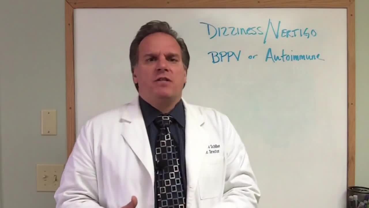

Is your vertigo or dizziness BPPV or autoimmune?

Learn a simple way to tell if you might be suffering from an undiagnosed autoimmune condition. Examples of autoimmune conditions include: • alopecia areata

Antiphospholipid (AN-te-fos-fo-LIP-id) syndrome occurs when your immune system attacks some of the normal proteins in your blood. It can cause blood clots in your arteries or veins. And it can cause pregnancy complications, such as miscarriage and stillbirth. Blood clots in your leg veins cause a condition known as deep vein thrombosis (DVT). Damage from blood clots in your organs, such as your kidneys, lungs or brain, depends on the extent and location of the clot. For instance, a clot in your brain can cause a stroke. There's no cure for antiphospholipid syndrome, but medications can reduce your risk of blood clots.

Pregnancy first Trimester
With so many antibiotics available, it isn't possible to list all of them here. But common antibiotics that are generally considered safe during pregnancy include penicillins (such as amoxicillin and ampicillin), cephalosporins (such as cephalexin), and erythromycin.

Therapeutic anticoagulation is recommended for all women with acute VTE; prophylactic anticoagulation is recommended for women at risk, such as those with a past history of thrombosis or thrombophilia or with a mechanical heart valve. The preferred anticoagulants during pregnancy are the heparin compounds.

Womens Issues and Blood Clotting
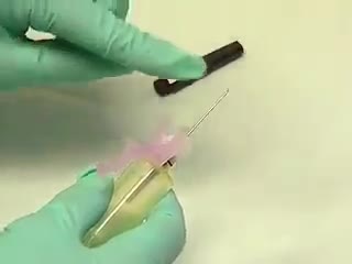

The venipuncture procedure is complex, requiring both knowledge and skill to perform. Each phlebotomist generally establishes a routine that is comfortable for her or him. Several essential steps are required for every successful collection procedure: Identify the patient. Assess the patient's physical disposition (i.e. diet, exercise, stress, basal state). Check the requisition form for requested tests, patient information, and any special requirements. Select a suitable site for venipuncture. Prepare the equipment, the patient and the puncture site. Perform the venipuncture. Collect the sample in the appropriate container. Recognize complications associated with the phlebotomy procedure. Assess the need for sample recollection and/or rejection. Label the collection tubes at the bedside or drawing area. Promptly send the specimens with the requisition to the laboratory.
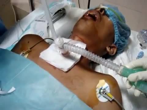
we use a single, tapered PDT dilator and kit . All the equipment and supplies listed must be present at the bed-side, because there is no time to go looking for supplies if an airway emergency occurs during the procedure. Two teams are used simultaneously. One team manages the endo-tracheal tube, and the other manages the placement of the tracheostomy tube

Multiple studies demonstrate the safety of propofol in pediatric EDPS. Each has identified a drop in blood pressure and transient hypoxemia as the most frequent complications. In all of the studies in which hypotension was identified there was no evidence of poor perfusion. The hypoxemia in all of these studies quickly responded to minimal intervention with no apparent lasting complications. Although these were pediatric studies, the results were very similar to ours in complication rates and sedation times. Our study did not demonstrate the frequency of decreased blood pressure seen in these pediatric studies but had similar hypoxemia rates.
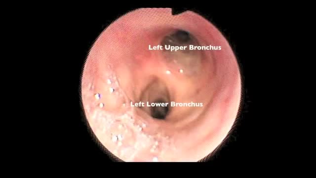
Flexible bronchoscopy is a procedure that allows a clinician to examine the breathing passages (airways) of the lungs (figure 1). Flexible bronchoscopy can be either a diagnostic procedure (to find out more about a possible problem) or a therapeutic procedure (to try to treat an existing problem or condition).
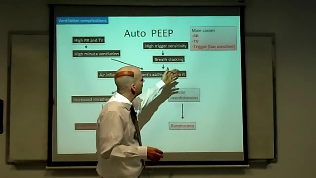
This is an introduction to ventilator settings like FIO2, PEEP, Flow rate,trigger,TV, and RR. I also discuss how these settings relate to CO2 and O2 control and to complications like oxygen toxicity and barotrauma with an emphasis on physiology.
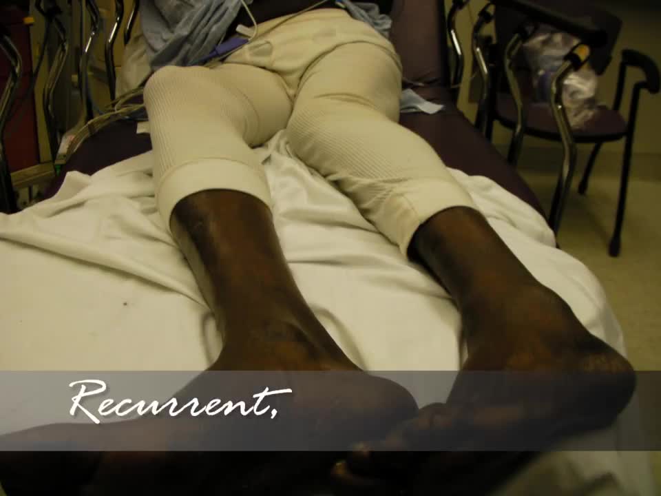
This video demonstrates a technique for reducing a dislocated hip. This patient had recurrent dislocations of his artificial hip.
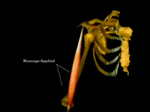
This is an animation of the biomechanics involved in relocating a dislocated shoulder.

Modified Milch method of relocating a dislocated shoulder
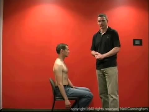
This is a demonstration of the scapula manipulation method of relocating a dislocated shoulder
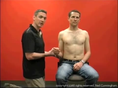
This is a demonstration of the Kocher's method of relocating a dislocated shoulder
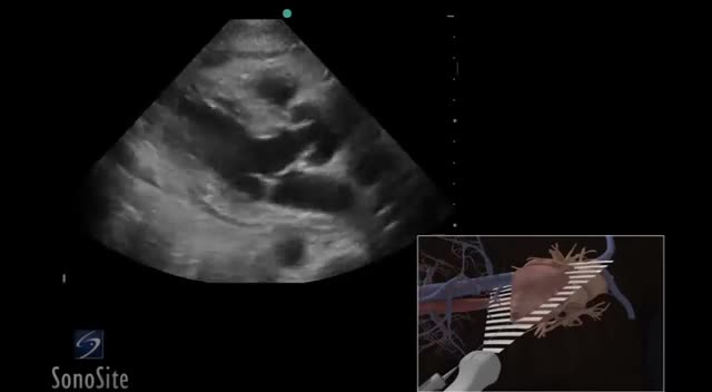
Using 3D animations we have come up with a new way of demonstrating how to perform portable ultrasound examinations
Central venous catheter. Diagram showing a tunneled central line inserted into the right subclavian vein. A central venous catheter (CVC), also known as a central line, central venous line, or central venous access catheter, is a catheter placed into a large vein.
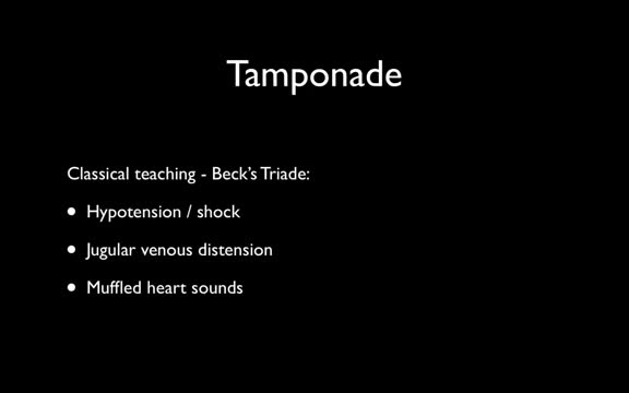
Cardiac tamponade is a medical emergency that requires urgent drainage of the pericardial fluid. Preferably, patients should be monitored in an intensive care unit. All patients should receive the following: Oxygen Volume expansion with blood, plasma, dextran, or isotonic sodium chloride solution, as necessary, to maintain adequate intravascular volume - Sagristà-Sauleda et al noted significant increase in cardiac output after volume expansion [24] (see the Cardiac Output calculator) Bed rest with leg elevation - This may help increase venous return Positive-pressure mechanical ventilation should be avoided because it may decrease venous return and aggravate signs and symptoms of tamponade. Inpatient care After pericardiocentesis, leave the intrapericardial catheter in place after securing it to the skin using sterile procedure and attaching it to a closed drainage system via a 3-way stopcock. Periodically check for reaccumulation of fluid, and drain as needed. The catheter can be left in place for 1-2 days and can be used for pericardiocentesis. Serial fluid cell counts can be useful for helping to discover an impending bacterial catheter infection, which could be catastrophic. If the white blood cell (WBC) count rises significantly, the pericardial catheter must be removed immediately. A Swan-Ganz catheter can be left in place for continuous monitoring of hemodynamics and to assess the effect of reaccumulation of pericardial fluid. A repeat echocardiogram and a repeat chest radiograph should be performed within 24 hours.
Medical Videos
Free medical and surgery videos for patients, students and professionals — trusted since 2007.
Founded by Dr. Mohamed Ibrahim, MD, PhD — physician-scientist.
Follow us on Facebook · Instagram