- Physical Examination
- Surgical Examination
- Ophthalmology
- Clinical Skills
- Orthopedics
- Surgery Videos
- Laparoscopy
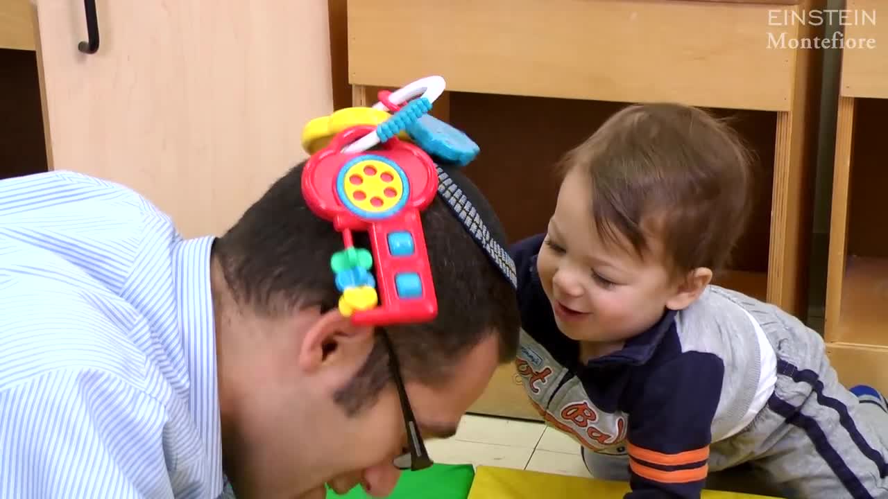
- Pediatrics
- Funny Videos
- Cardiothoracic Surgery
- Nursing Videos
- Plastic Surgery
- Otorhinolaryngology
- Histology and Histopathology
- Neurosurgery
- Dermatology
- Pediatric Surgery
- Urology
- Dentistry
- Oncology and Cancers
- Anatomy Videos
- Health and Fitness
- Radiology
- Anaesthesia
- Physical Therapy
- Pharmacology
- Interventional Radiology
- Cardiology
- Endocrinology
- Gynecology
- Emergency Medicine
- Psychiatry and Psychology
- Childbirth Videos
- General Medical Videos
- Nephrology
- Physiology
- Diet and Food Health
- Diabetes Mellitus
- Neurology
- Women Health
- Osteoporosis
- Gastroenterology
- Pulmonology
- Hematology
- Rheumatology
- Toxicology
- Nuclear Medicine
- Infectious Diseases
- Vascular Disease
- Reproductive Health
- Burns and Wound Healing
- Other
Latest videos
Developmental Milestones: Baby Talk from First Sounds to First Words
the motor milestones expected in typically developing babies, from head control to walking and what pediatricians look for during a well-baby visit. She also explains the specific types of motor control a baby must master before the next milestone can be achieved
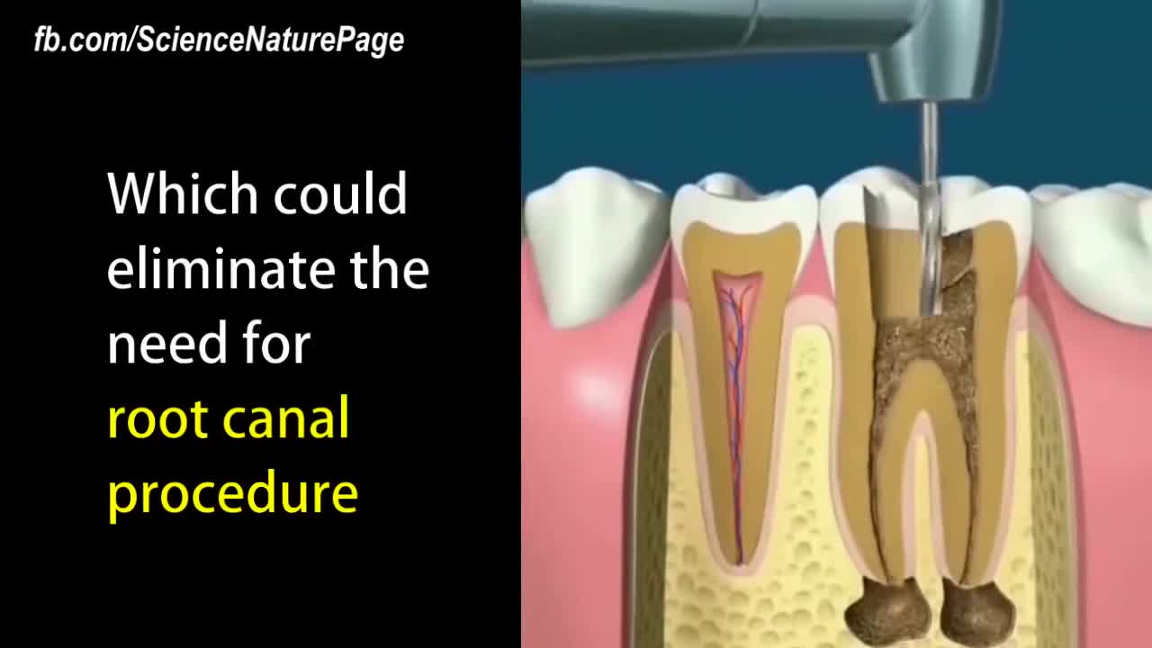
New dental fillings could allow your teeth to heal themselves.
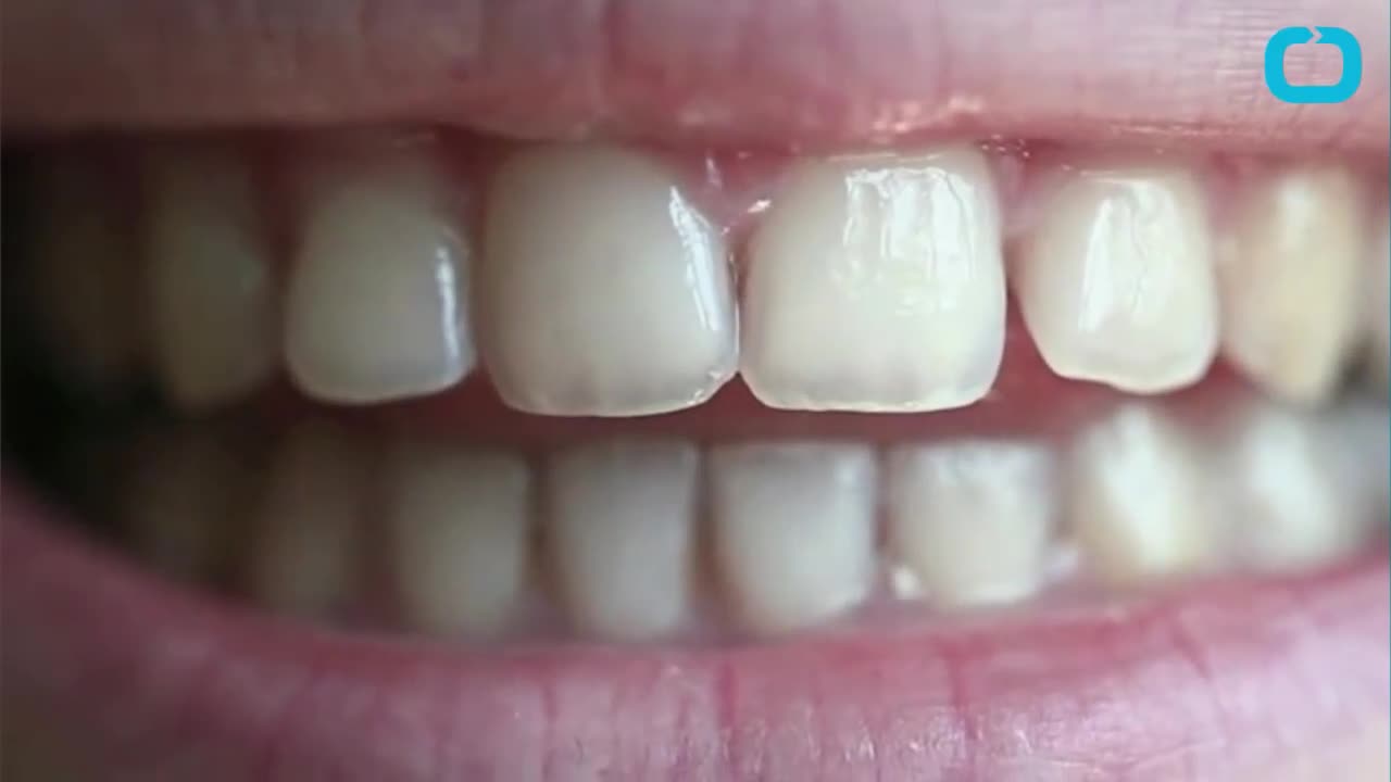
You May Be Able to Repair Cavities Without Getting a Filling
The Distal Femoral Osteotomy System utilizes the same principles of design featured in the Tibial Osteotomy System. Specifically designed femoral osteotomy plates take into account the anatomical differences between the distal femur and proximal tibia.
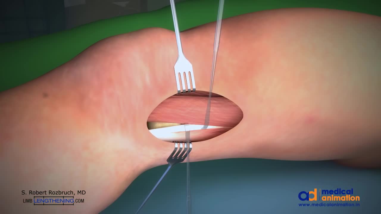
This is a technique of correcting knock knee (genu valgum) deformity by surgery. Highligh of the technique is that the bone is not cut, but merely weakened. The advantage is that it provides accuracy to the surgeon, and rapid healing. Once corrected, the bone is held in place with a special plate (Tomofix), which permits walking with crutches the very next day.
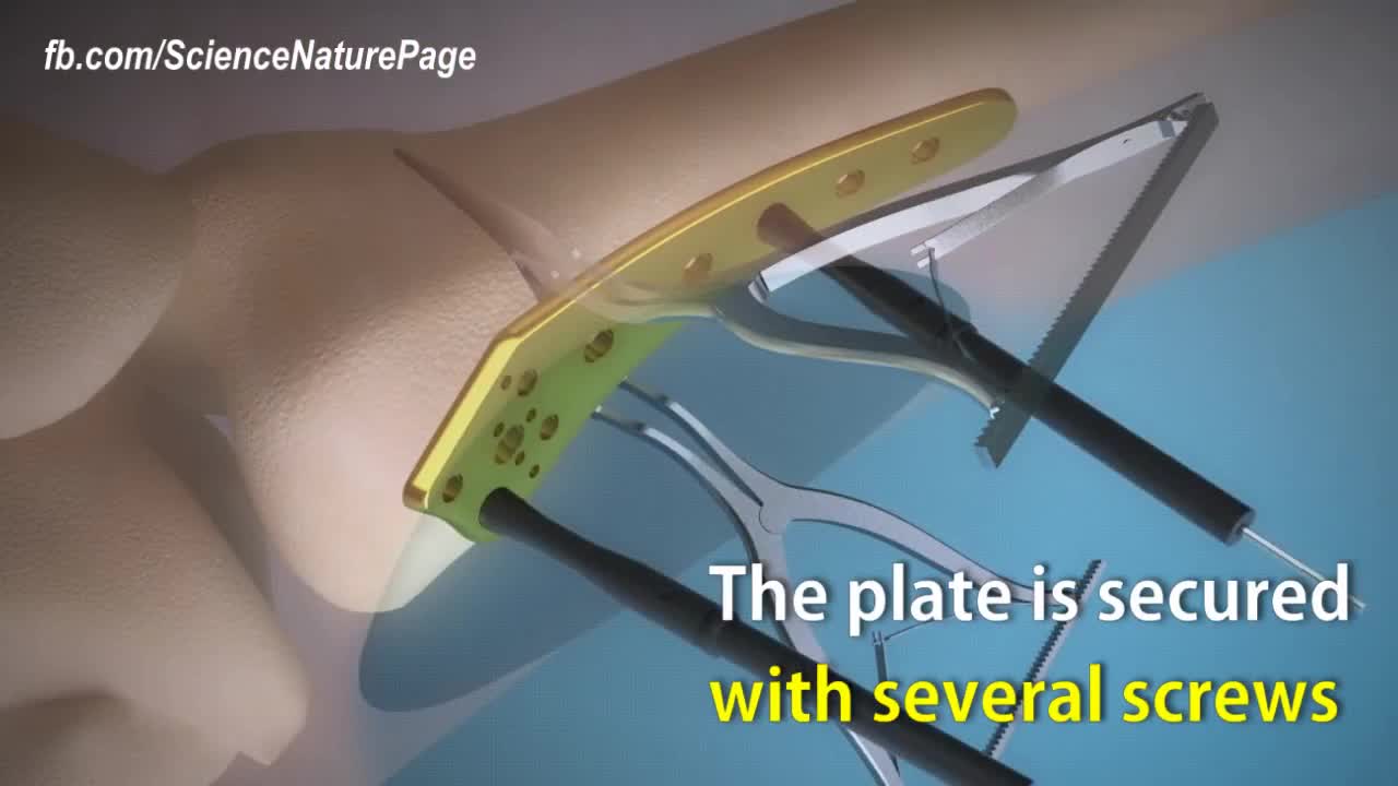
Here is how surgeons perform knock knee correction surgery. Titanium plate is used to stabilize the affected area. The femur is cut nearly through to help with the stability. Spreaders angle the cut align the leg. The plate is secured with several screws. Synthetic bone graft material is packed in the joint. The patient will be in crutches for 4 to 6 weeks.

Helping Babies Born with Drug Addiction. see to learn more
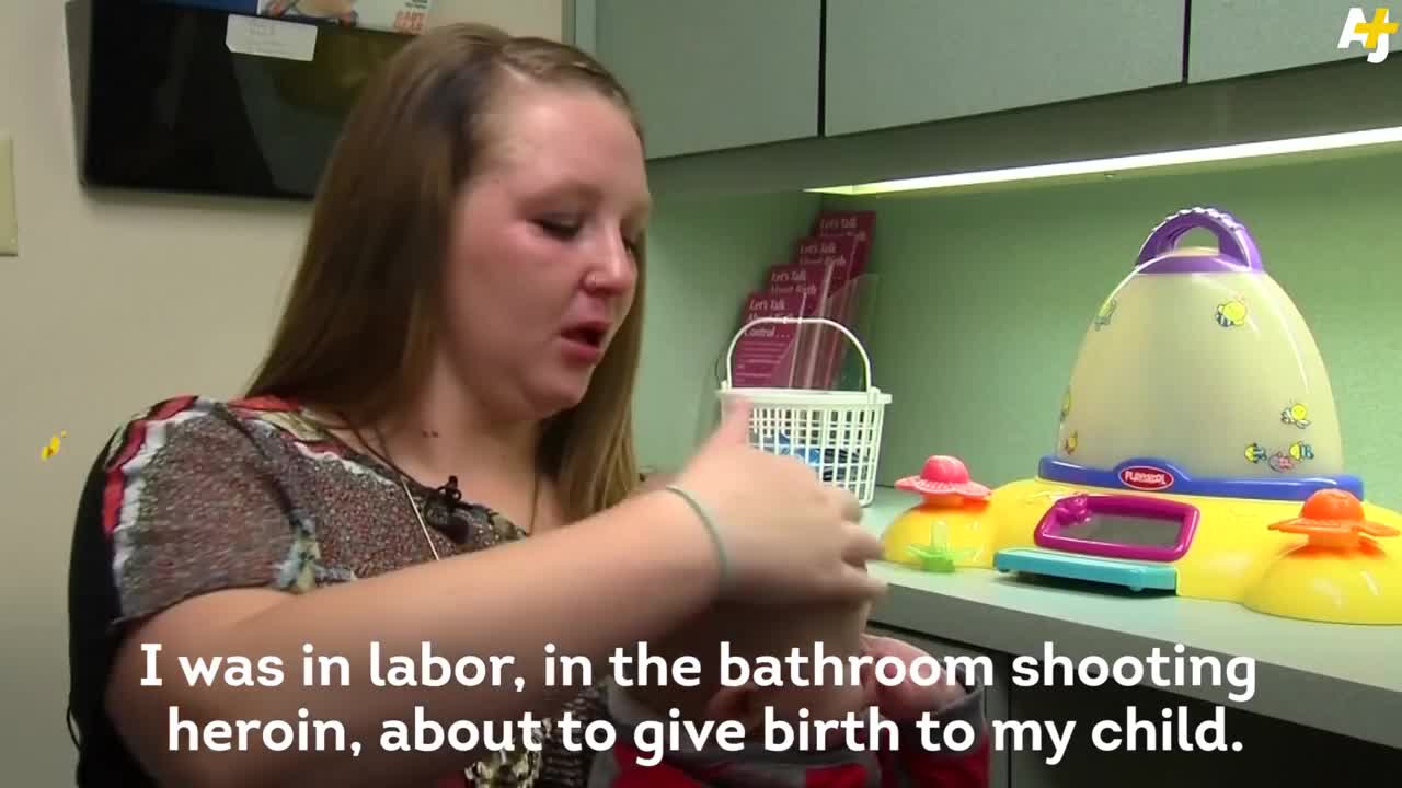
Each year, thousands of babies in the U.S. are born addicted to opiates. And the problem is getting worse.

Would you dare to explore your heritage? WIN a DNA kit and discover just how diverse you really are.
your DNA Journey

When United Airlines decides their employees flying to Kentucky is more important than a doctor or any passenger who paid for their ticket it is time to STOP FLYING UNITED!!! Here are United employees dragging the man off the plane like a criminal.
A bone marrow biopsy removes a small amount of bone and a small amount of fluid and cells from inside the bone (bone marrow). A bone marrow aspiration removes only the marrow. These tests are often done to find the reason for many blood disorders and may be used to find out if cancer or infection has spread to the bone marrow. Bone marrow aspiration removes a small amount of bone marrow fluid and cells through a needle put into a bone. The bone marrow fluid and cells are checked for problems with any of the blood cells made in the bone marrow. Cells can be checked for chromosome problems. Cultures can also be done to look for infection. A bone marrow biopsy removes bone with the marrow inside to look at under a microscope. The aspiration (taking fluid) is usually done first, and then the biopsy.
Stem Cell Injection Treatment - Stem Cell Therapy
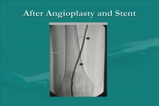
Roman Nowygrod, MD, a surgeon at NewYork-Presbyterian/Columbia University Medical Center, explains the different surgical approaches to treat Peripheral Arterial Disease (PAD).
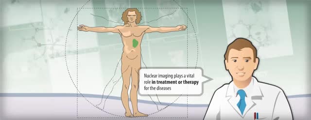
Nuclear medicine is a branch of medical imaging that uses small amounts of radioactive material to diagnose and determine the severity of or treat a variety of diseases, including many types of cancers, heart disease, gastrointestinal, endocrine, neurological disorders and other abnormalities within the body.
Simple interrupted suturing is the most basic and most important of the suturing techniques.
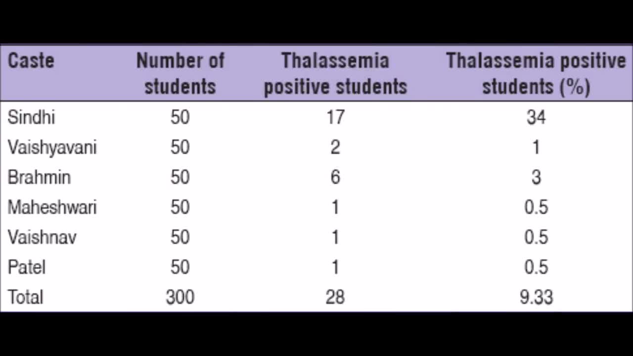
Thalassemia is a genetic blood disorder. People with Thalassemia disease are not able to make enough hemoglobin, which causes severe anemia. Hemoglobin is found in red blood cells and carries oxygen to all parts of the body. When there is not enough hemoglobin in the red blood cells, oxygen cannot get to all parts of the body. Organs then become starved for oxygen and are unable to function properly.
Sickle cell anemia (sickle cell disease) is a disorder of the blood caused by an inherited abnormal hemoglobin (the oxygen-carrying protein within the red blood cells). The abnormal hemoglobin causes distorted (sickled) red blood cells.

Pernicious anemia Email this page to a friend Print Facebook Twitter Google+ Anemia is a condition in which the body does not have enough healthy red blood cells. Red blood cells provide oxygen to body tissues. There are many types of anemia. Pernicious anemia is a decrease in red blood cells that occurs when the intestines cannot properly absorb vitamin B12.
