- Physical Examination
- Surgical Examination
- Ophthalmology
- Clinical Skills
- Orthopedics
- Surgery Videos
- Laparoscopy
- Pediatrics
- Funny Videos
- Cardiothoracic Surgery
- Nursing Videos
- Plastic Surgery
- Otorhinolaryngology
- Histology and Histopathology
- Neurosurgery
- Dermatology
- Pediatric Surgery
- Urology
- Dentistry
- Oncology and Cancers
- Anatomy Videos
- Health and Fitness
- Radiology
- Anaesthesia
- Physical Therapy
- Pharmacology
- Interventional Radiology
- Cardiology
- Endocrinology
- Gynecology
- Emergency Medicine
- Psychiatry and Psychology
- Childbirth Videos
- General Medical Videos
- Nephrology
- Physiology
- Diet and Food Health
- Diabetes Mellitus
- Neurology
- Women Health
- Osteoporosis
- Gastroenterology
- Pulmonology
- Hematology
- Rheumatology
- Toxicology
- Nuclear Medicine
- Infectious Diseases
- Vascular Disease
- Reproductive Health
- Burns and Wound Healing
- Other
Top videos

Iodine For Ringworm, Best Ointment For Ringworm, Where Do You Get Ringworm, How To Treat Ring Worms ---- http://ringworm-cure.plus101.com --- Ringworms, contrary to the common notion, do not come from worms. Tinea, which is the medical term for ringworms, is a fungal infection seen on the skin's surface. Knowing how to cure ringworm is important because ringworms can be highly contagious. It can be contracted from direct contact with the host (person or animal) as well as by other means such as having contact with the host's clothes. Swimming pools can also be a place where ringworms are transmitted from one person to another. How To Cure Ringworm - Understanding Aspects and Options Different means on how to cure ringworm are available and they sometimes vary in accordance with where the ringworm is located (it can appear in areas like the nails, fingers, toes, feet, scalp, stomach, chest, thighs, and scalp), and the particular type of ringworm. • Ringworms found in the scalp are usually treated with an antifungal shampoo to keep the area dry and clean. • Ringworms found in the feet can be treated through the application of ointments. • Oral medications can also be taken in especially when ringworms are on the nails. • Sprays, powders and creams are also forms by which anti-fungal drugs are bought. These medicines may take some time to work. The infection may persist for a few weeks to several months, depending on the severity and how the body responds to the medications. How To Cure Ringworm - OTC and Prescription Medications Ringworm appears on the skin's surface as an itchy, red, circular patch. As it progresses, it expands and smaller round patches can develop. It is important to immediately identify ringworms and know how to treat them properly. There are many over the counter topical creams (anti-fungal ones) and ointment that can be bought in the market. However, some people prefer to visit the doctor and ask for a prescription. Stronger formulations are generally available via prescriptions. William Oliver is a nutritionist, medical researcher, and author of the Fast Ringworm Cure e-book. To find out how to cure Ringworm in 3 days or less, click below: http://ringworm-cure.plus101.com
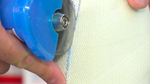
Split Skin Graft
How to Prepare, Apply & Remove a Total Contact Cast
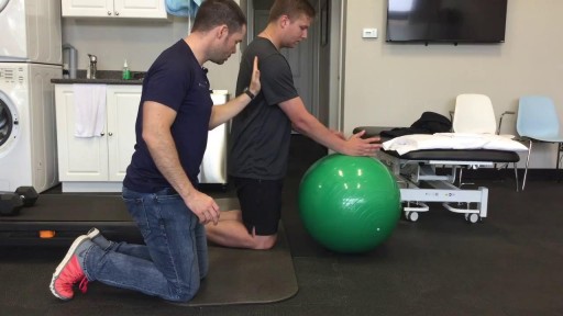
This is the first video of 5, where Mike teamed up with Graham from On Your Marks Fitness and Coaching to show us some exercises to strengthen our muscles, and improve our soccer game! Make sure your feet are planted safely or held by a friend, and keep your back straight, and over your knees. Use the swiss ball to keep you steady, and SQUEEZE those muscles! Check us out on Social Media! Facebook: https://www.facebook.com/striveptandperformance/ Instagram: https://www.instagram.com/striveptandperf/ Twitter: https://twitter.com/StrivePTandPerf Blog: http://www.strivept.ca/blog
The procedure is used most often to treat a condition called supraventricular tachycardia, or SVT, which occurs because of abnormal conduction fibers in the heart. Catheter ablation is also used to help control other heart rhythm problems such as atrial flutter and atrial fibrillation.

How to Imporve Sexual Health or Stamina Part 2 https://youtu.be/S17bCnwCLuI Dr. Aslam Naveed is a well known sexologist in Pakistan. He has treated more than 1 Lac patients since last 30 years of clinical Practice in sexology, he knows how to help the people facing sexual disorders. Contact: 021-34595050, 03432821919 sexologistpakistan.com facebook.com/menssexcareclinic/ Address: Men's Care Clinic, 2nd floor, The Modern Hospital Opposite Safari Park, University Road. Karachi.

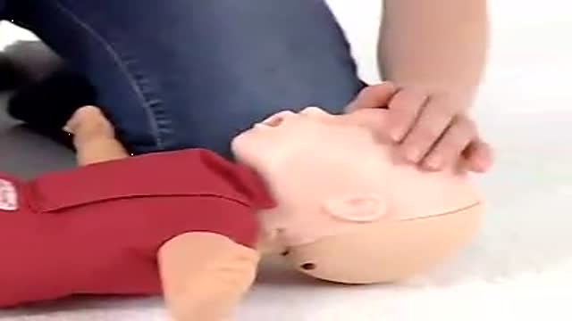
Baby CPR
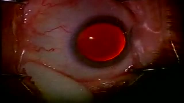
Unedited Cataract Surgery 3
ROTIGS medical device by Honolulu inventor Dr. Brad NaPier makes airway intubations easier for medical professionals. For more info, visit www.rotigs.com

More videos on my youtube channel
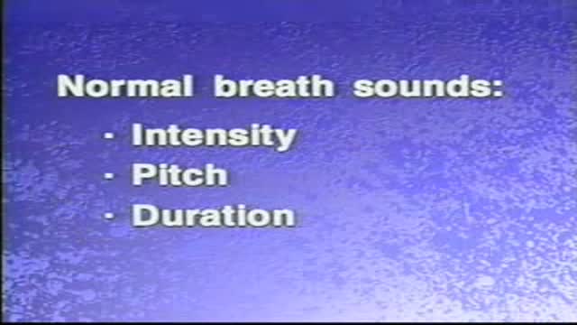
Normal and Adventitious Breath Sounds
The cornea occupies the front center part of the outer wall of the eye. It is made of collagen fibers in a very special arrangement so that the cornea is clear. One looks through the cornea to see the iris and pupil. The cornea bends light coming into the eye so that it is focused on the retina.
Cholesterol is a fat-like, waxy substance that can be found in all parts of your body. It helps your body make cell membranes, many hormones, and vitamin D. The cholesterol in your blood comes from two sources: the foods you eat and your liver. But your liver makes all the cholesterol your body needs.

The timing of the nausea or vomiting can indicate the cause. When appearing shortly after a meal, nausea or vomiting may be caused by food poisoning, gastritis (inflammation of the stomach lining), an ulcer, or bulimia. Nausea or vomiting one to eight hours after a meal may also indicate food poisoning.
Iatrogenic injury to the ureter is a potentially devastating complication of modern surgery. The ureters are most often injured in gynecologic, colorectal, and vascular pelvic surgery. There is also potential for considerable ureteral injury during endoscopic procedures for ureteric pathology such as tumor or lithiasis. While maneuvers such as perioperative stenting have been touted as a means to avoid ureteral injury, these techniques have not been adopted universally, and the available literature does not make a case for their routine use. Distal ureteral injuries are best managed with ureteroneocystostomy with or without a vesico-psoas hitch. Mid-ureteral and proximal ureteral injuries can potentially be managed with ureteroureterostomy. If the distal segment is unsuitable for anastomosis then a number of techniques are available for repair including a Boari tubularized bladder flap, transureteroureterostomy, or renal autotransplantation. In rare cases renal autotransplantation or ureteral substitution with gastrointestinal segments may be warranted to re-establish urinary tract continuity. Laparoscopic and minimally invasive techniques have been employed to remedy iatrogenic ureteral injuries.
