



Massive Pulmonary Embolus!

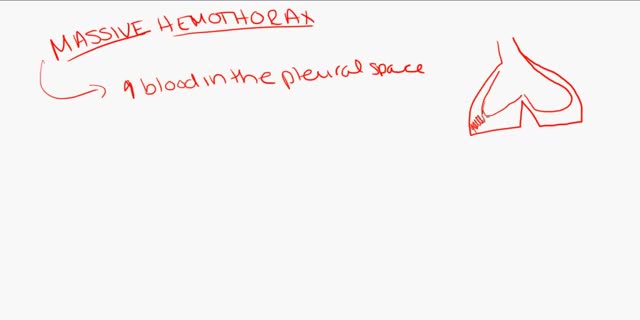
Massive PE causing hemodynamic instability (shock and/or low blood pressure, defined as a systolic blood pressure <90 mmHg or a pressure drop of 40 mmHg for >15 min if not caused by new-onset arrhythmia, hypovolemia or sepsis) is an indication for thrombolysis, the enzymatic destruction of the clot with medication.
Video Transcript
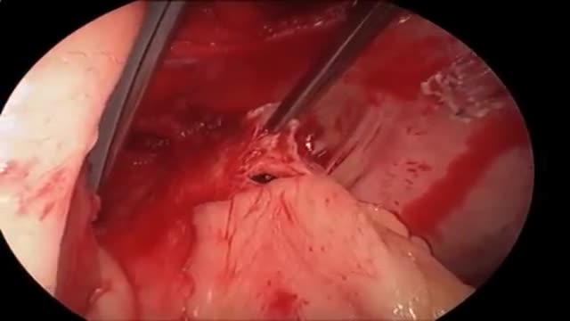
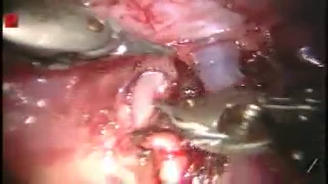
A cardiac surgeon narrates a pulmonary thromboembolectomy for a massive saddle embolus. On cardiopulmonary bypass he opens the pulmonary artery and extracts the large clot — traced to a fractured leg that had been in a cast — clears the branches with a catheter, then flushes the lungs backwards through the pulmonary veins to wash out small emboli and air before a final inspection. He reports that the patient did well.
Read the full transcript
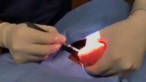
This is a case of an acute pulmonary embolism from a deep vein thrombosis. Here we're performing pulmonary thromboembolectomy using retrograde pulmonary vein perfusion. Here we've established the patient on cardiopulmonary bypass. A arterial line is placed in the right superior pulmonary vein as seen here. This is wide off of the arterial cannula and clamped. As you can see the clamp is in place. We are on full cardiopulmonary bypass support. The next step is to apply the order cross clamp and achieve cardiac arrest. Here we're performing the pulmonary arterialomy with a knife and then scissors and you can already get a glimpse of the
thrombus inside the main pulmonary artery as it branches. You can see a grasp is used to remove the thrombus. This is a very large saddle embolus that originated likely from the patient's femoral and iliac vein. She had a fracture of her right lower extremity. It was in a cast. This was likely sourced from the right leg. You can see the embolus is removed. A pulmonary catheter is used to clear any further clots which none are found. Then at this point in time we take the clamp off of the
right superior pulmonary vein cannula. This allows us to perform retrograde pulmonary perfusion. It goes from the pulmonary veins back through the lungs and out the pulmonary artery as you can see here. Small emboli and air are cleared. You can see
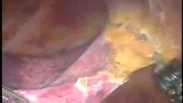
in this portion of the video. Once we're satisfied that the pulmonary arteries have been cleared of debris and we ventilate as well during this process. Once we're satisfied with that, we'll stop the retrograde pulmonary vein perfusion. We'll inspect with the thoracoscope. You can see here the small branches of the pulmonary veins are cleared. The main trunk of the pulmonary artery is also clear and we inspect the thrombus one more time. You can see this is a very well formed thrombus that is soft.
And that ends the case. The patient has done very well following the surgery and we anticipate a full recovery.
Transcript auto-generated from the video’s audio for accessibility and study. Educational content only — not medical advice.
Video Overview
From our General Medical Videos collection: General medical videos: a wide collection of procedures, conditions and explainers from every specialty — free medical education since 2007. Browse all General Medical Videos videos →
SORT BY-
Toppkommentarer
-
Senaste kommentarerna