










Transesophageal Echocardiogram of a Giant Thrombus in the Left Ventricle

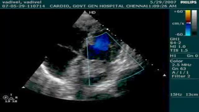
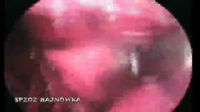
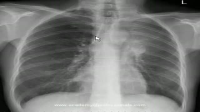
TEE of a giant LV thrombus as well as other, smaller thrombi in a 51 year-old man who came to the hospital complaining of chest pain. He was subsequently taken to the cardiac cath lab and was found to have clean coronary arteries. Surprisingly, he was clinically stable despite his TEE findings and an EF of 20%.
This video in 30 seconds
This video shows a transesophageal echocardiogram of a giant thrombus in the left ventricle, along with other smaller thrombi, in a 51 year old man who came to hospital with chest pain. The description notes he was found to have clean coronary arteries in the cardiac cath lab and was clinically stable despite an ejection fraction of 20 percent.
Key points
- Shows transesophageal echocardiogram imaging of a left ventricular thrombus
- Presents a 51 year old man with chest pain
- Notes clean coronary arteries and an ejection fraction of 20 percent in the case
Video Overview
This video presents “Transesophageal Echocardiogram of a Giant Thrombus in the Left Ventricle” — TEE of a giant LV thrombus as well as other, smaller thrombi in a 51 year-old man who came to the hospital complaining of chest pain. He was subsequently taken to the cardiac cath lab and was found to have clean coronary arteries. Whether you are preparing for a procedure, studying the technique, or learning how a condition is assessed, the recording shows the process as it is performed in practice.
From our Cardiology collection: ECG interpretation, echocardiograms, angioplasty, stents and heart conditions explained by cardiologists. Browse all Cardiology videos →
Educational content only — not a substitute for professional medical advice.
SORT BY-
Mga Nangungunang Komento
-
Pinakabagong komento