- Physical Examination
- Surgical Examination
- Ophthalmology
- Clinical Skills
- Orthopedics
- Surgery Videos
- Laparoscopy
- Pediatrics
- Funny Videos
- Cardiothoracic Surgery
- Nursing Videos
- Plastic Surgery
- Otorhinolaryngology
- Histology and Histopathology
- Neurosurgery
- Dermatology
- Pediatric Surgery
- Urology
- Dentistry
- Oncology and Cancers
- Anatomy Videos
- Health and Fitness
- Radiology
- Anaesthesia
- Physical Therapy
- Pharmacology
- Interventional Radiology
- Cardiology
- Endocrinology
- Gynecology
- Emergency Medicine
- Psychiatry and Psychology
- Childbirth Videos
- General Medical Videos
- Nephrology
- Physiology
- Diet and Food Health
- Diabetes Mellitus
- Neurology
- Women Health
- Osteoporosis
- Gastroenterology
- Pulmonology
- Hematology
- Rheumatology
- Toxicology
- Nuclear Medicine
- Infectious Diseases
- Vascular Disease
- Reproductive Health
- Burns and Wound Healing
- Other
Latest videos

Alcohol not broken down by the liver goes to the rest of the body, including the brain. Alcohol can affect parts of the brain that control movement, speech, judgment, and memory. These effects lead to the familiar signs of drunkenness: difficulty walking, slurred speech, memory lapses, and impulsive behavior.
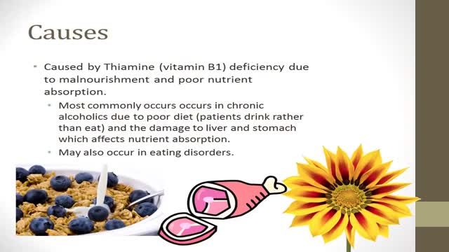
-Korsakoff's syndrome is a common and preventable sequel of Wernicke's encephalopathy. Thiamine, if given during the stage of Wernicke's encephalopathy, can prevent the onset of Korsakoff's psychosis. The administration of glucose prior to thiamine can precipitate Korsakoff's syndrome, as seen in this case. In such patients, brain MRI frequently shows abnormal enhancement of the mammillary bodies & thallamus
Thrombosis of the venous channels in the brain is an uncommon cause of cerebral infarction relative to arterial disease, but it is an important consideration because of its potential morbidity. (See Prognosis.) Knowledge of the anatomy of the venous system is essential in evaluating patients with cerebral venous thrombosis (CVT), since symptoms associated with the condition are related to the area of thrombosis. For example, cerebral infarction may occur with cortical vein or sagittal sinus thrombosis secondary to tissue congestion with obstruction. (See Presentation.) Lateral sinus thrombosis may be associated with headache and a pseudotumor cerebri–like picture. Extension into the jugular bulb may cause jugular foramen syndrome, while cranial nerve palsies may be seen in cavernous sinus thrombosis as a compressive phenomenon. Cerebral hemorrhage also may be a presenting feature in patients with venous sinus thrombosis. (See Presentation.) Imaging procedures have led to easier recognition of venous sinus thrombosis (see the images below), offering the opportunity for early therapeutic measures. (See Workup.) Left lateral sinus thrombosis demonstrated on magn Left lateral sinus thrombosis demonstrated on magnetic resonance venography (MRV). This 42-year-old woman presented with sudden onset of headache. Physical examination revealed no neurologic abnormalities. View Media Gallery Axial view of magnetic resonance (MR) venogram dem Axial view of magnetic resonance (MR) venogram demonstrating lack of flow in transverse sinus. View Media Gallery The following guidelines for CVT have been provided by the American Heart Association and the American Stroke Association [1] : In patients with suspected CVT, routine blood studies consisting of a complete blood count, chemistry panel, prothrombin time, and activated partial thromboplastin time should be performed. Screening for potential prothrombotic conditions that may predispose a person to CVT (eg, use of contraceptives, underlying inflammatory disease, infectious process) is recommended in the initial clinical assessment. Testing for prothrombotic conditions (including protein C, protein S, or antithrombin deficiency), antiphospholipid syndrome, prothrombin G20210A mutation, and factor V Leiden can be beneficial for the management of patients with CVT. Testing for protein C, protein S, and antithrombin deficiency is generally indicated 2-4 weeks after completion of anticoagulation. There is a very limited value of testing in the acute setting or in patients taking warfarin. In patients with provoked CVT (associated with a transient risk factor), vitamin K antagonists may be continued for 3-6 months, with a target international normalized ratio of 2.0-3.0. In patients with unprovoked CVT, vitamin K antagonists may be continued for 6-12 months, with a target international normalized ratio of 2.0-3.0. For patients with recurrent CVT, venous thromboembolism (VTE) after CVT, or first CVT with severe thrombophilia (ie, homozygous prothrombin G20210A; homozygous factor V Leiden; deficiencies of protein C, protein S, or antithrombin; combined thrombophilia defects; or antiphospholipid syndrome), indefinite anticoagulation may be considered, with a target international normalized ratio of 2.0-3.0. For women with CVT during pregnancy, low-molecular-weight heparin (LMWH) in full anticoagulant doses should be continued throughout pregnancy, and LMWH or vitamin K antagonist with a target international normalized ratio of 2.0-3.0 should be continued for ≥6 weeks postpartum (for a total minimum duration of therapy of 6 months). It is reasonable to advise women with a history of CVT that future pregnancy is not contraindicated. Further investigations regarding the underlying cause and a formal consultation with a hematologist or maternal fetal medicine specialist are reasonable. It is reasonable to treat acute CVT during pregnancy with full-dose LMWH rather than unfractionated heparin. For women with a history of CVT, prophylaxis with LMWH during future pregnancies and the postpartum period is reasonable. Next: Etiology What to Read Next on Medscape Related Conditions and Diseases Quiz: Do You Know the Complications, Proper Workup, and Best Treatment Practices for Ischemic Stroke? Quiz: How Much Do You Know About Hypothyroidism? Quiz: Do You Know the Risk Factors, Symptoms, and Potential Treatments for Alzheimer Disease? Quiz: How Much Do You Know About Hypertension? Quiz: Test Your Knowledge of Epilepsy and Seizure-related Conditions A 25-Year-Old Man With Painless Diplopia NEWS & PERSPECTIVE Temporal Trends and Factors Associated With Diabetes Mellitus Among Patients Hospitalized With Heart Failure Watchful Waiting Tied to Worse Outcomes in LVAD Patients With Hemolysis Age of Transfused Blood Impacts Perioperative Outcomes Among Patients Who Undergo Major Gastrointestinal Surgery TOOLS Drug Interaction Checker Pill Identifier Calculators Formulary SLIDESHOW Chronic Alcohol Abuse: Complications and Consequences Most Popular Articles According to Neurologists DHA Supplements Linked to Less Progression to Alzheimer's in APOE4 Carriers Heading in Soccer Linked to CNS Symptoms 'Transient Smartphone Blindness' Misdiagnosed as Multiple Sclerosis? New Advances in Traumatic Brain Injury FDA Clears Deflazacort (Emflaza) for DMD View More Overview Background
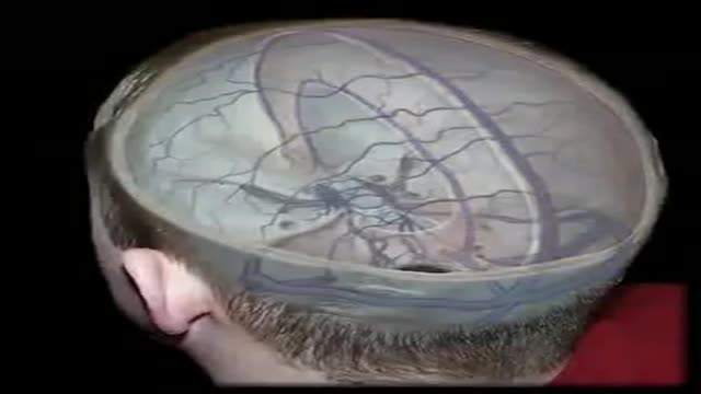
The dural venous sinuses are spaces between the endosteal and meningeal layers of the dura. They contain venous blood that originates for the most part from the brain or cranial cavity. The sinuses contain an endothelial lining that is continuous into the veins that are connected to them.

The superior sagittal sinus (also known as the superior longitudinal sinus), within the human head, is an unpaired area along the attached margin of falx cerebri. It allows blood to drain from the lateral aspects of anterior cerebral hemispheres to the confluence of sinuses.
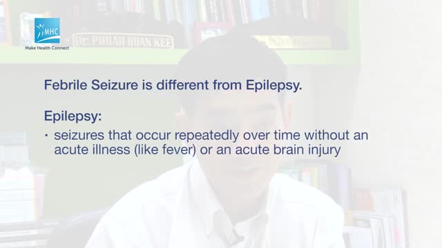
A febrile seizure is a convulsion in a child that may be caused by a spike in body temperature, often from an infection. Your child's having a febrile seizure can be alarming, and the few minutes it lasts can seem like an eternity. Febrile seizures represent a unique response of a child's brain to fever, usually the first day of a fever. Fortunately, they're usually harmless and typically don't indicate an ongoing problem. You can help by keeping your child safe during a febrile seizure and by comforting him or her afterward.

Pediatric febrile seizures, which represent the most common childhood seizure disorder, exist only in association with an elevated temperature. Evidence suggests, however, that they have little connection with cognitive function, so the prognosis for normal neurologic function is excellent in children with febrile seizures. [1] Epidemiologic studies have led to the division of febrile seizures into 3 groups, as follows: Simple febrile seizures Complex febrile seizures Symptomatic febrile seizures Essential update: Starting MMR/MMRV vaccination earlier may reduce seizure risk In a case-series analysis of a cohort of 323,247 US children born from 2004 to 2008, Hambidge et al found that delaying the first dose of measles-mumps-rubella (MMR) or measles-mumps-rubella-varicella (MMRV) vaccine beyond the age of 15 months may more than double the risk of postvaccination seizures in the second year of life. [2, 3] In infants, there was no association between vaccination timing and postvaccination seizures. [3] In the second year of life, however, the incident rate ratio (IRR) for seizures within 7-10 days was 2.65 (95% confidence interval [CI], 1.99-3.55) after first MMR doses at 12-15 months of age, compared with 6.53 (95% CI, 3.15-13.53) after first MMR doses at 16-23 months. For the MMRV vaccine, the IRR for seizures was 4.95 (95% CI, 3.68-6.66) after first doses at 12-15 months, compared with 9.80 (95% CI, 4.35-22.06) for first doses at 16-23 months.
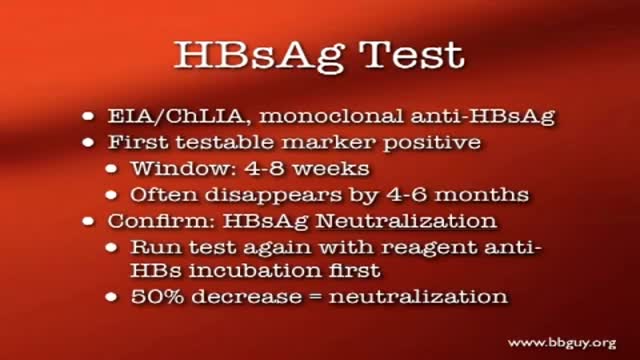
A blood transfusion is a routine medical procedure that can be lifesaving. During a blood transfusion, donated blood is added to your own blood. A blood transfusion may also be done to supplement various components of your blood with donated blood products. In some cases, a blood transfusion is done with blood that you've donated ahead of time before you undergo elective surgery. During a typical blood transfusion, certain parts of blood are delivered through an intravenous (IV) line that's placed in one of the veins in your arm. A blood transfusion usually takes one to four hours, though in an emergency it can be done much faster.
Blood Transfusion-Transmitted Diseases
A hemolytic transfusion reaction is a serious complication that can occur after a transfusion of blood. The red blood cells that were given in the transfusion are destroyed by the patient's immune system. There are other types of allergic transfusion reactions that do not cause hemolysis.

blood transfusion performance

Heparin is an anticoagulant (blood thinner) that prevents the formation of blood clots. Heparin is used to treat and prevent blood clots in the veins, arteries, or lung. It is also used before surgery to reduce the risk of blood clots.

Warfarin is an anticoagulant medication - it is used to slow down the blood-clotting process. Anticoagulants are used to prevent blood clots which may cause vein blockages, heart attack and stroke. Warfarin is known under the brand names Warfant, Jantoven, Coumadin, Lawarin, Marevan, and Waran.
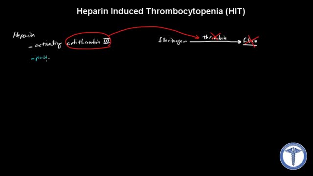
All forms of heparin (including low-molecular-weight heparin such as enoxaparin) must be stopped immediately in patients with suspected heparin-induced thrombocytopenia (HIT) while awaiting diagnostic confirmation. Patients with HIT remain at high risk of thrombosis even after discontinuation of heparin. Therefore, an alternate, rapidly acting, non-heparin anticoagulant such as direct thrombin inhibitor (eg, argatroban, bivalirudin) must be started immediately.
How to Reverse GERD and Leaky Gut
Many children receive MRIs at the hospital, and it can often be a scary experience if they are unprepared or don't know what to expect.

See the effects of cannabis first hand, unedited, on Parkinson's tremor dyskinesia, and voice.

Both selegiline and rasagiline can improve the symptoms of Parkinson's disease, although their effects are small compared with levodopa. They can be used alongside levodopa or dopamine agonists. MAO-B inhibitors are generally very well tolerated, but can occasionally cause side effects, including: nausea.

Experts do not know the exact cause of Zollinger-Ellison syndrome. About 25 to 30 percent of gastrinomas are caused by an inherited genetic disorder called multiple endocrine neoplasia type 1 (MEN1). MEN1 causes hormone-releasing tumors in the endocrine glands and the duodenum.
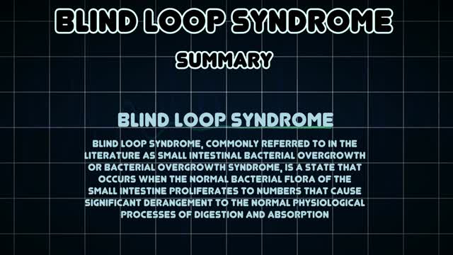
Blind loop syndrome (BLS), commonly referred to in the literature as small intestinal bacterial overgrowth (SIBO) or bacterial overgrowth syndrome (BOS), is a state that occurs when the normal bacterial flora of the small intestine proliferates to numbers that cause significant derangement to the normal physiological ...
