- Physical Examination
- Surgical Examination
- Ophthalmology
- Clinical Skills
- Orthopedics
- Surgery Videos
- Laparoscopy
- Pediatrics
- Funny Videos
- Cardiothoracic Surgery
- Nursing Videos
- Plastic Surgery
- Otorhinolaryngology
- Histology and Histopathology
- Neurosurgery
- Dermatology
- Pediatric Surgery
- Urology
- Dentistry
- Oncology and Cancers
- Anatomy Videos
- Health and Fitness
- Radiology
- Anaesthesia
- Physical Therapy
- Pharmacology
- Interventional Radiology
- Cardiology
- Endocrinology
- Gynecology
- Emergency Medicine
- Psychiatry and Psychology
- Childbirth Videos
- General Medical Videos
- Nephrology
- Physiology
- Diet and Food Health
- Diabetes Mellitus
- Neurology
- Women Health
- Osteoporosis
- Gastroenterology
- Pulmonology
- Hematology
- Rheumatology
- Toxicology
- Nuclear Medicine
- Infectious Diseases
- Vascular Disease
- Reproductive Health
- Burns and Wound Healing
- Other
Latest videos
When is endoscopy used? Endoscopes were first developed to look at parts of the body that couldn’t be seen any other way. This is still a common reason to use them, but endoscopy now has many other uses too. It’s often used in the prevention, early detection, diagnosis, staging, and treatment of cancer. To prevent and screen for cancer Some types of endoscopes are used to look for cancer in people who have no symptoms. For example, colonoscopy (KO-lun-AH-skuh-pee) and sigmoidoscopy (SIG-moid-AH-skuh-pee) are used to screen for colon and rectal cancer. These procedures can also help prevent cancer because they let doctors find and remove polyps (growths) that might become cancer if left alone. To find cancer early Endoscopy can sometimes be used to find cancer early, before it has had a chance to grow or spread. Looking for causes of symptoms When people go to the doctor with certain symptoms, endoscopy can sometimes be used to help find a cause. For instance: Laryngoscopy to look at the vocal cords in people with long-term hoarseness Upper endoscopy in people having trouble swallowing Colonoscopy in people with anemia (low red blood cell counts) with an unknown cause Colonoscopy in people with blood in their stool Looking at problems found on imaging tests Imaging tests such as x-rays and CT scans can sometimes show physical changes within the body. But these tests may only give information about the size, shape, and location of the problem. Doctors use endoscopes to see more details, like color and surface texture, when trying to find out what’s going on. Newer methods of endoscopy that include high magnification are being tested to find out whether they are more useful in detecting cancer and other abnormal cells on the inner surfaces of the body. To diagnose and find out the stage (extent) of cancer To get a tissue sample Going one step further, most types of endoscopes have tools on the end that the doctor can use to take out small tissue samples. This procedure is called a biopsy (BY-op-see). Samples can be taken from suspicious areas and then looked at under a microscope or tested in other ways to see if cancer is there. A biopsy is usually the best way to find out if a growth or change is cancer or something else. Getting a closer look In some cases endoscopes are used to help find out how far a cancer has spread. Thoracoscopy (THOR-uh-KAHS -kuh-pee) and laparoscopy (LAP-uh-RAHS-kuh-pee) can be very useful in finding out if cancer has spread into the thorax (chest) or abdomen (belly). The surgeon can look into these places making only a small incision (cut) in the skin.
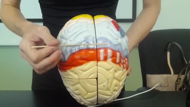
The brain is that part of the CNS contained within the cranial cavity (figure 13.1). It is the control center for many of the body's functions. The brain is much like a complex central computer but with additional functions that no computer can as yet match. Indeed, one goal in computer technology is to make computers that can function more like the human brain. The brain consists of the brainstem, the cerebellum, the diencephalon, and the cerebrum (table 13.1). The brainstem includes the medulla oblongata, pons, midbrain, and reticular formation. The structure of the brain is described in this chapter. Its functions are primarily discussed in chapter 14. Twelve pairs of cranial nerves, which are part of the PNS, arise directly from the brain. Two pairs arise from the cerebrum, nine pairs arise from the brainstem, and one pair arises from the spinal cord.
Each year in the United States, about 400 children and teens younger than age 20 are diagnosed. Osteosarcoma is the third most common cancer in teens, after lymphomas and brain tumors. It is extremely rare in children before age 5.
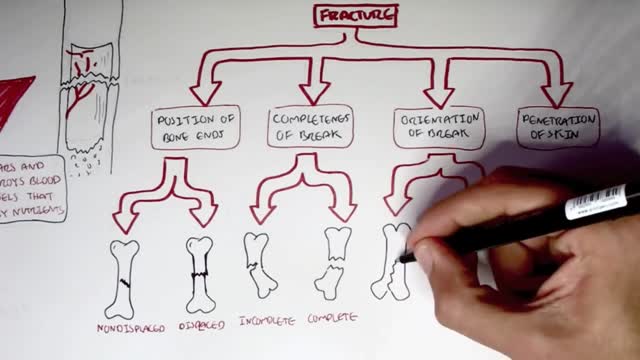
Common types of fractures include: Stable fracture. The broken ends of the bone line up and are barely out of place. Open, compound fracture. The skin may be pierced by the bone or by a blow that breaks the skin at the time of the fracture. ... Transverse fracture. ... Oblique fracture. ... Comminuted fracture.
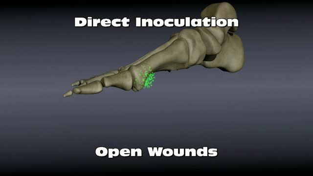
Osteomyelitis is an infection in a bone. Infections can reach a bone by traveling through the bloodstream or spreading from nearby tissue. Infections can also begin in the bone itself if an injury exposes the bone to germs. In children, osteomyelitis most commonly affects the long bones of the legs and upper arms. Adults are more likely to develop osteomyelitis in the bones that make up the spine (vertebrae). People who have diabetes may develop osteomyelitis in their feet if they have foot ulcers. Once considered an incurable condition, osteomyelitis can be successfully treated today. Most people require surgery to remove parts of the bone that have died — followed by strong antibiotics, often delivered intravenously, typically for at least four to six weeks.
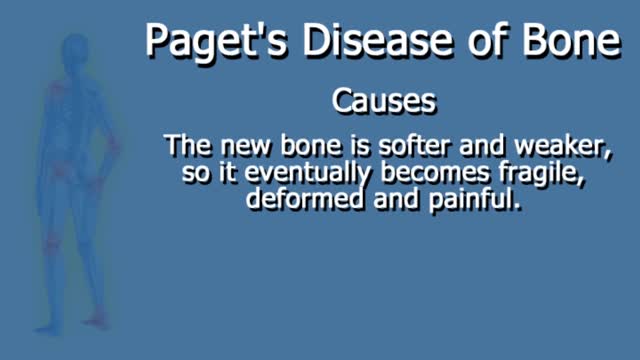
Paget's disease of bone disrupts your body's normal bone recycling process, in which old bone tissue is gradually replaced with new bone tissue. Over time, the affected bones may become fragile and misshapen. Paget's disease of bone most commonly occurs in the pelvis, skull, spine and legs.
Paget's disease of the breast or Paget disease of the breast (/ˈpædʒᵻt/, rhymes with "gadget") (also known as Paget's disease of the nipple) is a malignant condition that outwardly may have the appearance of eczema, with skin changes involving the nipple of the breast.
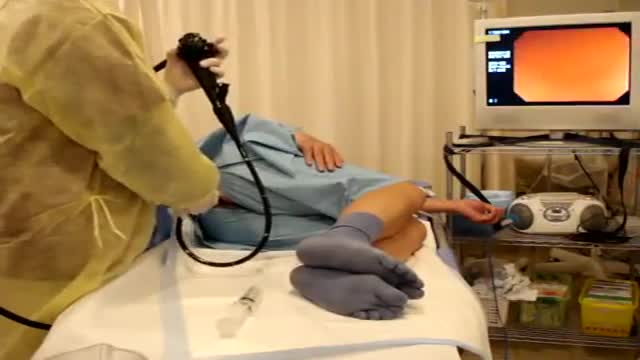
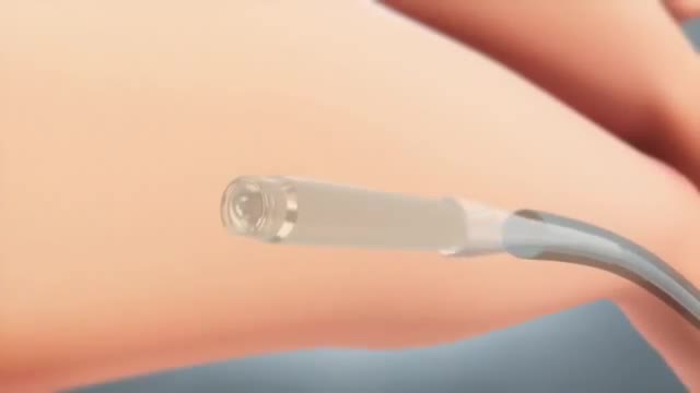
Colonoscopy is a test that allows your doctor to look at the inner lining of your large intestine (rectum and colon). He or she uses a thin, flexible tube called a colonoscope to look at the colon. A colonoscopy helps find ulcers, colon polyps, tumors, and areas of inflammation or bleeding.
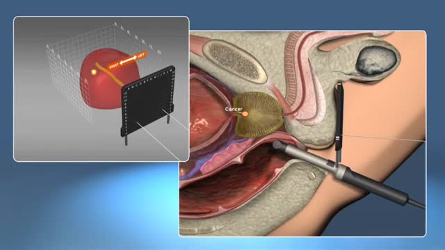
A prostate gland biopsy is a test to remove small samples of prostate tissue to be looked at under a microscope. ... For a prostate biopsy, a thin needle is inserted through the rectum (transrectal biopsy), through the urethra, or through the area between the anus and scrotum (perineum).

This video: The veins around your anus tend to stretch under pressure and may bulge or swell. Swollen veins (hemorrhoids) can develop from an increase in pressure in the lower rectum. Factors that might cause increased pressure include: Straining during bowel movements.
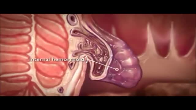
The veins around your anus tend to stretch under pressure and may bulge or swell. Swollen veins (hemorrhoids) can develop from an increase in pressure in the lower rectum. Factors that might cause increased pressure include: Straining during bowel movements.

Rectal bleeding can refer to any blood that passes from your anus, although rectal bleeding is usually assumed to refer to bleeding from your lower colon or rectum. Your rectum makes up the last few inches of your large intestine. Rectal bleeding may show up as blood in your stool, on the toilet paper or in the toilet bowl. Blood that results from rectal bleeding can range in color from bright red to dark maroon to a dark, tarry color.
Lip augmentation is a cosmetic procedure that can give you fuller, plumper lips. These days, an injectable dermal filler is the most commonly used method of lip augmentation. There are many types of dermal fillers that can be injected in your lips and around your mouth.
This tiny wireless pacemaker can be inserted into the body via a catheter instead of invasive surgery.
A colostomy is an operation that creates an opening for the colon, or large intestine, through the abdomen. A colostomy may be temporary or permanent. It is usually done after bowel surgery or injury.
This operation can be performed as an open or laparoscopic (keyhole procedure). During the operation the sigmoid colon is removed. This involves taking away the blood vessels and lymph nodes to that part of the bowel. The surgeon then re-makes the join (anastomosis) between the remaining left side of the colon and the top of the rectum. The surgeon may use either sutures or special staples to make this join.

Restrictive cardiomyopathy (RCM) is a rare form of heart muscle disease that is characterized by restrictive filling of the ventricles. In this disease the contractile function (squeeze) of the heart and wall thicknesses are usually normal, but the relaxation or filling phase of the heart is very abnormal.
Constrictive pericarditis is the result of scarring and consequent loss of the normal elasticity of the pericardial sac. This leads to impairment of ventricular filling in mid and late diastole. As a result, the majority of ventricular filling occurs rapidly in early diastole and the ventricular volume does not increase after the end of the early filling period. Restrictive cardiomyopathy is characterized by a nondilated rigid ventricle, resulting in severe diastolic dysfunction and restrictive filling that produces hemodynamic changes similar to those in constrictive pericarditis. Constrictive pericarditis and restrictive cardiomyopathy both lead to diastolic heart failure with normal (or near normal) systolic function, and characteristically abnormal ventricular filling that results in similar clinical and hemodynamic features. However, because of their markedly different treatments, differentiating between the two conditions is critical. In some patients, the correct diagnosis may be readily suggested from the history or routine diagnostic testing. In others, however, this differentiation cannot be diagnosed before biopsy or even surgical exploration.
Hypertrophic cardiomyopathy (HCM) is very common and can affect people of any age. It affects men and women equally. It is a common cause of sudden cardiac arrest in young people, including young athletes. Hypertrophic cardiomyopathy occurs if heart muscle cells enlarge and cause the walls of the ventricles (usually the left ventricle) to thicken. The ventricle size often remains normal, but the thickening may block blood flow out of the ventricle. If this happens, the condition is called obstructive hypertrophic cardiomyopathy. Sometimes the septum, the wall that divides the left and right sides of the heart, thickens and bulges into the left ventricle. This can block blood flow out of the left ventricle. Then the ventricle must work hard to pump blood. Symptoms can include chest pain, dizziness, shortness of breath, or fainting. Hypertrophic cardiomyopathy also can affect the heart's mitral valve, causing blood to leak backward through the valve. Sometimes, the thickened heart muscle doesn't block blood flow out of the left ventricle. This is referred to as non-obstructive hypertrophic cardiomyopathy. The entire ventricle may thicken, or the thickening may happen only at the bottom of the heart. The right ventricle also may be affected. In both obstructive and non-obstructive HCM, the thickened muscle makes the inside of the left ventricle smaller, so it holds less blood. The walls of the ventricle may stiffen, and as a result, the ventricle is less able to relax and fill with blood.
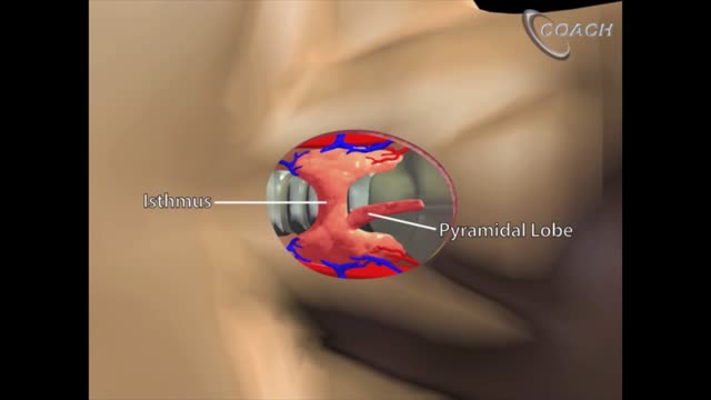
Minimally invasive open thyroidectomy (MIT) is similar to conventional thyroidectomy in its surgical approach. The major difference is the length of the neck incision. A smaller incision improves cosmesis and reduces discomfort. Typically, a skin incision less than 6 cm is considered minimally invasive. The remainder of the procedure is exactly the same as is used in conventional thyroidectomy. Adaptations to this technique include transection rather than lateral retraction of the strap muscles (the Sofferman technique). [1]
