
- Physical Examination
- Surgical Examination
- Ophthalmology
- Clinical Skills
- Orthopedics
- Surgery Videos
- Laparoscopy
- Pediatrics
- Funny Videos
- Cardiothoracic Surgery
- Nursing Videos
- Plastic Surgery
- Otorhinolaryngology
- Histology and Histopathology
- Neurosurgery
- Dermatology
- Pediatric Surgery
- Urology
- Dentistry
- Oncology and Cancers
- Anatomy Videos
- Health and Fitness
- Radiology
- Anaesthesia
- Physical Therapy
- Pharmacology
- Interventional Radiology
- Cardiology
- Endocrinology
- Gynecology
- Emergency Medicine
- Psychiatry and Psychology
- Childbirth Videos
- General Medical Videos
- Nephrology
- Physiology
- Diet and Food Health
- Diabetes Mellitus
- Neurology
- Women Health
- Osteoporosis
- Gastroenterology
- Pulmonology
- Hematology
- Rheumatology
- Toxicology
- Nuclear Medicine
- Infectious Diseases
- Vascular Disease
- Reproductive Health
- Burns and Wound Healing
- Kidney Zone Podcast
- Other

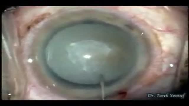

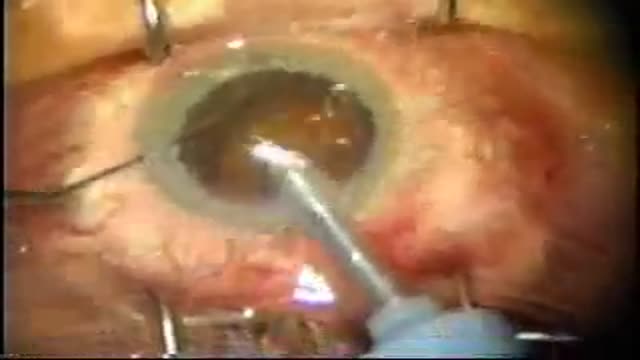


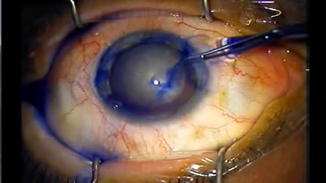

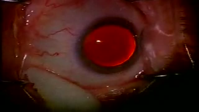

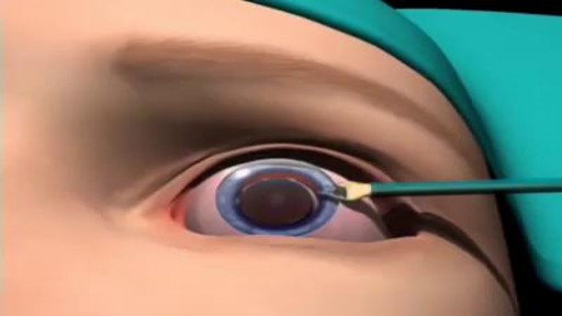
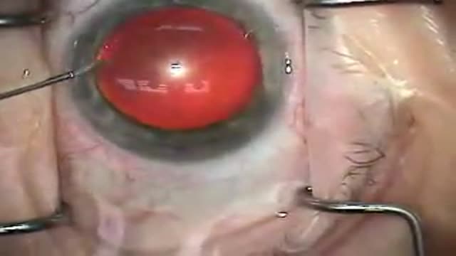

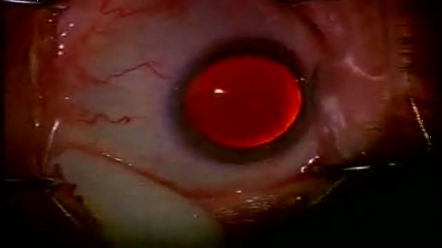
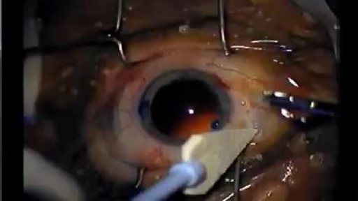
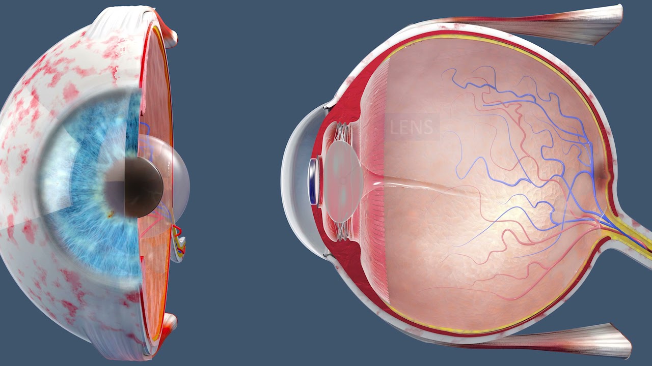
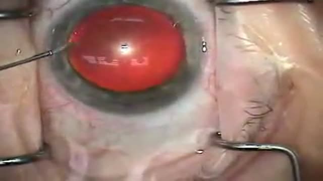
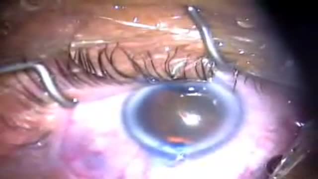
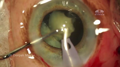
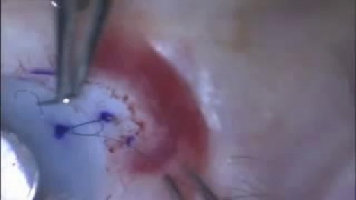
Great White Cataract Surgery Video

Educational only: videos on MedicalVideos.com are for general information and are not medical advice. Always consult your own doctor about your health.
A cataract is a clouding of the lens in the eye that affects vision. Most cataracts are related to aging. Cataracts are very common in older people. By age 80, more than half of all Americans either have a cataract or have had cataract surgery. A cataract can occur in either or both eyes. It cannot spread from one eye to the other.
This video in 30 seconds
A surgeon narrates phacoemulsification on a dense white cataract. He explains the difficulty of opening the capsule when the cortex has liquefied, his use of stain and a fine needle to decompress the lens first, the chopping technique used to remove it while protecting the back of the capsule, and insertion of a foldable acrylic lens.
Key points
- Trypan blue stain is used to visualize the anterior capsule
- A fine needle decompresses the liquefied lens before the capsule opening
- Phacoemulsification removes the central lens core
- A second instrument fractures the lens and shields the posterior capsule
- Remaining cortex is removed and a foldable acrylic lens is inserted
Chapters
- 0:00 — Dense white cataract challenge introduced
- 0:26 — Staining the anterior capsule
- 0:45 — Needle decompression of the lens
- 1:47 — Phacoemulsification of the lens core
- 3:18 — Fracturing lens into smaller pieces
- 5:14 — Foldable lens insertion preparation
Video Transcript
A surgeon's narrated case of phacoemulsification on a dense white cataract. He explains the difficulty of opening the capsule when the cortex has liquefied, his use of trypan blue stain and a fine needle to decompress the lens first, the chopping technique and machine settings he uses, protecting the back of the capsule, and finally inserting a foldable acrylic lens.
Read the full transcript
This is Dr. Stigent-Eisen. I would like to share with you a video of the fecal multiplication of a dense white cataract. These cases can be quite challenging and primarily challenging in respect of performing the capsule rexis, which can be challenging both from the visualization of the anterior capsule standpoint and also dealing with the liquefied cortex, which can emanate from the capsule. Once the anterior capsule is opened, here you can see we're using Tri-Pan Blue in order to stain the capsule and aid in visualization, after which it is evacuated with this goelastic.
What I like to do is use a 25 gauge needle to make an initial stab incision into the lens, right through the anterior capsule, and at this point one can aspirate all the liquefied cortex that will come forward. This decompresses the lens and prevents the upward pushing of the anterior capsule with the liquefied cortex and the possible disruption of the circular capsule rexis, which can then be performed quite easily. The remaining cortex and lens material is further liquefied and mobilized with BSS solution, and then fake emulsification is then performed.
I use a bowling technique where the central core of the lens is removed. You can see how mobile the lens is. Since it's lost its cortical moorings, by virtue of the fact that all the cortex has been liquefied, there's no more support structure for the lens within the bag. You can see that the lens is quite dense in a wittling way with the fake emulsification instrument. Then we go from a vacuum setting of 60 with an aspiration of 20 to a higher vacuum setting, roughly 400 millimeters of mercury, and an aspiration flow rate of 35 cc per minute.
I use the second instrument at this point to fracture the lens peripherally in a horizontal chopping technique in order to break it up into smaller, more manageable pieces, and then proceed to chew up the remaining lens for now in nice bite-sized pieces, if you will. At this point I'm also very aware of the fact that I have no protection of the posterior capsule since all the epineuclein material is already liquefied and gone. So I do use the second instrument as a barrier for the posterior capsule to keep it from bounding up into my fecotip with any surges that may occur.
You can see there's very little remaining cortex, basically in the capsaure bag fornaceae, and that is removed with a curved IATip. Find the curved IATip very helpful, especially for subincisional cortex, and it makes my job a lot easier. The bag is then re-inflated with this goelastic in preparation of lens insertion, and then a foldable acrylic lens is then placed into the capsaure bag. So the technique for this type of cataract is made much easier with the use of Tri-Pan Blue and a 25 gauge needle. So I hope you enjoyed the case, and thank you for watching.
Transcript auto-generated from the video’s audio for accessibility and study. Educational content only — not medical advice.
Video Overview
From our Ophthalmology collection: Eye examinations, cataract and LASIK surgery, retina procedures and common eye conditions explained by specialists. Browse all Ophthalmology videos →
SORT BY-
Top Comments
-
Latest comments