Neueste Videos
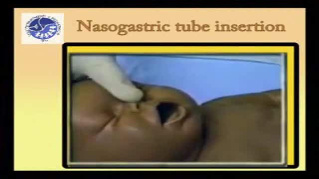
Insertion of pediatric nasogastric tube in children and babies
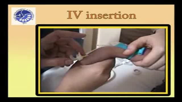
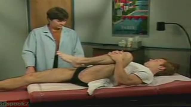
Pediatric IV insertion
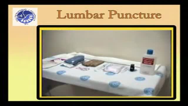
Pediatric Lumbar puncture
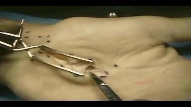
This is a video of a carpal tunnel release surgery

How to insert Endotracheal tube in children
Bone marrow examination refers to the pathologic analysis of samples of bone marrow obtained by bone marrow biopsy (often called a trephine biopsy) and bone marrow aspiration. Bone marrow examination is used in the diagnosis of a number of conditions, including leukemia, multiple myeloma, anemia, and pancytopenia. The bone marrow produces the cellular elements of the blood, including platelets, red blood cells and white blood cells. While much information can be gleaned by testing the blood itself (drawn from a vein by phlebotomy), it is sometimes necessary to examine the source of the blood cells in the bone marrow to obtain more information on hematopoiesis; this is the role of bone marrow aspiration and biopsy.
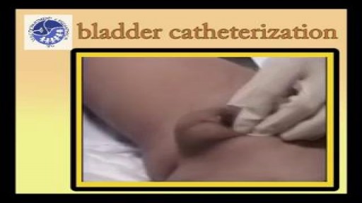
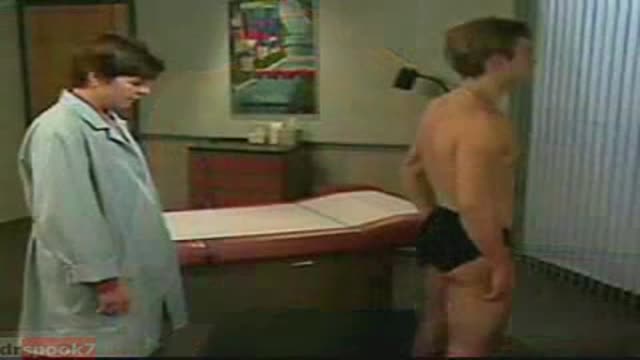
This video shows how to insert a catheter in a baby girl
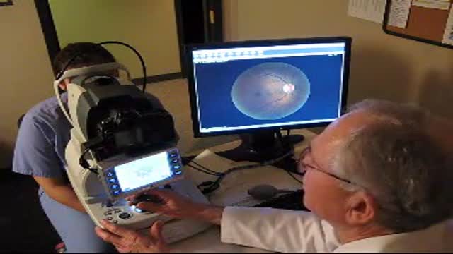
new fundus camera for examining the retina without dilating the pupil
With an Ophthalmoscope, light is shone into the eye and the retina and the optic nerve is examined. This is called as Examination of the Fundus. This is what the eye-doctor sees when he peeps into your eye! Through the transparent cornea, into the dark interior. The Fundus Exam When he looks into the eye with the Ophthalmoscope, he sees a orange glowing interior. That is the retina. The retina is actually transparent. It appears bright because of blood vessels in the choroid layer below. It is like looking at your ear against the bright sunlight. The yellow circle is the Optic Nerve, the cable of vision! A red, shiny dot attracts attention. That is the macula. If indicated, the exam of periphery of the retina is done with an Indirect ophthalmoscope. The ophthalmologist wears this instrument on the head and focuses the light into the eye with a lens held in his hand. This is usually done in a dark room.

Examination of neck veins and arteries - French Subtitled
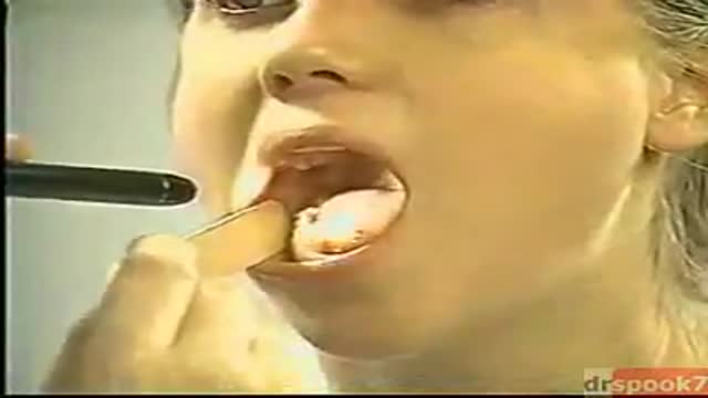
The exam should be performed in an orderly fashion as follows: 1. Have the patient stick out their tongue so that you can examine the posterior pharynx (i.e. the back of the throat). Ask the patient to say "Ah", which elevates the soft palate, giving you a better view. If you are still unable to see, place the tongue blade � way back on the tongue and press down while the patient again says "Ah," hopefully improving your view. This causes some people to gag, particularly when the blade is pushed onto the more proximal aspects of the tongue. It may occasionally be important to determine whether the gag reflex is functional (e.g. after a stroke that impairs CNs 9 or 10; or to determine if a patient with depressed level of consciousness is able to protect their airway from aspiration). This is done by touching a q-tip against the posterior pharynx, uvula or tongue. It is not necessary to do this during your routine exam as it can be quite noxious!
2. Note that the uvula hangs down from the roof of the mouth, directly in the mid-line. With an "Ah," the uvula rises up. Deviation to one side may be caused by CN 9 palsy (the uvula deviates away from the affected side), a tumor or an infection. CN9 Pasly Cranial Nerve 9 Dysfunction: Patient has suffered stroke, causing loss of function of left CN 9. As a result, uvula is pulled towards the normally functioning (ie right) side. 3. The normal pharynx has a dull red color. In the setting of infection, it can become quite red, frequently covered with a yellow or white exudate (e.g. with Strep. Throat or other types of pharyngitis).
4. The tonsils lie in an alcove created by arches on either side of the mouth. The apex of these arches are located lateral to and on a line with the uvula. Normal tonsils range from barely apparent to quite prominent. When infected, they become red, are frequently covered by whitish/yellow discharge. In the setting of a peritonsilar abscess, the tonsils appear asymmetric and the uvula may be pushed away from the affected side. When this occurs, the tonsil may actually compromise the size of the oral cavity, making breathing quite difficult.
5. Look carefully along the upper and lower gum lines and at the mucosa in general, which can appear quite dry if the patient is dehydrated.
6. Examine the teeth to get a sense of general dentition, particularly if the patient has a dental complaint. Pain produced by tapping on a tooth is commonly caused by a root abscess. Tooth Abscess: Tooth abscess involving left molar region. Associated inflammation of left face can clearly be seen. 7. Have the patient stick their tongue outside their mouth, which allows evaluation of CN 12. If there is nerve impairment, the tongue will deviate towards the affected side. Any obvious growths or abnormalities? Ask them to flip their tongue up so that you can look at the underside. If you see something abnormal, grasp the tongue with gauze so that you can get a better look. Left CN 12 Dysfunction: Stroke has resulted in L CN 12 Palsy. Tongue therefore deviates to the left.
8. Make note of any growths along the cheeks, hard palate (the roof of the mouth between the teeth), soft palate, or anywhere else. In particular, patients who smoke or chew tobacco are at risk for oral squamous cell cancer. Any areas which are painful or appear abnormal should also be palpated. Put on a pair of gloves to better explore these regions. What do they feel like? Are they hard? To what extent does a growth involve deeper structures? If the patient feels something that you cannot see, try to get someone else to hold the light source, freeing both your hands to explore the oral cavity with two tongue depressors.
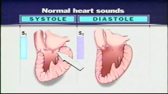
Normal Heart Sounds With the aid of a stethoscope you can hear the characteristic sounds of the normal heartbeat, typically described as a "lub-dub." These sounds are produced by the closure of the heart valves. The first heart sound or "lub" results from closure of the tricuspid and mitral valves. It is a rather low-pitched and a relatively long sound which, as indicated in, represents the beginning of ventricular systole. The second heart sound, or "dub," marks the beginning of ventricular diastole. It is produced by closure of the aortic and pulmonary (pulmonic) semilunar vanes when the intraventricular pressure begins to fall. This "dub" sound is typically heard as a sharp snap because the semilunar valves tend to close much more rapidly than the AV valves. Because diastole occupies more time than systole, a brief pause occurs after the second heart sound when the heart is beating at a normal rate. Therefore, the pattern that one hears is one of: "lub-dub" pause, "lub-dub" pause, and so on. Sometimes, especially in young normal individuals, a third heart sound can be heard. This sound is produced by the very rapid influx of blood into the partially filled ventricle. It is typically very faint and as such difficult to hear.

Shoulder Exam
I think that the most daunting aspect of the shoulder exam is appreciating the functional anatomy of this incredibly mobile joint. The primary benefit of the ball and socket arrangement is that it allows the hand to be positioned precisely in space, maximizing our ability to function. In terms of functionality, the shoulder might be best described as having a golf ball-on-a-tee design.
Location Of The Muscle Groups Is Approximated In The Pictures Above.
Start by looking at the normal (or more normal) side. Note any scars, obvious asymmetry, discoloration, swelling, or muscle asymmetry.
Palpation
Gently palpate around the shoulder, touching each of the landmarks noted above. Make note of pain.

Function and Anatomy:
Hinge type joint formed by the articulation of the Ulna and Radius (bones of the forearm), and Humerus (upper arm). Full extension is equal to 0 degrees, full flexion to ~ 150 degrees. Maximum supination (turning hand palm up so that it can hold a bowl of "soup") and pronation (palm down) are both 90 degrees
Facial Tenderness
1. Ask the patient to tell you if these maneuvers causes excessive discomfort or pain. ++
2. Press upward under both eyebrows with your thumbs.
3. Press upward under both maxilla with your thumbs.
4. Excessive discomfort on one side or significant pain suggests sinusitis.
Sinus Trans illumination 1. Darken the room as much as possible. ++
2. Place a bright otoscope or other point light source on the maxilla.
3. Ask the patient to open their mouth and look for an orange glow on the hard palate.
4. A decreased or absent glow suggests that the sinus is filled with something other than air.
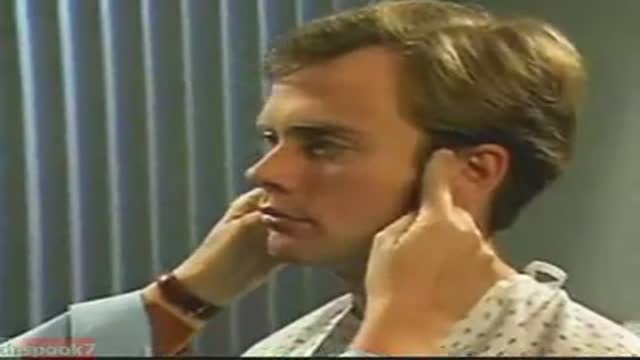
Temporomandibular Joint 1. Place the tips of your index fingers directly in front of the tragus of each ear. ++
2. Ask the patient to open and close their mouth.
3. Note any decreased range of motion, tenderness, or swelling.
The Knee Exam
Observation:
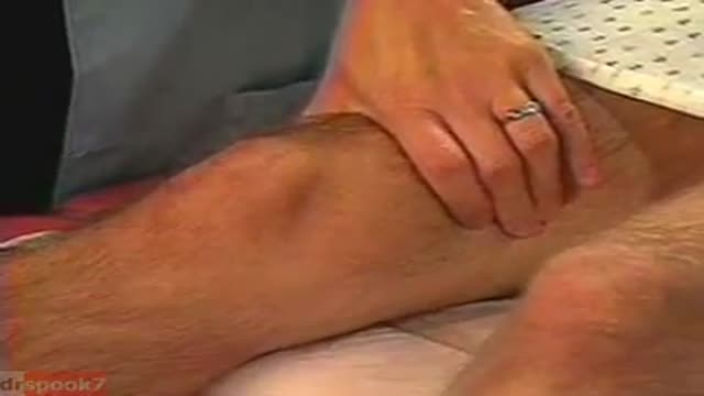
1. Make sure that both knees are fully exposed. The patient should be in either a gown or shorts. Rolled up pant legs do not provide good exposure!
2. Watch the patient walk. Do they limp or appear to be in pain? When standing, is there evidence of bowing (varus) or knock-kneed (valgus) deformity? There is a predilection for degenerative joint disease to affect the medical aspect of the knee, a common cause of bowing. Varus Knee Deformity, more marked on the left leg. 3. Make note of any scars or asymmetry. Chronic/progressive damage, as in degenerative joint disease, may lead to abnormal contours and appearance. Is there obvious swelling as would occur in an effusion? Redness suggesting inflammation? 4. Is there evidence of atrophy of the quadriceps, hamstring, or calf muscle groups? Knee problems/pain can limit the use of the affected leg, leading to wasting of the muscles.
While both legs have well developed musculature,
the left calf and hamstring are bulkier than the right. 5. Look at the external anatomy, noting structures above and below the knee itself: 1. Patella 2. Patellar tendon 3. Quadriceps/Hamstring/Calf muscles 4. Medial and lateral joint lines. 5. Femur and Tibia 6. Tibial tuberosity
Ballotment (helpful if the effusion is large) 1. Slightly flex the knee which is to be examined.
2. Place one hand on the supra-pateallar pouch, which is above the patella and communicates with the joint space. Gently push down and towards the patella, forcing any fluid to accumulate in the central part of the joint.
3. Gently push down on the patella with your thumb.
4. If there is a sizable effusion, the patella will feel as if it's floating and "bounce" back up when pushed down.
Common Benign Pain Syndromes--Symptoms and Etiology:
1. Non-specific musculoskeletal pain: This is the most common cause of back pain. Patients present with lumbar area pain that does not radiate, is worse with activity, and improves with rest. There may or may not be a clear history of antecedent over use or increased activity. The pain is presumably caused by irritation of the paraspinal muscles, ligaments or vertebral body articulations. However, a precise etiology is difficulty to identify.
2. Radicular Symptoms: Often referred to as "sciatica," this is a pain syndrome caused by irritation of one of the nerve roots as it exits the spinal column. The root can become inflamed as a result of a compromised neuroforamina (e.g. bony osteophyte that limits size of the opening) or a herniated disc (the fibrosis tears, allowing the propulsus to squeeze out and push on the adjacent root). Sometimes, it's not precisely clear what has lead to the irritation. In any case, patient's report a burning/electric shock type pain that starts in the low back, traveling down the buttocks and along the back of the leg, radiating below the knee. The most commonly affected nerve roots are L5 and S1.
3. Spinal Stenosis: Pain starts in the low back and radiates down the buttocks bilaterally, continuing along the backs of both legs. Symptoms are usually worse with walking and improve when the patient bends forward. Patient's may describe that they relieve symptoms by leaning forward on their shopping carts when walking in a super market. This is caused by spinal stenosis, a narrowing of the central canal that holds the spinal cord. The limited amount of space puts pressure on the nerve roots when the patient walks, causing the symptoms (referred to as neurogenic claudication). Spinal stenosis can be congenital or develop over years as a result of djd of the spine. As opposed to true claudication (pain in calfs/lower legs due to arterial insufficiency), pain resolves very quickly when person stops walking and assumes upright position. Also, peripheral pulses should be normal.
4. Mixed symptoms: In some patients, more then one process may co-exist, causing elements of more then one symptom syndrome to co-exist.
Function and Anatomy: The hip is a ball and socket type joint, formed by the articulation of the head of the femur with the pelvis. Normal range of motion includes: abduction 45 degrees, adduction 20-30 degrees, flexion 135 degrees, extension 30 degrees, internal and external rotation. Hip pathology can cause symptoms anywhere around the joint, though frequently pain is anterior and radiates to the groin region. Additionally, pathology outside of the hip can be referred to this region. History and exam obviously help in making these distinctions.

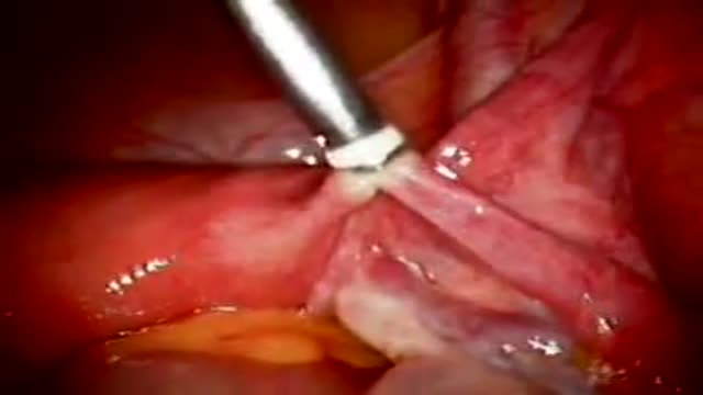
Laparoscopic Tubal Reversal of fallopian tubes after ligation
Tubal ligation using Fallope Ring
