- Physical Examination
- Surgical Examination
- Ophthalmology
- Clinical Skills
- Orthopedics
- Surgery Videos
- Laparoscopy
- Pediatrics
- Funny Videos
- Cardiothoracic Surgery
- Nursing Videos
- Plastic Surgery
- Otorhinolaryngology
- Histology and Histopathology
- Neurosurgery
- Dermatology
- Pediatric Surgery
- Urology
- Dentistry
- Oncology and Cancers
- Anatomy Videos
- Health and Fitness
- Radiology
- Anaesthesia
- Physical Therapy
- Pharmacology
- Interventional Radiology
- Cardiology
- Endocrinology
- Gynecology
- Emergency Medicine
- Psychiatry and Psychology
- Childbirth Videos
- General Medical Videos
- Nephrology
- Physiology
- Diet and Food Health
- Diabetes Mellitus
- Neurology
- Women Health
- Osteoporosis
- Gastroenterology
- Pulmonology
- Hematology
- Rheumatology
- Toxicology
- Nuclear Medicine
- Infectious Diseases
- Vascular Disease
- Reproductive Health
- Burns and Wound Healing
- Other
Latest videos
laparoscopy for repair of rupture of urinary bladder
Bladder and prostate injection- Botox

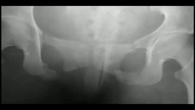
Endoscopic crushing of a bladder stone
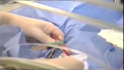
Catheters and Long Lines are introduced in Neonates to administer fluid and Total Parentral Nutrition. The proceedure is not easy to perform and is prone to get infections.
Strict Aseptic technique is mandatory
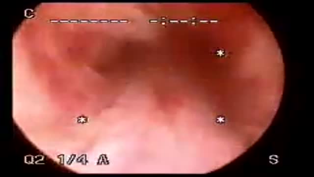
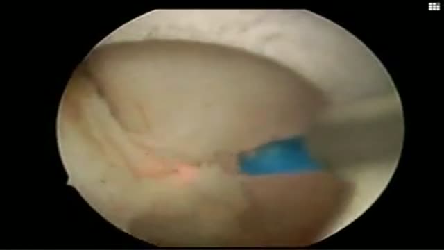
Cystoscopy
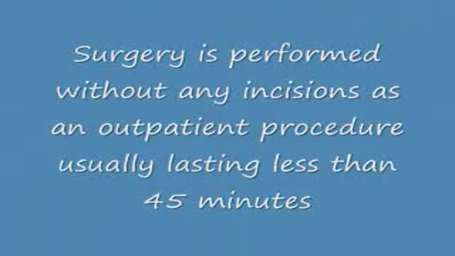
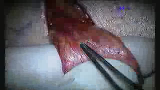
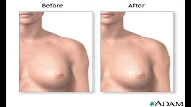
New York Plastic Surgery ,Dr. Robert Vitolo ,board certified plastic surgeon , brings you into the operating room for a glimpse at how his transumbilical breast augmentation procedure is performed. Dr. Vitolo, a pioneer in the 'no visible scar' breast enlargement surgery, has been using this technique since 1994. Dr. Vitolo use Allergan Natrelle saline breast implants and Mentor saline implants. Dr. Vitolo also performs a removal of silicone gel implants and replacement with saline implants using the transumbilical method.
Minimally invasive kidney and ureteral stone surgery using holmium laser performed at El Camino Urology Medical Group,
Microsurgical varicocelectomy is performed for patients with a varicocele and impaired semen parameters, testicular atrophy or pain due to the varicocele.

Basic Surgical Instruments- Forceps, scissors.
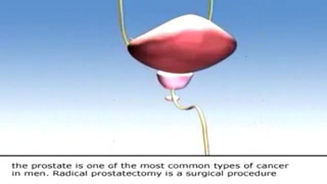
This is a educational video for the prostate cancer patient and their family. Depending on the individual patient, a radical prostatectomy, might a procedure that your urologist could recommend as treatment.
HoLEP (Holmium laser enucleation of prostate)

Incision of the bladder neck for a small prostate
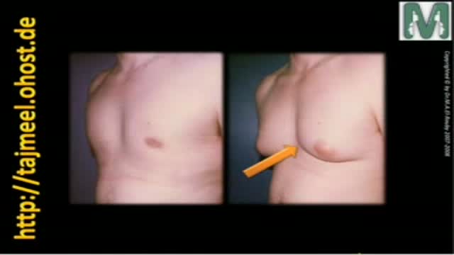
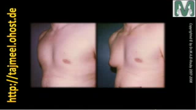
Gynecomastia means enlargement of male breast to resample female breast that is a common problem between males and causes many psychological problem
Dr. Mohamed El-Rouby
Consltant of Plastic surgery - Faculty of Medicine - Ain Shams University
تضخم الثدى عند الرجال من المشاكل المنشرة جدا بين الشباب و تسبب الكثير من المشاكل النفسية و الصحية
د. محمد الروبى
استشارى جراحات التجميل - جامعة عين شمس
عملية تجميل أو اعادة شكل الانف
د. محمد الروبى
استشارى جراحات التجميل - جامعة عين شمس
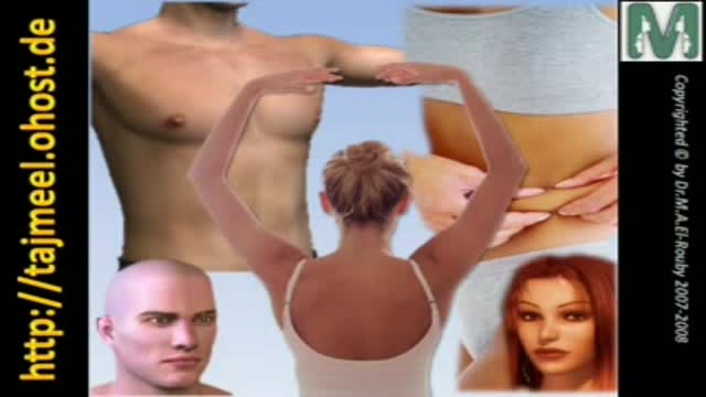
تناسق القوام مطلب كل أنسان سواء رجل أو أمرأة ولذلك يجب تحديد معدل تراكم الدهون بالجسم و تحديد نوع تناسق القوام و كيفيته
د. محمد الروبي
استشارى جراحات التجميل بجامعة عين شمس
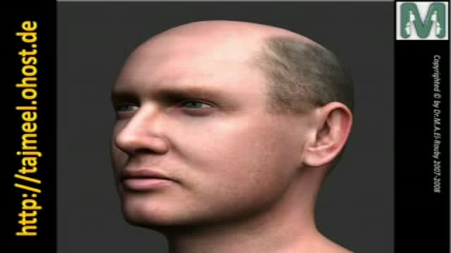
كيفية منع تساقط الشعر و علاج الصلع
د. محمد الروبي
استشارى جراحات التجميل - جامعة عين شمس
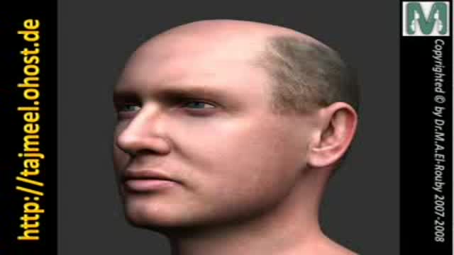
How Hair can be retsored and transplanted? natural versus biofibers?
Dr. Mohamed El Ruby
Consultant of Plastic Surgery - Ain Shams University
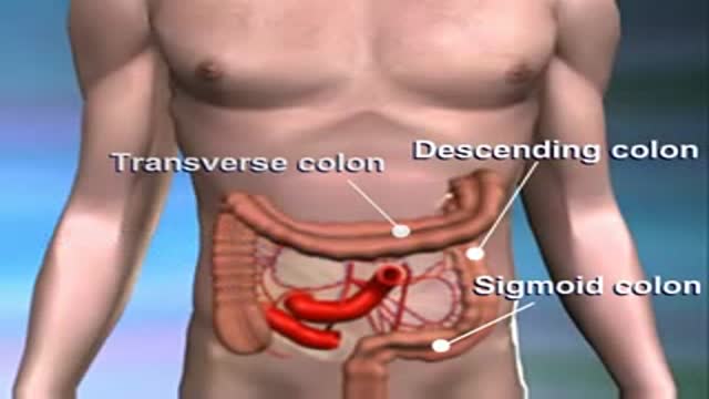
An animation illustrating carcinoma of the colon
Symptoms of carcinoma of the breast
