- Physical Examination
- Surgical Examination
- Ophthalmology
- Clinical Skills
- Orthopedics
- Surgery Videos
- Laparoscopy
- Pediatrics
- Funny Videos
- Cardiothoracic Surgery
- Nursing Videos
- Plastic Surgery
- Otorhinolaryngology
- Histology and Histopathology
- Neurosurgery
- Dermatology
- Pediatric Surgery
- Urology
- Dentistry
- Oncology and Cancers
- Anatomy Videos
- Health and Fitness
- Radiology
- Anaesthesia
- Physical Therapy
- Pharmacology
- Interventional Radiology
- Cardiology
- Endocrinology
- Gynecology
- Emergency Medicine
- Psychiatry and Psychology
- Childbirth Videos
- General Medical Videos
- Nephrology
- Physiology
- Diet and Food Health
- Diabetes Mellitus
- Neurology
- Women Health
- Osteoporosis
- Gastroenterology
- Pulmonology
- Hematology
- Rheumatology
- Toxicology
- Nuclear Medicine
- Infectious Diseases
- Vascular Disease
- Reproductive Health
- Burns and Wound Healing
- Other
Top videos

MRCPCH Clinical Revision - more videos at http://mrcpch.paediatrics.co.uk
Revise for your MRCPCH Clinical exam, with videos and high quality content created by the London Paediatrics Trainees Committee.
Video Credits: Dr Caroline Fertleman, Dr Hermione Race, Dr Camilla Sen, Dr Chloe Macaulay, Dr Emma McLaren, Chris Knapp

#final #fumc #mbbs #medicalstudents #mbbsabroad #doctor #fcps #fcpspart #surgeryeducation #surgeryreview #trainee #exampreparation

Doctor Ricky Brown reacts to this surgery simulation of an inguinal hernia repair where they repair the hernia sack and create a mesh for the organ to comfortably rest on.
3D Animation powered by:
3DMedWorld - 3dmedworld.com
#shorts #doctor #education #surgery #medical

The anatomy of the direct and indirect inguinal hernia.
Music:
Berries and Lime by Gregory David
https://www.epidemicsound.com/track/z6iCiiyCPm/

This gentleman has a significant lumbar herniated disc with a positive well straight leg raise test. In this evaluation I test his deep tendon reflexes, sensation, muscle strength, and perform a straight leg raise test, Braggards's test and Well straight leg raise test.
✅ Support OEP: https://paypal.me/OrthoEvalPal?locale.x=en_US
✅ OEP Website: https://orthoevalpal.com/
✅ Online Coaching: https://orthoevalpal.com/coaching
✅ OEP Podcasts: https://orthoevalpal.com/podcast
▶▶ Like us on facebook: https://www.facebook.com/OrthoEvalPal
▶▶ Follow on Instagram: https://www.instagram.com/
▶▶ Follow on Twitter: https://twitter.com/home
✔ Get our NEW downloadable 1.5 hour shoulder anatomy with cadaver dissection lecture: http://www.meorthopedicseminar....s.com/shop/shoulder-
✔Get our NEW downloadable 7.5 hour cervical and lumbar continuing ed course: http://www.meorthopedicseminar....s.com/shop/rehabilit
✔Get our NEW downloadable 6.0 hour shoulder continuing ed course: http://www.meorthopedicseminar....s.com/shop/comprehen
Interested in our Sponsor Product
EZ Slant (http://ezslant.com/)
Check out our new OEP merchandise: 👚👕☕️https://www.youtube.com/channe....l/UC76MsdkAQaBkCb35K
#wellstraightlegraise #herniateddisc #LBP #lowbackpain #OrthoEvalPal #SpecialTests #Orthopedics #physicaltherapy #physicaltherapytests #athletictraining

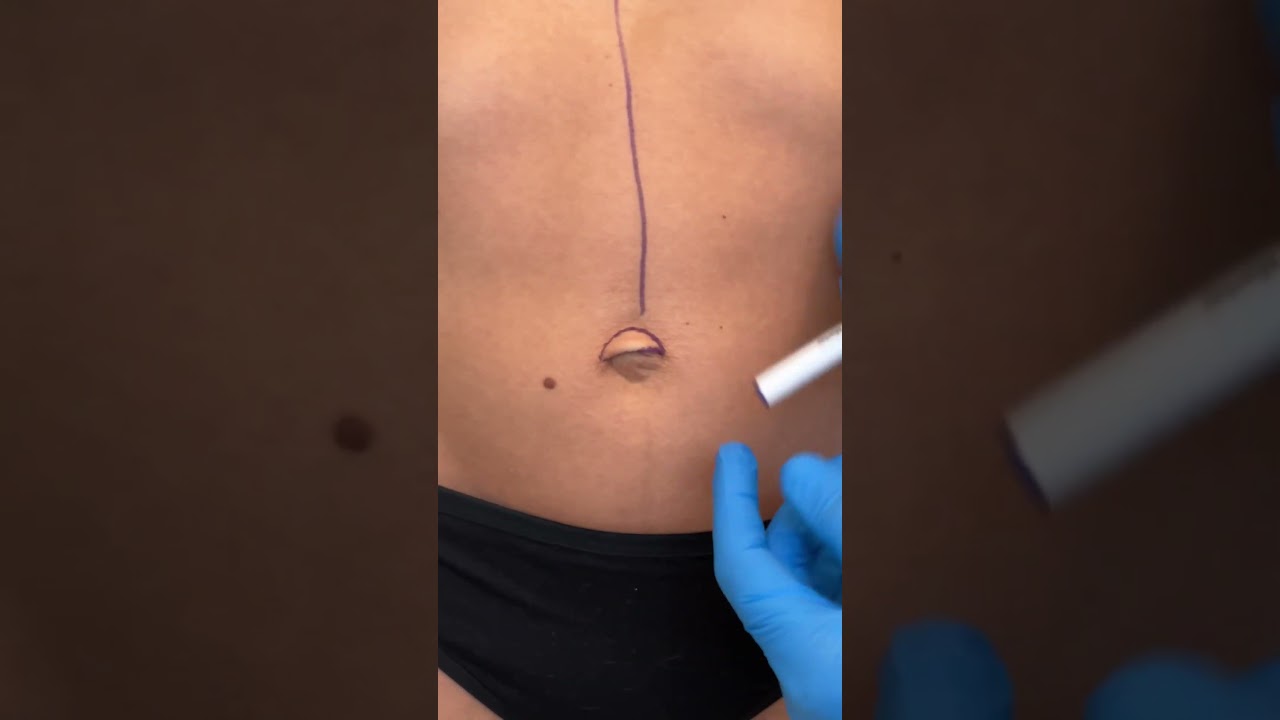
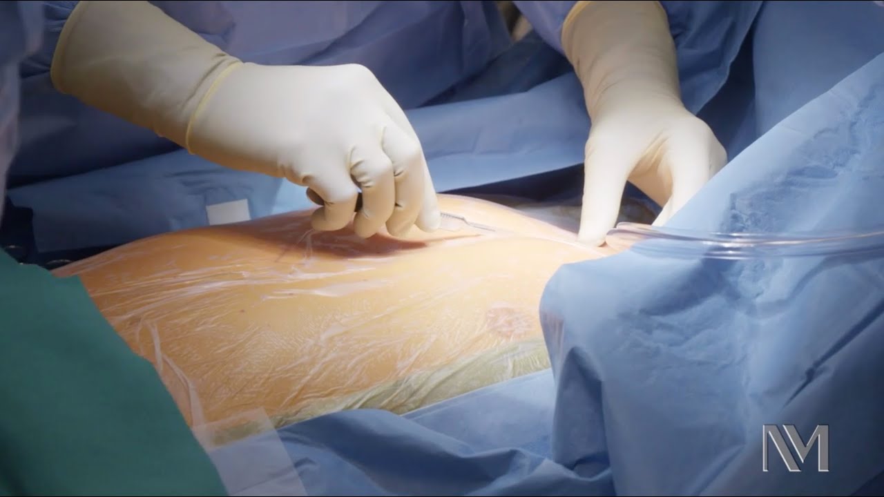
This is how Paraumbilical hernia looks like and how it is examined although it looks very simple but in exam it can be very difficult to perform all steps in small amount of time. This can be short case or even long of #cpsp #fcps #mbbs #medicalstudent #mbbsexams #plab2 #plab #plab1 and MS #genernalknowledge #surgery exams
#para-umbilical hernia
#umbilical hernia #paraumbilical #hernia repair#laparoscopic paraumbilical hernia repair. #umbilical defect, #vetral hernia surgery. #herniatreatment #herniatreatment #ventral hernia hernia,#laparoscopic ventral hernia repair,umbilicus,carl lowe jr,hernia repair,training,north carolina,hernia repair surgery,charlotte,operation,laparoscopic,bulge,surgery,surgeon,dr. lowe,ipom repair,live surgery,mesh,
#mesh #ipom repair
Check out @barrettplasticsurgery on TikTok!
Like and subscribe for more! #shorts #medical #plasticsurgery
More information:
www.drdanielbarrett.com

This video shows Prof Dan Reinstein, MD MA(Cantab) FRCSC DABO FRCOphth FEBO performing a ReLEx SMILE keyhole LASIK procedure using the latest surgical instrument that he helped to develop (Malosa MMSU1297 - Reinstein Lenticule Separator: http://www.malosa.com/en/reinstein-le...). This instrument enables the procedure to be performed with one instrument, through one 2mm incision, using only one sweep per plane, and taking about 30 seconds to separate and withdraw the lenticule, improving day 1 uncorrected vision over other lenticule extraction techniques that require more corneal manipulation.
Dr. Leo Maguire, a Mayo Clinic ophthalmologist, explains how laser-assisted in situ keratomileusis (LASIK) eye surgery can correct common vision problems.
This interview originally aired Jan. 26, 2019.
To learn more about LASIK surgery, visit: https://www.mayoclinic.org/tests-procedures/lasik-eye-surgery/about/pac-20384774?mc_id=us&utm_source=newsnetwork&utm_medium=l&utm_content=content&utm_campaign=mayoclinic&geo=national&placementsite=enterprise&cauid=100721&_ga=2.112234244.1227307149.1547427243-1780934405.1469629163
Ever considered getting laser eye surgery, but didn’t know how it worked? Allow us to help!
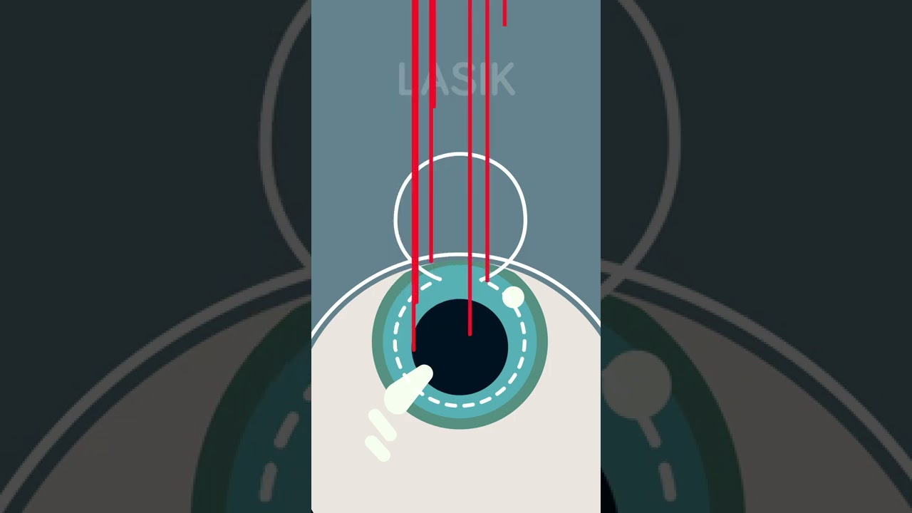
There are three different main types of laser eye surgery: LASIK, SMILE, and Surface Laser Treatments, and each can be explained pretty easily.
LASIK uses two lasers to open up a thin flap on the surface of the cornea, and then reshapes the cornea underneath. The flap is then placed back over the reshaped cornea, and heals independently with time.
SMILE uses one laser to reshape the cornea through a small, self-healing hole.
And Surface Eye Treatments remove the clear skin over the eye, to then reshape the cornea underneath with - you guessed it - a laser!

If you are tired of dealing with glasses or squinting to read signs in the distance, then you should consider LASIK Eye Surgery. In this outpatient refractive procedure, lasers are used to correct vision issues by changing the structure of the cornea. This may entirely eliminate reliance upon glasses or contacts. In this interactive LASIK Eye Surgery, you will assist in numbing the patient’s eye and cleaning the area for the procedure. With a speculum, you will hold the eye open, mark the cornea using a water-soluble ink, then attach a suction ring to it. After that, a specialized blade device is used to cut into the corneal flap and peel it back so that the laser can clear away corneal tissue underneath. This process corrects the shape of the cornea in less than a minute before putting the corneal flap back in place. After the procedure, we will go over LASIK Eye Surgery recovery instructions. Scrub in and let’s get started!
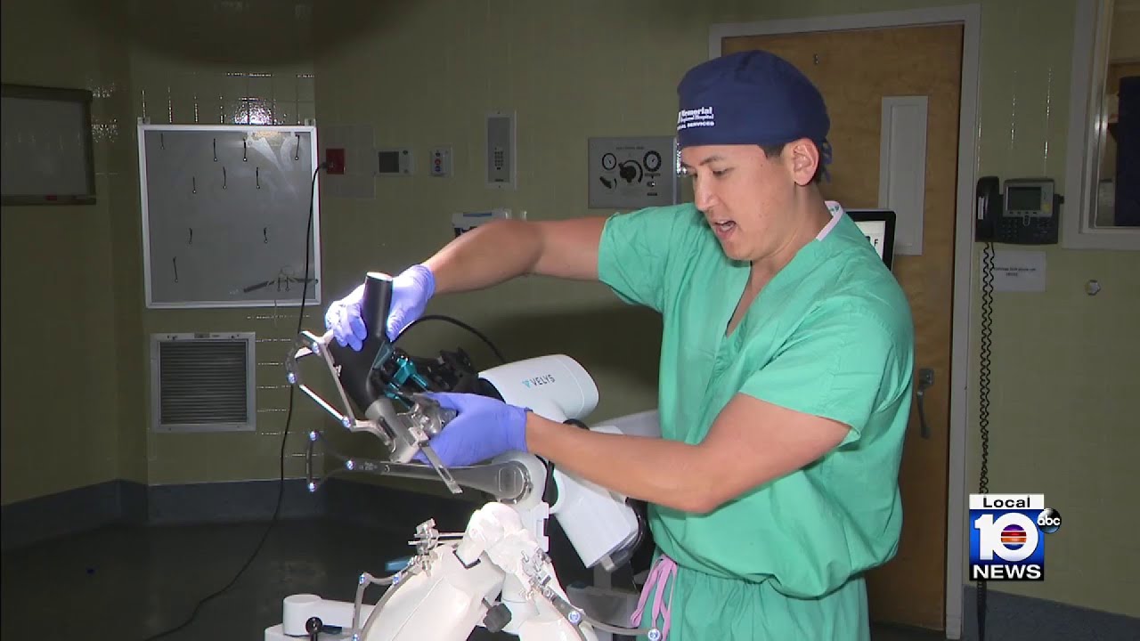
An estimated 900,000 knee replacements are performed in the U.S. every year, but experts say about 15% of patients aren’t totally pleased with the outcome. An advancement in technology is focused on improving those outcomes.

Each person's knee is different. This is why UC San Diego Health offers several surgical options for knee replacements to tailor care to each person's injury and health. Each surgical approach has benefits for the right surgical candidate. Our surgeons can discuss what option is most appropriate for each person.
To learn more about knee replacement options at UC San Diego Health, visit:
https://health.ucsd.edu/specia....lties/orthopedics/jo
Francis Gonzales, MD, is a board-certified orthopedic surgeon who specializes in adult hip and knee joint replacement surgery. Learn more about Dr. Gonzales: https://providers.ucsd.edu/det....ails/11935/orthopedi
UC San Diego Health is repeatedly ranked among the nation's best in orthopedic care by U.S. News & World Report. We are also a Blue Distinction Center recognized for our treatment expertise and better overall patient results for knee replacement, as well as a designated Center of Excellence for orthopedic care by Optum. This means you'll receive expert, safe and cost-effective care.
UC San Diego Health's orthopedic surgeons are the first and only in San Diego to offer customized knee replacements with the ROSA knee system — for a faster recovery and more natural feeling knee. Talk to one of our surgeons about whether a ROSA knee replacement is right for you. https://health.ucsd.edu/specia....lties/orthopedics/jo#a

Outpatient -- or same-day -- knee replacement surgery is more convenient than traditional knee replacement surgery and often can help you recover faster.
Outpatient -- or same-day -- knee replacement surgery is more convenient than traditional knee replacement surgery and often can help you recover faster. At Duke Ambulatory Surgery Center Arringdon, your knee replacement will be followed immediately by physical therapy to get you moving and start your recovery process right away. Our expert joint replacement team ensures your knee replacement surgery is safe and effective so you can return to the comfort of your home as soon as possible.
Tough to beat! Head #InsideTheOR with S. Christopher Malaisrie, MD, and witness open heart surgery by one of the best cardiology and heart surgery programs in the nation as ranked by US News and World Report.

Join Dr. Parsia Vagefi, Chief of Surgical Transplantation and Dr. Steven Hanish, Surgical Director of Liver Transplantation, as they grant unprecedented access to the OR while performing a #Liver #Transplant #Surgery.
To find out more about UT Southwestern's transplant programs visit:
https://www.utswmed.org/transplant
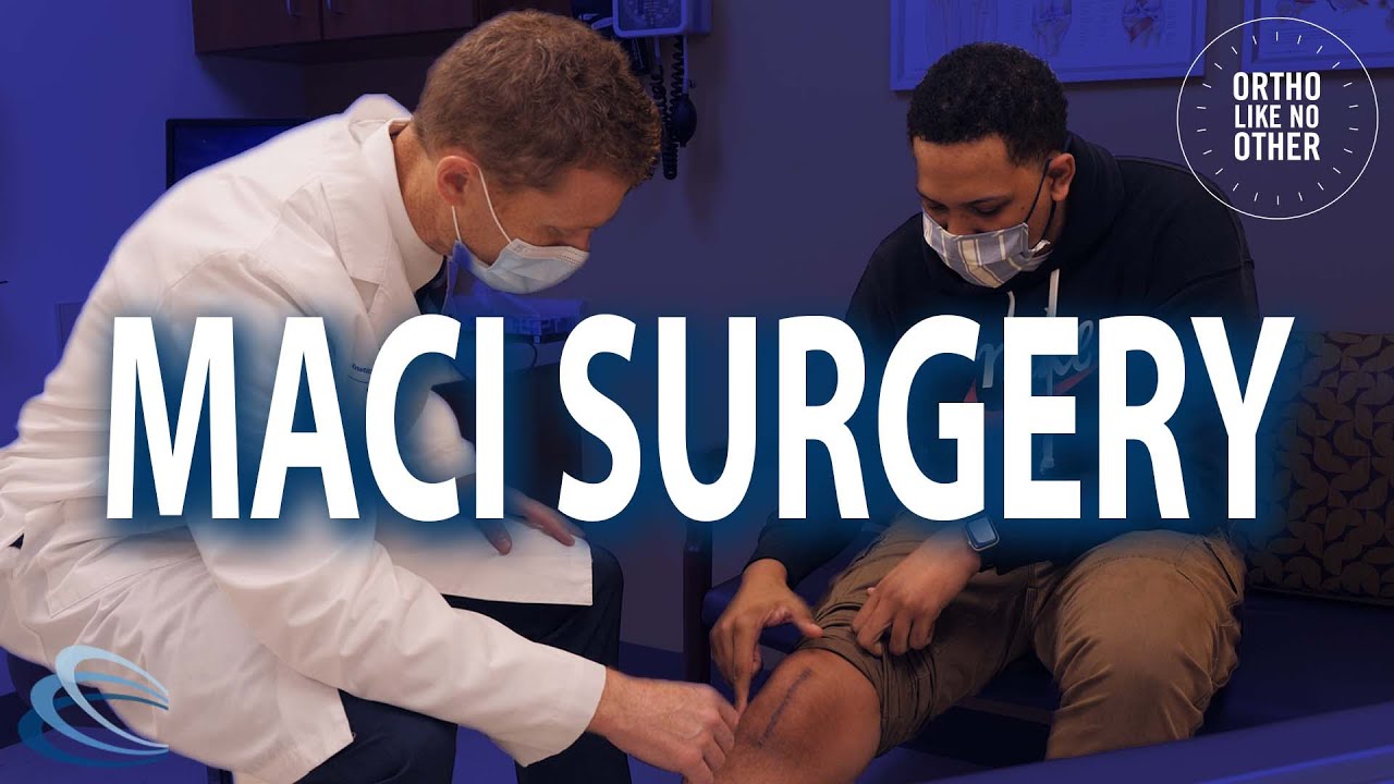
Lattrell Wells was a perfect candidate for the MACI procedure. Dr. Michael O'Malley is a sports medicine surgeon at Carilion Clinic, "It’s a two stage procedure. So what we do is we actually harvest a small portion of the patient's cartilage and bone cells and we send it to a lab where the lab then that grows additional cartilage cells. It comes back to us in a little sheet and six weeks after that initial surgery, we re-implant the cartilage in a second surgery where we implant that sheet depending on the size of lesion right where his defect. This the only option where there’s virtually no risk of any kind of graft rejection or anything of that nature.

In this video, Dr. Robert Rozbruch, chief of Limb Lengthening and Complex Reconstruction at Hospital for Special Surgery performs an osseointegration after a primary amputation. The patient, a 40 year old woman, had chronic nerve pain and compromised function of her residual limb.
For more information, visit: https://www.limblengthening.com/
https://www.hss.edu/limblengthening
https://www.hss.edu/LSARC
https://www.facebook.com/limblengtheningNYC
https://www.instagram.com/limblengthening
https://www.twitter.com/limblengthen
https://www.youtube.com/channe....l/UC-JL_X6ALjZXiXtcP
key words: Osseointegration, Amputee, Amputation, Limb Replacement, Tibia, Osseointegration
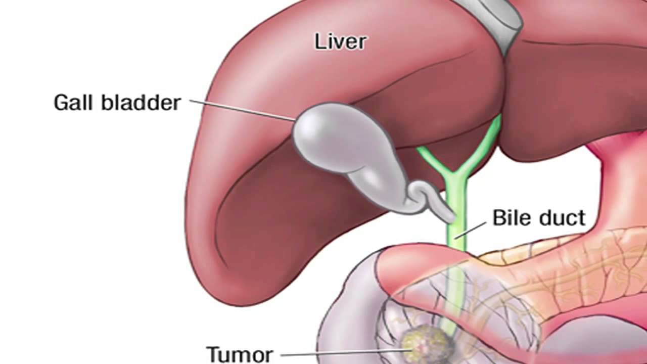
Dr. Horacio Asbun, Mayo Clinic in Florida, explains the Whipple procedure using this animated graphic of a pancreas. Cancer of the pancreas affects 45,000 people every year in the U.S., and it is the fourth leading cause of cancer-related deaths. The five-year overall survival rate if a tumor is detected early and surgically removed is 22 percent, versus 6 percent without early detection and surgery. To learn more, visit http://mayocl.in/2zk7FDi.
This video in Spanish/español: https://www.youtube.com/watch?v=N_zWboNMKWk
Cleft palate is among the most common birth defects affecting children in North America. The incomplete formation of the roof of the mouth can occur individually, or in addition to cleft lip. Cleft palate repair is a type of plastic surgery to correct this abnormal development both to restore function and a more normal appearance. This video explains what to expect for families scheduled for cleft palate surgery at the Craniofacial Anomalies Program at University of Michigan C.S. Mott Children's Hospital.
Learn more about our program at http://www.mottchildren.org/craniofacial
