
- Physical Examination
- Surgical Examination
- Ophthalmology
- Clinical Skills
- Orthopedics
- Surgery Videos
- Laparoscopy
- Pediatrics
- Funny Videos
- Cardiothoracic Surgery
- Nursing Videos
- Plastic Surgery
- Otorhinolaryngology
- Histology and Histopathology
- Neurosurgery
- Dermatology
- Pediatric Surgery
- Urology
- Dentistry
- Oncology and Cancers
- Anatomy Videos
- Health and Fitness
- Radiology
- Anaesthesia
- Physical Therapy
- Pharmacology
- Interventional Radiology
- Cardiology
- Endocrinology
- Gynecology
- Emergency Medicine
- Psychiatry and Psychology
- Childbirth Videos
- General Medical Videos
- Nephrology
- Physiology
- Diet and Food Health
- Diabetes Mellitus
- Neurology
- Women Health
- Osteoporosis
- Gastroenterology
- Pulmonology
- Hematology
- Rheumatology
- Toxicology
- Nuclear Medicine
- Infectious Diseases
- Vascular Disease
- Reproductive Health
- Burns and Wound Healing
- Other
Top videos
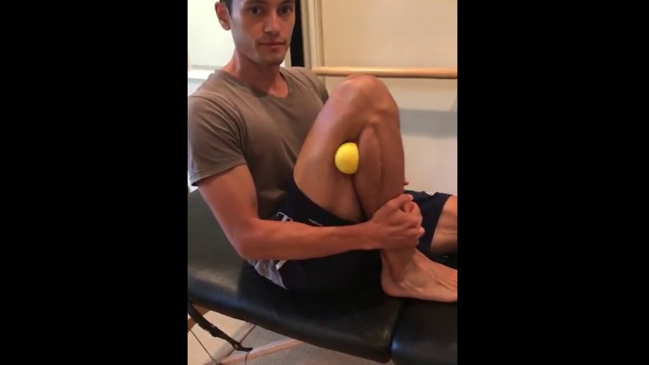

A displaced fibular head can create tightness, pain, and even numbness or tingling along the outside of your knee and down your leg. This most often occurs after a modest hyperextension knee injury, such as landing on one leg after jumping. If you have lingering knee pain and are searching for an answer, try this move

This 3D animation video explains airway clearance anatomy & physiology in the lungs.
Learn more about Baxter Respiratory Health products at www.hillrom.com/en/products-ca....tegory/non-invasive-
Rx Only. For safe and proper use of product mentioned herein, please refer to the Instructions for Use or Operator manual.
The information contained in these videos is provided for educational purposes only and is not intended nor implied to be a substitute for professional medical advice. You assume full responsibility for how you choose to use this information. Please speak with your healthcare provider about any questions you may have regarding a medical condition.
Baxter retains all right, title, and interest in and to the video, and retains the right to demand that you immediately cease use of the video and unembed the video. Baxter may discontinue or disable videos you have embedded at any time for any reason. You will not misrepresent the content contained in the video or use it in conjunction with price comparisons, in derogatory comparisons or in negative comparisons, with Baxter's competitor's products, or in derogatory or negative commentaries about Baxter's products - doing so may subject you to liability. Any and all claims made by you regarding the use, operation, quality, etc. of Baxter's products are your own, and you shall be responsible for ensuring that all such claims comply fully with all applicable federal, state and local laws.
US-FLC174-230024 v1

Make sure to subscribe to our channel to get regular updates on other interesting legal animations.
Interested in other medical videos? Click the link below to watch a surgery animation!
https://www.youtube.com/watch?v=Cc6-Y_CIHx4&t=53s&ab_channel=CourtroomAnimation
Or, if you want to watch a video about a diskectomy, a common but major surgery, watch this video:
https://www.youtube.com/watch?v=PVoOVUPhdaM&t=8s&ab_channel=CourtroomAnimation
--------------------------------------------------------------------------------------------------------
Learn more about medical animations from our in-depth guide:
https://courtroomanimation.com..../qa-everything-about
Learn more about medical graphics in general from our free eBook:
https://info.courtroomanimatio....n.com/ebook-complete
Check out our blog page for other topics on legal graphics and 3D medical illustrations:
https://courtroomanimation.com/blog/
--------------------------------------------------------------------------------------------------------
Connect with us online!
Facebook: https://www.facebook.com/courtroomanimation
LinkedIn: https://www.linkedin.com/compa....ny/courtroom-animati
Twitter: https://twitter.com/courtanimation
Instagram: https://www.instagram.com/courtroomanimation/
Or give us a call to see how we can create a trial animation for your plaintiff's civil case, and help you land a more favorable verdict or settlement!
(424) 321-7488
--------------------------------------------------------------------------------------------------------
#discectomy #medicalanimation #surgery #forensicanimation #legaltechnology #legaltech #medicalvideos #lumbar #lumbarpain

To learn more about licensing this video for content marketing or patient education purposes, visit: http://www.nucleushealth.com/?utm_source=youtube&utm_medium=video-description&utm_campaign=hiv-112513
This video, created by Nucleus Medical Media, shows the function of white blood cells in normal immunity. It also portrays how the human immunodeficiency virus (HIV) affects the immune system and causes acquired immunodeficiency syndrome (AIDS). Common types of antiretroviral medications used to treat HIV and AIDS are also shown.
#HIV #AIDS #HumanImmunodeficiencyVirus
ANH13111

https://bit.ly/3HIStRc #shorts
Tracheotomy and tracheostomy are surgical procedures that create an opening in the trachea (windpipe) to help patients breathe when they have difficulty doing so through the nose or mouth. Though they are similar in purpose, there are some key differences between them.
Tracheotomy is a temporary procedure that involves creating a small incision in the trachea to insert a breathing tube. The tube is typically removed once the patient no longer requires it, and the incision heals on its own. Tracheostomy, on the other hand, is a more permanent solution that involves creating a hole in the trachea and inserting a tracheostomy tube, which remains in place for an extended period.
Indications for these procedures include:
Airway obstruction due to trauma, tumors, or infection
Severe respiratory distress or failure
Prolonged mechanical ventilation
Inability to protect the airway due to neurological disorders or impaired consciousness
Steps for performing a tracheotomy and tracheostomy:
Preparation: The patient is positioned, and the neck area is cleaned and draped. Local anesthesia is often administered, although general anesthesia may be used in some cases.
Incision: A small incision is made in the neck, and the muscles and tissues are carefully separated to expose the trachea.
Tracheal opening: A small opening is made in the trachea, typically between the second and third tracheal rings.
Tube insertion: A tracheotomy tube is inserted through the incision and into the trachea for a tracheotomy, while a tracheostomy tube is inserted for a tracheostomy. Both tubes are secured in place.
Confirmation: Proper placement of the tube is confirmed by listening for breath sounds and checking for adequate ventilation.
Pre-operative care typically involves a thorough assessment of the patient's medical history, as well as any necessary imaging studies or lab tests to ensure the procedure is appropriate and safe. Informed consent should be obtained from the patient or their legal representative.
Post-operative care includes monitoring the patient's vital signs, ensuring the tube remains secure and patent, and managing any pain or discomfort. For tracheostomy patients, regular cleaning and maintenance of the stoma (the opening in the trachea) and the tracheostomy tube are essential to prevent infection and other complications. Long-term care may involve speech therapy, respiratory therapy, and support from a multidisciplinary team to address any ongoing needs.
It's crucial to remember that these procedures should only be performed by trained medical professionals in a clinical setting.
for additional information about this procedure check our article @ www.medicalartsshop.com
For more free resources, find us on Pinterest & Facebook pages:
https://www.pinterest.ca/medicalartsofficial/
https://www.facebook.com/Medicalartsofficial
https://www.youtube.com/@medic....alarts?sub_confirmat
https://www.instagram.com/medicalartsofficial/
https://www.tiktok.com/@medicalarts
This video and associated content are for entertainment and educational purposes only!!

Follow one family's journey through the agony of waiting for a donor, a 31-hour surgery, and the prospect of a long road to recovery.
➡ Watch the full documentary here: https://youtu.be/fFua9-Rc4pw
➡ Access our digital archive by becoming a member of National Geographic: https://on.natgeo.com/39wAStK
#NationalGeographic #KatieStubblefield #FaceTransplant
About National Geographic:
National Geographic is the world's premium destination for science, exploration, and adventure. Through their world-class scientists, photographers, journalists, and filmmakers, Nat Geo gets you closer to the stories that matter and past the edge of what's possible.
Get More National Geographic:
Official Site: http://bit.ly/NatGeoOfficialSite
Facebook: http://bit.ly/FBNatGeo
Twitter: http://bit.ly/NatGeoTwitter
Instagram: http://bit.ly/NatGeoInsta
TikTok: http://www.tiktok.com/@natgeo
Tenor: http://on.natgeo.com/31b3Koc
To learn more, please read on here:
https://on.natgeo.com/2vQE1SO
Youngest Face Transplant Recipient in U.S. | National Geographic https://youtu.be/quU9s7I1NLI
National Geographic
https://www.youtube.com/natgeo

This particular video is intended as a demonstration of Neurologic Examination. This demonstration is intended as an example of a neurologic exam which may be used as part of the initial evaluation of patients with complaints that may have an underlying neurologic origin. This video is solely for educational purposes and intended for use to prepare for OSCEs incorporating standardized patient encounters. It is not intended as a demonstration of a comprehensive neurologic examination and is not intended as medical advice or medical guidelines.
It is not intended as a complete instructional video and should not be considered a source of complete physical examination instruction.
Instead, it should be treated as a supplement to independent learning using primary Osteopathic Clinical Skills instructional resources. Clinical skills are best learned and developed with support from faculty in the context of a complete Osteopathic Medical School Curriculum.
Osteopathic Clinical Skills is a channel dedicated to discussing and exploring Osteopathic Clinical Skills concepts for medical students, residents, and clinicians and presenting them in an easy to understand manner.
Attributions:
Many thanks to the University of North Texas Health Science Center Texas College of Osteopathic Medicine (UNTHSC - TCOM) for permitting use of the Simulation facilities and equipment during the production of this video.
Additional thanks to the UNTHSC-TCOM standardized patient and faculty volunteers who participated in this production and provided permission for the use of their image in this video.

Watch this clinical examination video to learn how to diagnose cervical spine pathology.
This video clip is part of the FIFA Diploma in Football Medicine and the FIFA Medical Network. To enrol or to find our more click on the following link http://www.fifamedicalnetwork.com
The Diploma is a free online course designed to help clinicians learn how to diagnose and manage common football-related injuries and illnesses. There are a total of 42 modules created by football medicine experts. Visit a single page, complete individual modules or finish the entire course.
The network provides the opportunity for clinicians around the world to meet and share ideas relating to football medicine. Ask about an interesting case, debate current practice and discuss treatment strategies. Create a profile and log on to interact with other health professionals from around the globe.
This is not medical advice. The content is intended as educational content for health care professionals and students. If you are a patient, seek care of a health care professional.
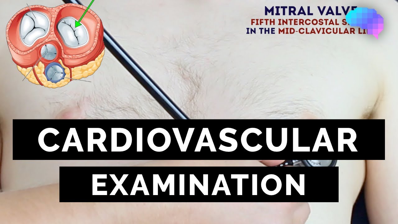
This video demonstrates how to perform a cardiovascular examination in an OSCE station.
You can access our step-by-step OSCE guide to accompany this video here: https://geekymedics.com/cardio....vascular-examination
Check out our other awesome clinical skills resources including:
• 🔥 Geeky Medics Bundles (discounted products): https://app.geekymedics.com/purchase/bundles/
• ✨ 1000+ OSCE Stations: https://app.geekymedics.com/pu....rchase/osce-stations
• 🏥 Geeky Medics OSCE Revision Book: https://app.geekymedics.com/purchase/book/
• 📝 150+ PDF OSCE Checklists: https://geekymedics.com/pdf-osce-checklists/
• 🗂️ 3000+ OSCE Flashcards: https://app.geekymedics.com/pu....rchase/flashcard-col
• 📱 Geeky Medics OSCE App: https://geekymedics.com/geeky-medics-app/
• 🩺 Medical Finals SBA Question Pack: https://app.geekymedics.com/pu....rchase/medical-stude
• 💊 PSA Question Pack: https://app.geekymedics.com/pu....rchase/prescribing-s
Chapters:
- Introduction 00:00
- General inspection 00:35
- Hands 00:46
- Schamroth's window test 01:07
- Capillary refill 01:27
- Pulses 01:35
- Carotid auscultation 02:21
- Carotid pulse 02:43
- Jugular venous pressure 02:55
- Hepatojugular reflux 03:09
- Inspection of the face 03:21
- Inspection of the chest 03:49
- Apex beat 04:12
- Heaves and thrills 04:28
- Heart valve ausculation 04:49
- Accentuation manoeuvres 05:45
- Lung base auscultation 06:23
- Sacral and pedal oedema 06:43
- Summary 07:10
Subscribe to our newsletter to be the first to know about our latest content: https://geekymedics.com/newsletter/ ✉️
Join the Geeky Medics community: 👩👩👧👧
Twitter: http://www.twitter.com/geekymedics
Instagram: https://instagram.com/geekymedics
Facebook: http://www.facebook.com/geekymedics
Always adhere to your medical school/local hospital guidelines when performing examinations or clinical procedures. DO NOT perform any examination or procedure on patients based purely upon the content of these videos. Geeky Medics accepts no liability for loss of any kind incurred as a result of reliance upon the information provided in this video.
Normal heart sounds and aortic regurgitation/stenosis sounds
Recorded on a Thinklabs Digital Stethoscope (https://www.thinklabs.com)
Some people have found this video useful for ASMR purposes.

The cardiac examination is one of the earliest clinical skills that medical students learn. As a junior doctor, the examination of the cardiovascular system can be almost a dreaded examination, as cardiac murmurs can literally take years of exposure in order to gain confidence with their identification through cardiac auscultation.
This video demonstrates not merely the examination of the heart, but the complete cardiovascular system including peripheries.
I hope these clinical skill revision videos are helpful, please like and subscribe and join the community so that we can create more effective videos to help with your journey through medical school
#CardiacExam #ClinicalExamination #asmr

This video shows you how to conduct a clinical examination of the foot and how to identify common causes of foot pain.
This video clip is part of the FIFA Diploma in Football Medicine and the FIFA Medical Network. To enrol or to find our more click on the following link http://www.fifamedicalnetwork.com
The Diploma is a free online course designed to help clinicians learn how to diagnose and manage common football-related injuries and illnesses. There are a total of 42 modules created by football medicine experts. Visit a single page, complete individual modules or finish the entire course.
The network provides the opportunity for clinicians around the world to meet and share ideas relating to football medicine. Ask about an interesting case, debate current practice and discuss treatment strategies. Create a profile and log on to interact with other health professionals from around the globe.
This is not medical advice. The content is intended as educational content for health care professionals and students. If you are a patient, seek care of a health care professional.
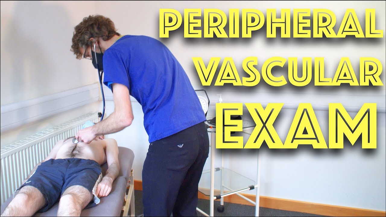
Peripheral Vascular Examination OSCE - Clinical Skills - Dr Gill
In the cardiovascular examination, particularly in the case of an OSCE station, we conclude the examination often by stating that the examiner would want to perform:
- An ECG
- Check full blood count
- and "do a peripheral vascular examination
In this video, we demonstrate that oft-talked about, but comparatively less common examination.
Starting off, with the examination of the hands, the radial, brachial and carotid pulses. before moving down to assess for a AAA, checking the femoral and popliteal pulses, before wrapping up around the ankle with the posterior tibial and dorsalis pedis pulses
For completeness, the cardiovascular examination is demonstrated here
https://www.youtube.com/watch?v=ECs9O5zl6XQ&t=2s
#PeripheralVascular #ClinicalSkills #DrGill
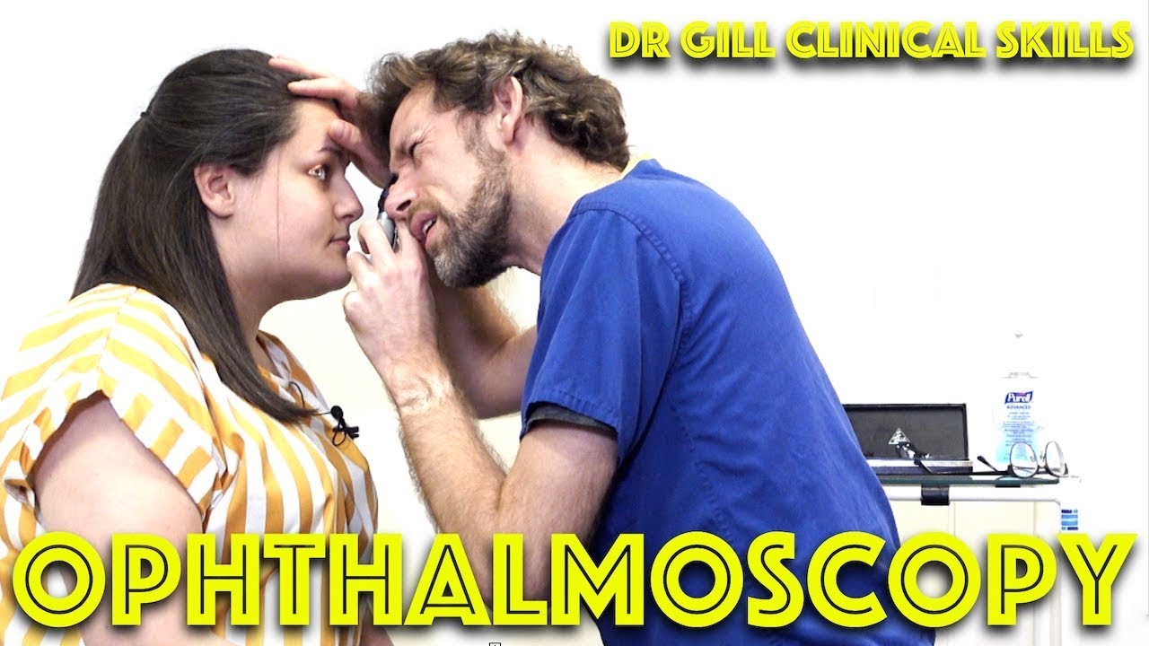
Ophthalmoscopy - Eye Clinical Examination - OSCE - Dr Gill
Direct Ophthalmoscopy use of the eyes is a very challenging clinical skill, incorporating both the examiner's knowledge of the retina, but also understanding the use of the ophthalmoscope
In this clinical skills tutorial, we look at the use of the direct ophthalmoscope as part of an ophthalmic examination
it should be noted that in the ideal circumstances, the room lights will be dimmed during the examination, and dilating eye drops used to improve the visualisation of the fundus
Some people may notice an ASMR effect from this clinical examination
#DrGill #Ophthalmoscopy #ClinicalSkills #EyeExam

Guest surgeon Dr. Nicole Callan gives us an introduction to hernias. She discusses what a hernia is, the different types of hernias, and an introduction to treatment.
Please see the complete user agreement at talkingwithdocs.com
Medical emergency
Do NOT use the Video for medical emergencies. If you have a medical emergency, call a physician or qualified healthcare provider, or CALL 911 immediately. Under no circumstances should you attempt self-treatment based on anything you have seen or read on the Video.
General information is not medical advice
The general information provided on the Video is for informational purposes only and is not professional medical advice, diagnosis, treatment, or care, nor is it intended to be a substitute therefore. Always seek the advice of your physician or other qualified health provider properly licensed to practise medicine or general healthcare in your jurisdiction concerning any questions you may have regarding any information obtained from this Video and any medical condition you believe may be relevant to you or to someone else. Never disregard professional medical advice or delay in seeking it because of something you have read on this Video. Always consult with your physician or other qualified healthcare provider before embarking on a new treatment, diet, or fitness program. Information obtained on the Video is not exhaustive and does not cover all diseases, ailments, physical conditions, or their treatment.
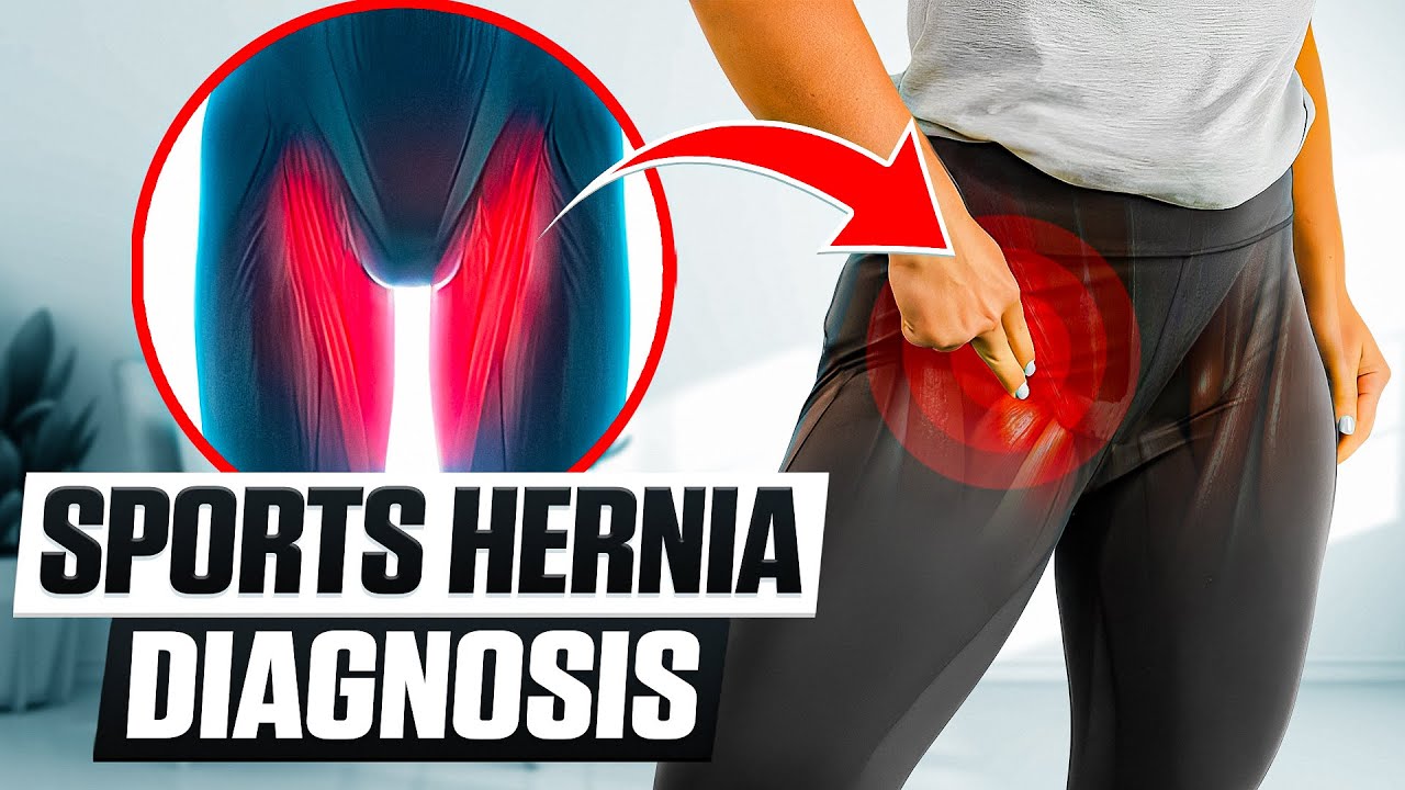
In this video, we show a sports hernia self treatment we give many of our clients. It is not the only part of treatment. Grabbing the skin around the region of the groin strain can reduce pain and stiffness with turning and twisting. Sports hernias are often misdiagnosed with hip labrum tears, hip impingement, adductor tendonitis and abdominal strains.
Want more information? We have a more detailed free webinar on our page here. https://bit.ly/37thtNF
Want some treatment or suggestions of exercises or stretches? Contact us! We have in-person and virtual sessions.
Costa Mesa CA 715-502-4243 www.p2sportscare.com
Sports Hernia Diagnosis
What Is A Sports Hernia?
A sports hernia is tearing of the transversalis fascia of the lower abdominal or groin region. A common misconception is that a sports hernia is the same as a traditional hernia. The mechanism of injury is rapid twisting and change of direction within sports, such as football, basketball, soccer and hockey.
The term “sports hernia” is becoming mainstream with more professional athletes being diagnosed. The following are just to name a few:
Torii Hunter
Tom Brady
Ryan Getzlaf
Julio Jones
Jeremy Shockey
If you follow any of these professional athletes, they all seem to have the same thing in common: Lingering groin pain. If you play fantasy sports, this is a major headache since it seems so minor, but it can land a player on Injury Reserve on a moments notice. In real life, it is a very frustrating condition to say the least. It is hard to pin point, goes away with rest and comes back after activity, but is hardly painful enough to make you want to stop. It lingers and is always on your mind. And if you’re looking for my step-by-step sports hernia rehab video course here it is.
One the best definitions of Sport hernias is the following by Harmon:
The phenomena of chronic activity–related groin pain that it is unresponsive to conservative therapy and significantly improves with surgical repair.”
This is truly how sports hernias behave in a clinical setting. It is not uncommon for a sports hernia to be unrecognized for months and even years. Unlike your typical sports injury, most sports medicine offices have only seen a handful of cases. It’s just not on most doctors’ radar. The purpose of this article is not only to bring awareness about sports hernias, but also to educate.
Will you find quick fixes in this article for sports hernia rehab?
Nope. There is no quick fix for this condition, and if someone is trying to sell you one, they are blowing smoke up your you-know-what.
Is there a way to decrease the pain related to sports hernias?
Yes. Proper rehab and avoidance of activity for a certain period of time will assist greatly, but this will not always stop it from coming back. Pain is the first thing to go and last thing to come. Do not be fooled when you become pain-free by resting it. Pain is only one measure of improvement in your rehab. Strength, change of direction, balance and power (just to name a few) are important, since you obviously desire to play your sport again. If you wanted to be a couch potato, you would be feeling better in no time. Watching Sports Center doesn’t require any movement.
Why is this article so long?
There is a lot of information on sports hernias available to you on the web. However, much of the information is spread out all over the internet and hard for athletes to digest due to complicated terminology. This article lays out the foundational terminology you will need to understand what options you have with your injury. We will go over anatomy, biomechanics, rehab, surgery, and even the fun facts. The information I am using is from the last ten years of medical research, up until 2016. We will be making updates overtime when something new is found as well. So link to this page and share with friends. This is the best source for information on sports hernias you will find.
Common Names (or Aliases?) for Sports Hernias
Sportsman’s Hernia
Athletic Pubalgia
Gilmore’s Groin
How Do You Know If You Have A Sports Hernia?
Typical athlete characteristics:
Male, age mid-20s
Common sports: soccer, hockey, tennis, football, field hockey
Motions involved: cutting, pivoting, kicking and sharp turns
Gradual onset
How A Sports Hernia Develops
Chronic groin pain typically happens over time, which is why with sports hernias, we do not hear many stories of feeling a “pop” or a specific moment of injury. It is the result of “overuse” mechanics stemming from a combination of inadequate strength and endurance, lack of dynamic control, movement pattern abnormalities, and discoordination of motion in the groin area.
#sportsherniadiagnosisselftreatment #sportshernia #california


Dr. Kathryn Baerman is a Board Certified General Surgeon specializing in Women's Health and Breast Care. She shares with us that in women, hernias present differently than in men. If you are experiencing groin pain, it may be a hernia.
To visit Dr. Baerman in Apex, Chapel Hill, or Durham, North Carolina, call 919-281-1699 to schedule an appointment with her at EmergeOrtho.

The anatomy of the direct and indirect inguinal hernia.
Music:
Berries and Lime by Gregory David
https://www.epidemicsound.com/track/z6iCiiyCPm/

Thinking about laser eye surgery? Watch our live stream and learn what the procedure involves.
Find out more about laser eye surgery
on our website: www.opticalexpress.co.uk
in our online magazine: www.opticalexpress.co.uk/magazine
on our Facebook page: https://www.facebook.com/opticalexpress/
or on our Instagram page: https://www.instagram.com/opticalexpressuk

Recovery Tips
LASIK eye surgery is the best known and most commonly performed laser refractive surgery to correct vision problems. The total recovery time is 1 to 2 weeks. However, vision may fluctuate slightly over the next 2 months.
Avoid watching television or reading for the first few days.
You can get back to work after 1 week.
Avoid applying pressure on the eyes for 7 days.
Avoid dust, smoke, yard and garden work, and eye make-up.
Wear eye shields at night given by a surgeon for 1 week.
Driving is allowed after 4 to 5 days.
Avoid swimming or using a hot tub for 2 weeks after surgery.
While using the computer, take frequent breaks and lubricate your eyes with artificial tears.
For treatment assistance in your country or abroad:
Email: hello@vaidam.com
Phone/WhatsApp/Viber: +91-9650001746
Website: www.vaidam.com
Vaidam is an ISO and NABH accredited medical assistance company. Patients from 100+ countries have used our services.
Useful Links:
India
Doctors: https://www.vaidam.com/doctors/opthalmology/lasik-procedure/india
Hospitals: https://www.vaidam.com/hospitals/opthalmology/lasik-procedure/india
Cost of Lasik Eye Surgery: https://www.vaidam.com/cost/lasik-procedure-cost-in-india
Turkey
Doctors: https://www.vaidam.com/doctors/opthalmology/lasik-procedure/turkey
Hospitals: https://www.vaidam.com/hospitals/opthalmology/lasik-procedure/turkey
