
সেরা ভিডিও


This patient presented to the ER for umbilical pain and had a history of umbilical hernia. He was concerned about the possibility of incarceration of the hernia.
In this video we explain how the clinical exam helps to differentiate a simple painful hernia from an incarcerated one.
***Thanks to the patient for sharing his history and exam with YouTube world***

#final #fumc #mbbs #medicalstudents #mbbsabroad #doctor #fcps #fcpspart #surgeryeducation #surgeryreview #trainee #exampreparation

Learn all about the differences between these important hernias!

Are you worried about getting a sports hernia exam? In this video, we'll show you exactly what to expect when you get your hernia exam.
We'll take you through the various steps that are taken during the hernia exam, so you can have a more comfortable and informative experience. After watching this video, you'll have a better idea of what to expect and be prepared for your hernia exam!
#sportshernia #groinpain #california
جروب الداتا على #تليجرام 👇🏻
https://t.me/IslamKhalaf
أي استفسار 👇🏻
twitter
https://twitter.com/Islamkhalaf0
Insta
https://www.instagram.com/islamkhalaf1
FB
https://www.facebook.com/islamkhalaf.A


Dr. Kathryn Baerman is a Board Certified General Surgeon specializing in Women's Health and Breast Care. She shares with us that in women, hernias present differently than in men. If you are experiencing groin pain, it may be a hernia.
To visit Dr. Baerman in Apex, Chapel Hill, or Durham, North Carolina, call 919-281-1699 to schedule an appointment with her at EmergeOrtho.


MRCPCH Clinical Revision - more videos at http://mrcpch.paediatrics.co.uk
Revise for your MRCPCH Clinical exam, with videos and high quality content created by the London Paediatrics Trainees Committee.
Examiner: Jonathan Round
Candidate: Amitav Parida
Filming: Mary Chesshyre, Huey Miin Lee, Chris Kelly
Thank you to the Evelina Children's Hospital for allowing us to film during their MRCPCH Revision Course (https://www.guysandstthomaseve....nts.co.uk/mrcpch-cli

This video provides a demonstration of how to assess for transillumination when assessing scrotal swelling.
Read our step-by-step guide here: https://geekymedics.com/testic....ular-examination-osc
Check out our other awesome clinical skills resources, including:
• 🔥 Geeky Medics Bundles (discounted products): https://app.geekymedics.com/purchase/bundles/
• ✨ 1000+ OSCE Stations: https://app.geekymedics.com/pu....rchase/osce-stations
• 🏥 Geeky Medics OSCE Revision Book: https://app.geekymedics.com/purchase/book/
• 📝 150+ PDF OSCE Checklists: https://geekymedics.com/pdf-osce-checklists/
• 🗂️ 3000+ OSCE Flashcards: https://app.geekymedics.com/pu....rchase/flashcard-col
• 📱 Geeky Medics OSCE App: https://geekymedics.com/geeky-medics-app/
• 🩺 Medical Finals SBA Question Pack: https://app.geekymedics.com/pu....rchase/medical-stude
• 💊 PSA Question Pack: https://app.geekymedics.com/pu....rchase/prescribing-s
Subscribe to our newsletter to be the first to know about our latest content: https://geekymedics.com/newsletter/ ✉️
Join the Geeky Medics community: 👩👩👧👧
Twitter: http://www.twitter.com/geekymedics
Instagram: https://instagram.com/geekymedics
Facebook: http://www.facebook.com/geekymedics
Always adhere to your medical school/local hospital guidelines when performing examinations or clinical procedures. DO NOT perform any examination or procedure on patients based purely on the content of these videos. Geeky Medics accepts no liability for loss of any kind incurred as a result of reliance upon the information provided in this video.

this video about identifying a hernia vs a cyst
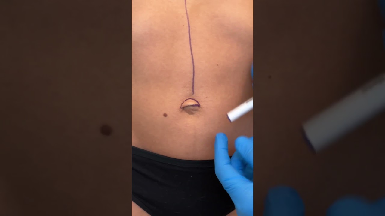
Check out @barrettplasticsurgery on TikTok!
Like and subscribe for more! #shorts #medical #plasticsurgery
More information:
www.drdanielbarrett.com

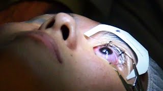
Business Insider's Michelle Yan has been nearsighted since she was 9 years old. After laser eye surgery, she has 20/20. She walks us through the pre-surgery steps, the actual surgery, as well as the recovery process.
MORE MEDICAL TECH:
8 Medical Procedures That Are Improving Lives
https://www.youtube.com/watch?v=kTMMrAP6DNI
13 Medical Procedures Changing The Health World
https://www.youtube.com/watch?v=VAR44vnxWis
Lifelike Medical Robot Actually Bleeds
https://www.youtube.com/watch?v=IjnhmcCQLsc
------------------------------------------------------
#Lasik #Surgery #TechInsider
Tech Insider tells you all you need to know about tech: gadgets, how-to's, gaming, science, digital culture, and more.
Visit us at: https://www.businessinsider.com
TI on Facebook: https://www.facebook.com/techinsider
TI on Instagram: https://www.instagram.com/tech_insider/
TI on Twitter: https://twitter.com/techinsider
TI on Amazon Prime: http://read.bi/PrimeVideo
INSIDER on Snapchat: https://insder.co/2KJLtVo
------------------------------------------------------
What It's Like To Get Laser Eye Surgery
#lasik #eyesurgery #BusinessInsiderIndia #lasereyesurgery
Business Insider's Michelle Yan has been nearsighted since she was 9 years old. After laser eye surgery, she has 20/20. She walks us through the pre-surgery steps, the actual surgery which the doctor referred to as a spa for eyes, as well as the recovery process.
Don't miss the suggestions and recommendations she has for all those who are planning to undergo laser eye surgery for themselves, towards the end.
-----------------------------------------------
Business Insider India features the country's business heavyweights, ranging from start-ups to veterans across industries. It offers amazing synergy between India and its foreign counterpart - developed, analyzed and presented in hallmark BI Style. It covers latest news & trends on Tech, Business, Careers, Startups and Finance.
For more such content, Visit us at: https://www.businessinsider.in/
Subscribe at: https://www.youtube.com/channe....l/UC62AhAC25Ukuscqux
BI on Facebook: https://www.facebook.com/BusinessInsiderIndia/
BI on Twitter:https://twitter.com/BiIndia
BI on Instagram: https://www.instagram.com/businessinsiderin/
BI on Tiktok: https://vm.tiktok.com/GNpHXg/
What It's Like To Get Laser Eye Surgery
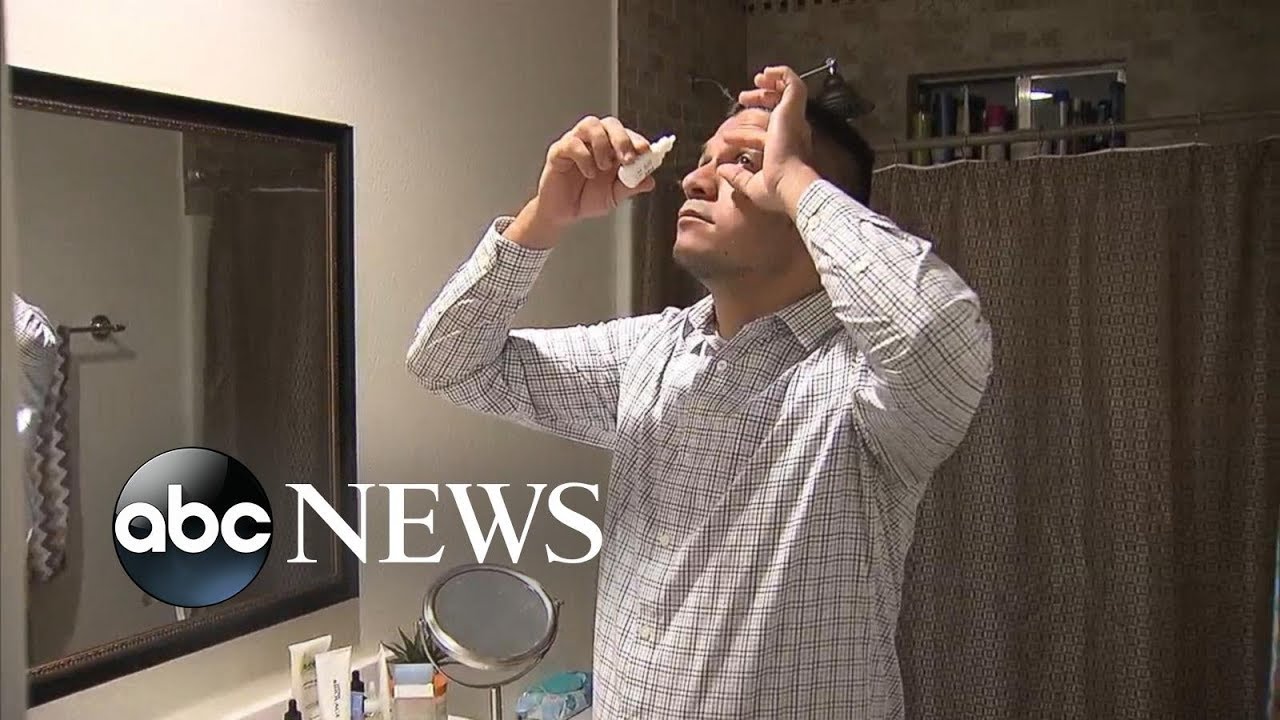
One man is speaking out about the potential risks of laser eye surgery, after he says the procedure left his vision permanently impaired.
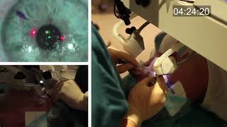

In this video, Professor Dan Reinstein performs a bilateral LASIK procedure filmed in real-time to demonstrate the full 8 and-a-half minute procedure from multiple angles. The superior design and experience of the Carl Zeiss Meditec Visumax femtosecond Laser for flap creation is seen, where the patient is only in contact with the device for about 30 seconds with extremely low contract force such that the patient feels effectively nothing, there are no red splodges (subconjunctival haemorages) left behind. From the surgeons' standpoint there is no device that is easier to use or faster for LASIK flap creation. The Carl Zeiss Meditec MEL80 excimer laser portion of the procedure is seamlessly integrated and incorporates all the features that make clinical outcomes so reproducible including the unique cone-for-controlled-atmosphere (CCA) and high efficiency, high sensitivity calibration test which can be performed for each individual patient to compensate for minor changes in energy that occur with excimer laser devices during the course of a day.
For reference to the clinical outcomes for LASIK with the MEL80 in presbyopia using PRESBYOND Laser Blended Vision see:
Reading glasses presbyopia (ageing eyes) only:
LASIK for presbyopia correction in emmetropic patients using aspheric ablation profiles and a micro-monovision protocol with the Carl Zeiss Meditec MEL 80 and VisuMax.
J Refract Surg. 2012 Aug;28(8):531-41. Reinstein DZ, Carp GI, Archer TJ, Gobbe M.
http://www.ncbi.nlm.nih.gov/pubmed/22869232
Short sighted, astigmatism and presbyopia (ageing eyes)
LASIK for Myopic Astigmatism and Presbyopia Using Non-Linear Aspheric Micro-Monovision with the Carl Zeiss Meditec MEL 80 Platform.
J Refract Surg. 2011 Jan;27(1):23-37. Epub 2010 Mar 1.
Reinstein DZ, Archer TJ, Gobbe M.
http://www.ncbi.nlm.nih.gov/pubmed/20205360
Long-sighted, astigmatism and presbyopia (ageing eyes)
LASIK for hyperopic astigmatism and presbyopia using micro-monovision with the Carl Zeiss Meditec MEL80 platform.
J Refract Surg. 2009 Jan;25(1):37-58. Reinstein DZ, Couch DG, Archer TJ.
http://www.ncbi.nlm.nih.gov/pubmed/19244952
For more information about laser eye surgery and PRESBYOND Laser Blended Vision, please contact the London Vision Clinic on 020 7224 1005.
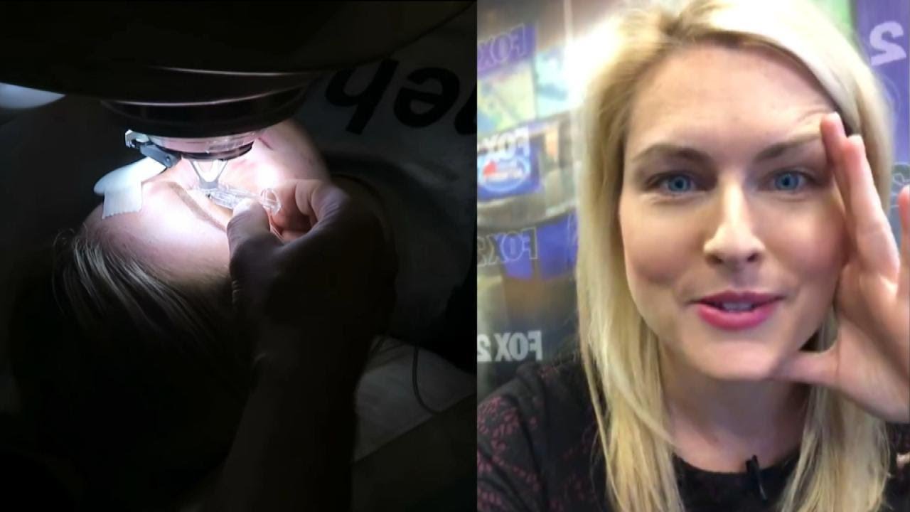
Detroit TV meteorologist Jessica Starr posted a heart-wrenching video on social media a month before dying by suicide this week. She had told viewers she was struggling in the aftermath of undergoing Lasik surgery. After learning of her death, her heartbroken colleagues on WJBK fought back tears live on TV. Twelve people have died by suicide after suffering pain and even blindness after the operation. Inside Edition also spoke to a doctor who wants the surgery banned. #InsideEdition

St. Luke's originally broadcast this live in a webcast and later re-purposed it for air on KCRG-TV9 as an educational video. It is hosted by Ashley Hinson, KCRG-TV9 anchor and Dr. Sandeep Munjal. Dr. Jeff Nassif performs the knee replacement surgery on an eastern Iowa woman. St. Luke's has a rapid recovery joint replacement program, which gets people back to life quickly after surgery.
