
- Physical Examination
- Surgical Examination
- Ophthalmology
- Clinical Skills
- Orthopedics
- Surgery Videos
- Laparoscopy
- Pediatrics
- Funny Videos
- Cardiothoracic Surgery
- Nursing Videos
- Plastic Surgery
- Otorhinolaryngology
- Histology and Histopathology
- Neurosurgery
- Dermatology
- Pediatric Surgery
- Urology
- Dentistry
- Oncology and Cancers
- Anatomy Videos
- Health and Fitness
- Radiology
- Anaesthesia
- Physical Therapy
- Pharmacology
- Interventional Radiology
- Cardiology
- Endocrinology
- Gynecology
- Emergency Medicine
- Psychiatry and Psychology
- Childbirth Videos
- General Medical Videos
- Nephrology
- Physiology
- Diet and Food Health
- Diabetes Mellitus
- Neurology
- Women Health
- Osteoporosis
- Gastroenterology
- Pulmonology
- Hematology
- Rheumatology
- Toxicology
- Nuclear Medicine
- Infectious Diseases
- Vascular Disease
- Reproductive Health
- Burns and Wound Healing
- Other
Top videos


Thinking about laser eye surgery? Watch our live stream and learn what the procedure involves.
Find out more about laser eye surgery
on our website: www.opticalexpress.co.uk
in our online magazine: www.opticalexpress.co.uk/magazine
on our Facebook page: https://www.facebook.com/opticalexpress/
or on our Instagram page: https://www.instagram.com/opticalexpressuk

Preeclampsia is a pregnancy complication characterized by high blood pressure and signs of damage to another organ system, often the kidneys. Preeclampsia usually begins after 20 weeks of pregnancy in a woman whose blood pressure had been normal. Even a slight rise in blood pressure may be a sign of preeclampsia. Left untreated, preeclampsia can lead to serious — even fatal — complications for both you and your baby. If you have preeclampsia, the only cure is delivery of your baby. If you're diagnosed with preeclampsia too early in your pregnancy to deliver your baby, you and your doctor face a challenging task. Your baby needs more time to mature, but you need to avoid putting yourself or your baby at risk of serious complications.
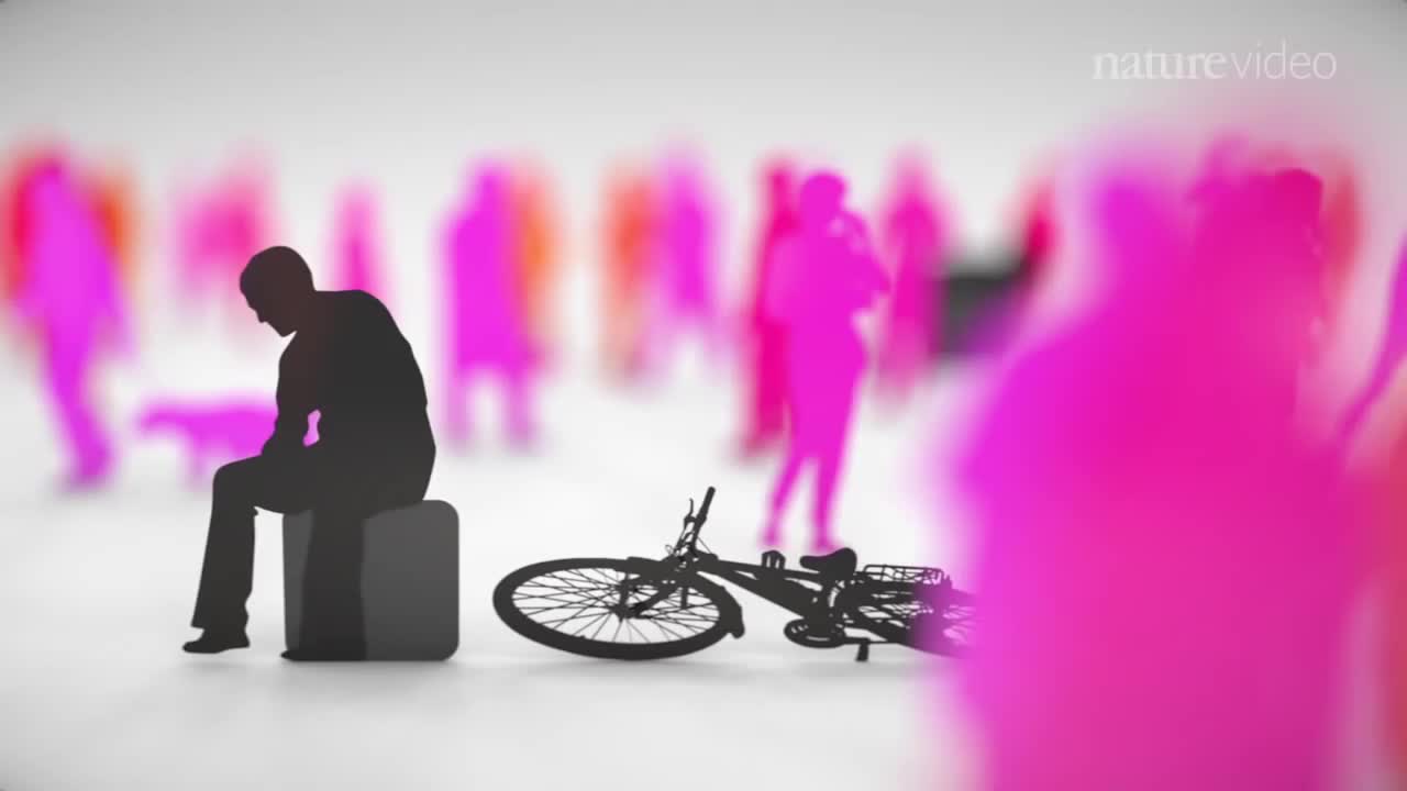
When you’re depressed, it can feel like you’ll never get out from under a dark shadow. However, even the most severe depression is treatable. So, if your depression is keeping you from living the life you want to, don’t hesitate to seek help. Learning about your depression treatment options will help you decide what approach is right for you. From therapy to medication to healthy lifestyle changes, there are many effective treatments that can help you overcome depression and reclaim your life.
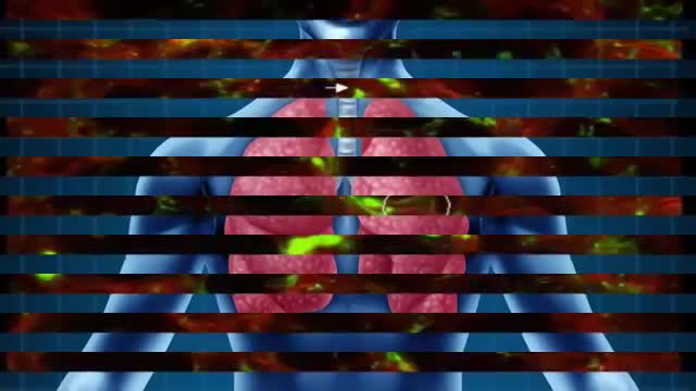
A good starting point for any scientist in any field is to recognize that there is much we do not know. We do not know, for example, why there is more matter than antimatter in the universe. We do not know very well how the evolution of the dinosaurs filtered out. And, perhaps most surprising of all is that we do not know very well how many organs the human body has or what all its functions are.

The cause for TS is unknown. Early research suggested that TS is an inherited condition (often, the person's near or distant relatives have had some form of transient or chronic tic disorder or associated symptoms). Recent studies point to a combination of environmental and genetic factors as a cause of the disorder. The specific genes involved in the development of TS are still being investigated. Studies suggest that TS has a neurological basis and results from an abnormality which affects the brain's metabolism of certain neurotransmitters (chemicals in the brain that regulate behavior.) Current research being funded by the Tourette Syndrome Association (TSA) will help provide more information about the causes and genetic factors of TS.
Pilonidal disease with lateral extension. Follicle removal (Bascon's technique)
Get the best medical animation videos made at https://www.b2w.tv/healthcare-video-production
Check out more animated healthcare videos from out blog here https://www.b2w.tv/blog/health....care-marketing-video
Medical device manufacturers need to find new and innovative ways to explain their products to potential buyers.
It can be difficult for potential buyers to understand how a medical device works, and even more difficult to visualize how it would be used in a clinical setting.
Medical animation videos are the perfect way to showcase your medical devices.
They are engaging, easy to understand, and help potential buyers see how your product would fit into their workflow.
Jump to the video you like:
8. Pharming Healthcare 0:09
7. ThermoFisher Scientific 2:46
6. Fibrogen 5:49
5. OrthAlign 9:29
4. Edwards LifeSciences 11:34
3. Edwards LifeSciences 12:51
2. Edwards LifeSciences 13:43
1. Edwards LifeSciences 18:14
Check out more Healthcare Videos we have made for our clients:
1. Healthcare Explainer Video for WelbeHealth: https://on.b2w.tv/3OFRaWo
2. Healthcare Product Explainer Video for Edwards Lifesciences: https://on.b2w.tv/3OSdMDb
3. Healthcare Commercial Video for Coopervision: https://on.b2w.tv/45muvpf
4. Healthcare Marketing Video for OrthAlign: https://on.b2w.tv/3P8KBgD
5. Healthcare Video Marketing with The Video-First Approach: https://on.b2w.tv/3LiNDfW
6. 12 Best Brand Archetypes for Healthcare Videos: https://on.b2w.tv/3EIQ0Vu
Want to learn more about Healthcare Videos? Check out our blogs:
1. 10 Best Healthcare Marketing Videos: https://on.b2w.tv/47LxhpJ
2. 5 Animated Healthcare Commercial Videos: https://on.b2w.tv/47IgpAd
3. 11 Animated Healthcare Explainer Videos: https://on.b2w.tv/3Zd7fYM
4. How Long Does It Take To Make an Healthcare Explainer Video: https://on.b2w.tv/45nasak
5. Script for Healthcare Explainer Videos: https://on.b2w.tv/47IY1af
6. Guide to Making Your Own Healthcare Explainer Video: https://on.b2w.tv/3P6FKMR
#medicaldevice #medicalanimation #medicalanimations
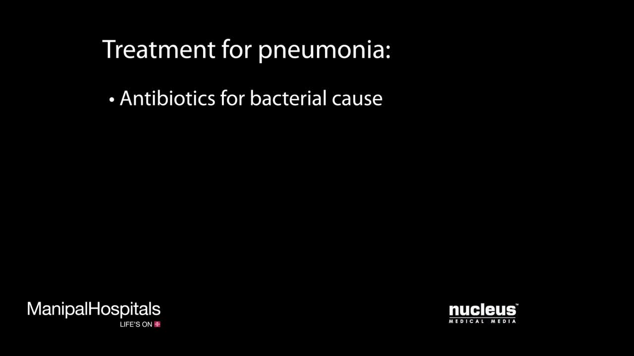
Pneumonia is an infection that inflames the air sacs in one or both lungs. The air sacs may fill with fluid or pus (purulent material), causing cough with phlegm or pus, fever, chills, and difficulty breathing. A variety of organisms, including bacteria, viruses and fungi, can cause pneumonia.

The exam should be performed in an orderly fashion as follows: 1. Have the patient stick out their tongue so that you can examine the posterior pharynx (i.e. the back of the throat). Ask the patient to say "Ah", which elevates the soft palate, giving you a better view. If you are still unable to see, place the tongue blade � way back on the tongue and press down while the patient again says "Ah," hopefully improving your view. This causes some people to gag, particularly when the blade is pushed onto the more proximal aspects of the tongue. It may occasionally be important to determine whether the gag reflex is functional (e.g. after a stroke that impairs CNs 9 or 10; or to determine if a patient with depressed level of consciousness is able to protect their airway from aspiration). This is done by touching a q-tip against the posterior pharynx, uvula or tongue. It is not necessary to do this during your routine exam as it can be quite noxious!
2. Note that the uvula hangs down from the roof of the mouth, directly in the mid-line. With an "Ah," the uvula rises up. Deviation to one side may be caused by CN 9 palsy (the uvula deviates away from the affected side), a tumor or an infection. CN9 Pasly Cranial Nerve 9 Dysfunction: Patient has suffered stroke, causing loss of function of left CN 9. As a result, uvula is pulled towards the normally functioning (ie right) side. 3. The normal pharynx has a dull red color. In the setting of infection, it can become quite red, frequently covered with a yellow or white exudate (e.g. with Strep. Throat or other types of pharyngitis).
4. The tonsils lie in an alcove created by arches on either side of the mouth. The apex of these arches are located lateral to and on a line with the uvula. Normal tonsils range from barely apparent to quite prominent. When infected, they become red, are frequently covered by whitish/yellow discharge. In the setting of a peritonsilar abscess, the tonsils appear asymmetric and the uvula may be pushed away from the affected side. When this occurs, the tonsil may actually compromise the size of the oral cavity, making breathing quite difficult.
5. Look carefully along the upper and lower gum lines and at the mucosa in general, which can appear quite dry if the patient is dehydrated.
6. Examine the teeth to get a sense of general dentition, particularly if the patient has a dental complaint. Pain produced by tapping on a tooth is commonly caused by a root abscess. Tooth Abscess: Tooth abscess involving left molar region. Associated inflammation of left face can clearly be seen. 7. Have the patient stick their tongue outside their mouth, which allows evaluation of CN 12. If there is nerve impairment, the tongue will deviate towards the affected side. Any obvious growths or abnormalities? Ask them to flip their tongue up so that you can look at the underside. If you see something abnormal, grasp the tongue with gauze so that you can get a better look. Left CN 12 Dysfunction: Stroke has resulted in L CN 12 Palsy. Tongue therefore deviates to the left.
8. Make note of any growths along the cheeks, hard palate (the roof of the mouth between the teeth), soft palate, or anywhere else. In particular, patients who smoke or chew tobacco are at risk for oral squamous cell cancer. Any areas which are painful or appear abnormal should also be palpated. Put on a pair of gloves to better explore these regions. What do they feel like? Are they hard? To what extent does a growth involve deeper structures? If the patient feels something that you cannot see, try to get someone else to hold the light source, freeing both your hands to explore the oral cavity with two tongue depressors.

What is idiopathic intracranial hypertension? Idiopathic intracranial hypertension (IIH) is a disorder that results from an increase in the pressure of the Cerebro-Spinal Fluid (CSF) that cushions and protects the brain and spinal cord. The CSF is constantly produced in the brain and reabsorbed back into the bloodstream at a fairly constant rate. This allows the fluid pressure around the brain to remain constant. What are the symptoms of idiopathic intracranial hypertension? Headaches that are generally nonspecific in location, type and frequency and can be associated with nausea and vomiting. Pulsatile tinnitus is a rhythmic or pulsating ringing heard in one or both ears. Horizontal double vision can be a sign of pressure on the 6th cranial nerve(s). Nonspecific radiating pain in the arms or legs (radicular pain). Transient obscurations of vision (TOV), which are temporary dimming or complete blacking out of vision. Visual field defects. These defects can occur in the central as well as the peripheral vision. Loss of color vision. What causes idiopathic intracranial hypertension? The cause is usually not known. A common explanation for increased pressure is a problem with the reabsorption of this fluid back into the body, which causes the pressure to increase. Sometimes the cause is determined and is referred to as “secondary” intracranial hypertension.
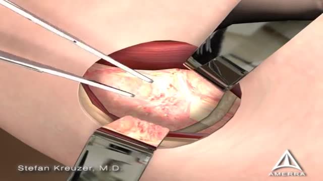
Femoroacetabular impingement (FAI) is a condition in which extra bone grows along one or both of the bones that form the hip joint — giving the bones an irregular shape. Because they do not fit together perfectly, the bones rub against each other during movement. Over time this friction can damage the joint, causing pain and limiting activity.
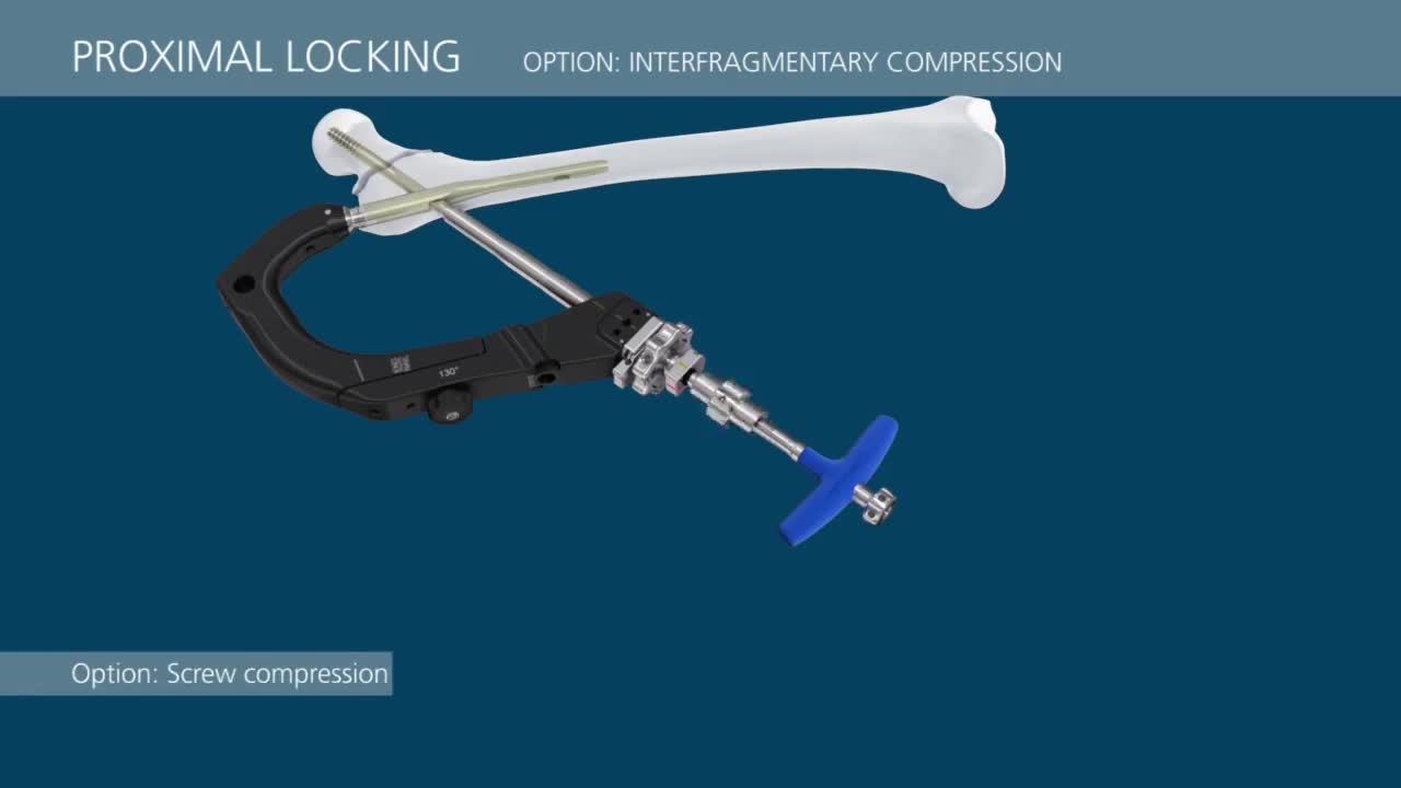
This video demonstrates a step-by-step technique for using the TFN-Advanced™ Proximal Femoral Nailing System (TFNA).
New Drugs Improve Osteoporosis Treatment
An arthroscopic meniscectomy is a procedure to remove some or all of a meniscus from the tibio-femoral joint of the knee using arthroscopic (aka 'keyhole') surgery. In a complete meniscectomy the meniscus including the meniscal rim is removed. A partial meniscectomy involves partial removal of the meniscus. This may vary from minor trimming of a frayed edge to anything short of removing the rim. This is a minimally invasive procedure often done as day suas an outpatient in a one-day clinic [1] This procedure is performed when a meniscal tear is too large to be corrected by a surgical meniscal repair.[1] When non-operative therapy provides some degree of symptom relief over the long-term, these benefits may wane with continued meniscal degeneration. In such patients, arthroscopic partial meniscectomy can be effective in improving patient quality of life.
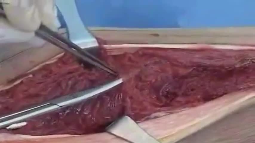
Our results in this study of MIPO treated with conventional plates are comparable to the results of the femoral shaft fractures treated with intramedullary nailing. The technique can be used for all femoral shaft fractures. Although the biomechanics of the plate fixation are less stable compared to the intamedullary nail, the mechanical stability is stable enough for bone healing. Healing was rapid, and postoperative care was simplified. The two major complications were malalignment and screw breakage. We recommend using at least three separated screws in each fragment to prevent stress on the screw and screw breakage. Intraoperative limb length, axial alignment, and rotation must be carefully assessed to prevent malalignment. The limitations of our study include lack of a comparison group, retrospective data collection, and no randomisation in outcome evaluation
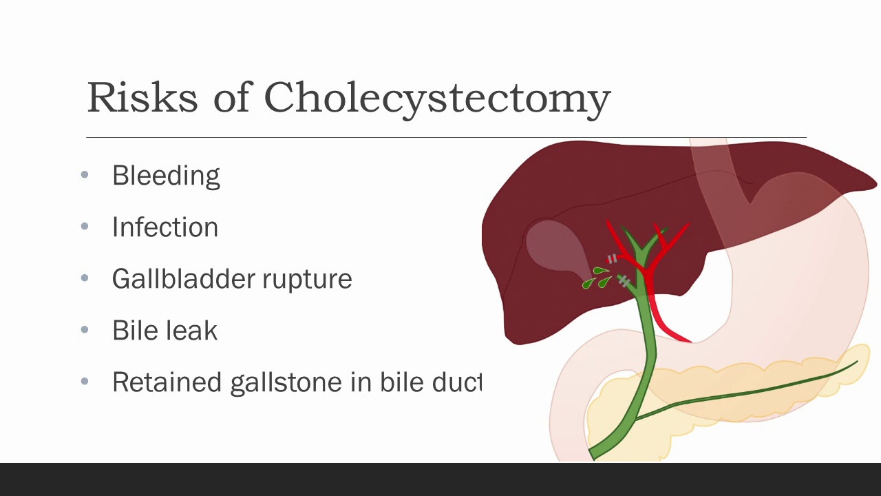
Cholecystectomy means removal of the gallbladder. The most common reasons
your doctor might recommend a cholecystectomy are biliary colic, cholecystitis,
choledocolithiasis, or gallstone pancreatitis. Biliary colic, also known as symptomatic
cholelithiasis, is caused by gallstones, which are hardened deposits of bile. Gallstones are
common in the general population, and gallstones alone are not a reason for gallbladder
removal if they do not cause symptoms. However, sometimes gallstones can get caught at the
neck of the gallbladder, causing pain when the gallbladder contracts against them trying to
release its bile, especially after a fatty meal. With biliary colic, the pain typically resolves within
an hour or so. Occasionally, a stone or some other blockage may prevent the gallbladder from
emptying over a long period of time, causing an increase in pressure and trapped fluid within the
gallbladder. This can cause inflammation and infection of the gallbladder, which we call
cholecystitis. Choledocholithiasis is when there are one or more stones in the bile ducts, which
can cause back up of bile into the liver, and depending on the location of the stones, could
cause pancreatitis, which is inflammation of the pancreas. Other reasons for gallbladder
removal, though less common, are gallbladder polyps and cancer. All of these are reasons for
gallbladder removal.
Rhabdomyolysis is a condition in which damaged skeletal muscle (Ancient Greek: rhabdomyo-) tissue breaks down rapidly (Greek –lysis). This damage may be caused by physical (e.g. crush injury), chemical, or biological factors. Breakdown products of damaged muscle cells are released into the bloodstream; some of these, such as the protein myoglobin, are harmful to the kidney and may lead to kidney dysfunction. The severity of the symptoms (which may include muscle pains, vomiting and confusion) depends on the extent of the muscle damage, and whether kidney failure develops. The mainstay of treatment is generous intravenous fluids, but could include dialysis or hemofiltration.
Rhabdomyolysis and its complications are significant problems for those injured in disasters such as earthquakes and bombing. Relief efforts in areas struck by earthquakes often include medical teams with skills and equipment for treatment of survivors with rhabdomyolysis. The disease and its mechanisms were first fully elucidated during the Blitz of London in 1941.
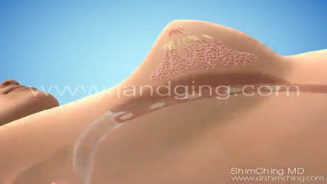
http://www.landging.com/breast-augmentation-animation.html4
This breast augmentation animation demonstrates the procedure of saline breast implants.
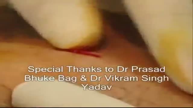
The procedure of Suprapubic Cystostomy
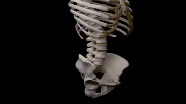
abdominal cavity
