סרטונים מובילים

Histology of Ureter
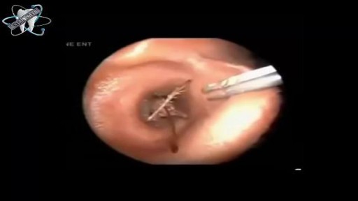
Bugs Removal from Ear Canal

Hemodialysis Introduction for Kidney

Absence of a woman's monthly menstrual period is called amenorrhea. Secondary amenorrhea is when a woman who has been having normal menstrual cycles stops getting her periods for 6 months or longer. Causes Secondary amenorrhea can occur due to natural changes in the body. For example, the most common cause of secondary amenorrhea is pregnancy. Breastfeeding and menopause are also common, but natural, causes. Women who take birth control pills or who receive hormone shots such as Depo-Provera may not have any monthly bleeding. When they stop taking these hormones, their periods may not return for more than 6 months. You are more likely to have absent periods if you: Are obese Exercise too much and for long periods of time Have very low body fat (less than 15 to 17%) Have severe anxiety or emotional distress Lose a lot of weight suddenly (for example, from strict or extreme diets or after gastric bypass surgery) Other causes include: Brain (pituitary) tumors Drugs for cancer treatment Drugs to treat schizophrenia or psychosis Overactive thyroid gland Polycystic ovarian syndrome Reduced function of the ovaries
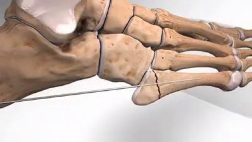
Fracture of meta-diaphyseal junction of the fifth metatarsal of the foot
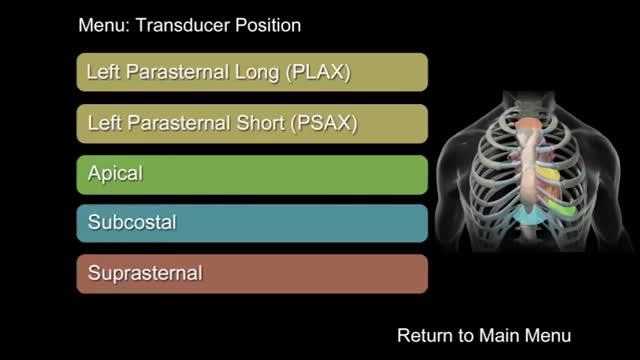
"How to Perform a Transthoracic Echocardiographic Study Volume 1: Transducer Position and Anatomy" is an instructional video, offered by ASE, and can be used for professional lectures and offers an interactive section for flexible presentations. The video includes an overview of relevant cardiac anatomy, a step by step presentation of all Transducer Positions, and the sequential transducer movements to acquire standard echo images needed to complete a Transthoracic Echocardiographic Study.

-Intrapartum antibiotic prophylaxis for mothers colonized with group B Streptococcus can prevent early-onset neonatal disease. Adequate prophylaxis consists of ampicillin, penicillin, or cefazolin for ;::4 hours before delivery. Regardless of intrapartum treatment, all high-risk infants must be observed for ;::49 hours. A complete blood count with differential and blood culture are indicated if the infant is preterm <37 weeks or was exposed to prolonged rupture of membranes.>18 hrs.

Congestive Heart Failure 3D Animation

This video demonstrates how a broken nose is fixed using only local anesthesia and without sedation. Of course, this can also be performed while asleep.

A new bionic body part that talks to your phone is the next frontier in knee replacements. It's called a smart knee, a new technology designed to improve recovery after surgery. Stephanie Stahl reports.

Why You’re Attracted To Certain People

Veryyyyy funny!

Tracheal Deviation Technique

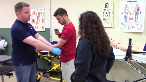
Dealing with choking

There’s a strange, mysterious world inside us, an alien-looking environment that turns the food we eat into nutrients that keep us alive. Michael Mosley swallows a camera to take a closer look.

Skin cancer is the most common of all cancer types, accounting for an estimated one third of all new cases. It’s important to take the right steps to ensure proper protection and adopt good sun care habits no matter what your age or stage in life.

Cosmetic facial plastic surgery is surgery performed to enhance visual appearance of the facial structures and features. Common procedures include facelifts, eye lifts, rhinoplasty, chin and cheek implants, liposuction, and procedures to correct facial wrinkles.
