- Physical Examination
- Surgical Examination
- Ophthalmology
- Clinical Skills
- Orthopedics
- Surgery Videos
- Laparoscopy
- Pediatrics
- Funny Videos
- Cardiothoracic Surgery
- Nursing Videos
- Plastic Surgery
- Otorhinolaryngology
- Histology and Histopathology
- Neurosurgery
- Dermatology
- Pediatric Surgery
- Urology
- Dentistry
- Oncology and Cancers
- Anatomy Videos
- Health and Fitness
- Radiology
- Anaesthesia
- Physical Therapy
- Pharmacology
- Interventional Radiology
- Cardiology
- Endocrinology
- Gynecology
- Emergency Medicine
- Psychiatry and Psychology
- Childbirth Videos
- General Medical Videos
- Nephrology
- Physiology
- Diet and Food Health
- Diabetes Mellitus
- Neurology
- Women Health
- Osteoporosis
- Gastroenterology
- Pulmonology
- Hematology
- Rheumatology
- Toxicology
- Nuclear Medicine
- Infectious Diseases
- Vascular Disease
- Reproductive Health
- Burns and Wound Healing
- Other
Top videos

Mr Brian MacCormack talking about Paediatric Surgery Emergencies. This talk is part of the Paediatric Emergencies 2022 event. To get your CME certificate for watching the video please visit https://www.paediatricemergenc....ies.com/conference/p
#PaediatricEmergencies #PaediatricEmergencies2022 #PaediatricSurgery
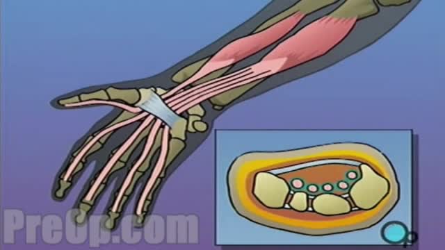
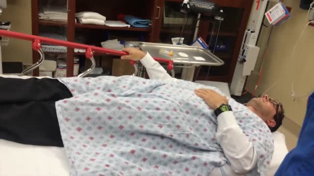
During endoscopic carpal tunnel release surgery , the transverse carpal ligament is cut. This releases pressure on the median nerve, relieving carpal tunnel syndrome symptoms. The small incisions in the palm are closed with stitches. The gap where the ligament was cut will eventually fill with scar tissue.
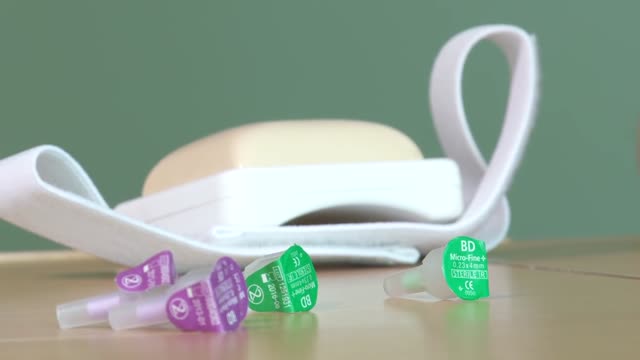
Insert the needle into the rubber stopper of the insulin bottle. Push the plunger down to inject air into the bottle (this allows the insulin to be drawn more easily). Leave the needle in the bottle. Turn the bottle and syringe upside-down.
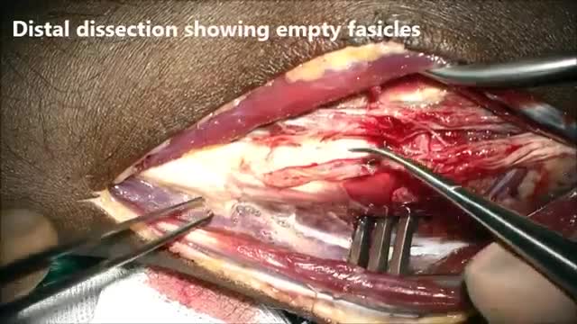
A nerve transfer is a surgical technique that may be used when a patient has a nerve injury resulting in complete loss of muscle function or sensation. Median to radial transfer. Distal AIN to median recurrent motor branch transfer.
Encourage your child to drink lots of fluids to prevent dehydration. Milk and water are both fine. However, if your child refuses solids, give your child just milk, rather than water. ... Keep giving your child table foods while he has diarrhea. Diarrhea is most often spread through fecally contaminated food, hands or surfaces touched by objects or hands put into the mouth (fecal-oral route).Water contaminated by human or animal feces (e.g., swimming pools) or trips to sites with animals (e.g., farms, pet stores, petting zoos) are also possible routes of ... The best foods for your child are easily digestible foods, such as rice cereal, pasta, breads, cooked beans, mashed potatoes, cooked carrots, applesauce, and bananas. Pretzels or salty crackers can help your child replace the salt lost from diarrhea. Foods containing large amounts of sugar or fat should be avoided.

Blood cells travel through the circulatory system suspended in a yellowish fluid called plasma. Plasma is 90% water and contains nutrients, proteins, hormones, and waste products. Whole blood is a mixture of blood cells and plasma.
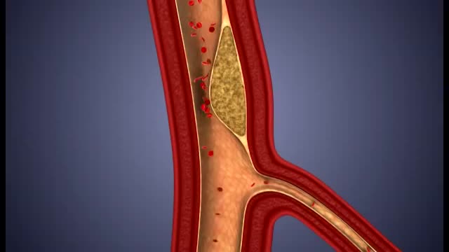
What damage does atherosclerosis cause? Plaque may partially or totally block the blood's flow through an artery in the heart, brain, pelvis, legs, arms or kidneys. Some of the diseases that may develop as a result of atherosclerosis include coronary heart disease, angina (chest pain), carotid artery disease, peripheral artery disease (PAD) and chronic kidney disease.
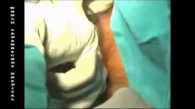
Haemorrhoids is one of the most common problems seen in surgical OPD. Open haemorrhoidectomy has remained the gold standard for a long time with a high post-operative morbidity. The quest for a better understanding of the pathology of haemorrhoids resulted in the evolvement of stapler haemorrhoidopexy. Our aim is to study the efficacy of stapler haemorrhoidopexy with regards to role of immediate post-operative morbidity. A prospective study of 50 patients (n = 50) with the second- and third-degree symptomatic haemorrhoids was done. The mean age of the patients was 44.1 years. Fourteen patients had co-morbid conditions. The average duration of the operation was 29 min. Patients with the second-degree haemorrhoids had higher rate of complication. The complication rate was 32%. Three patients had urinary retention. Two patients had minor bleeding, and one patient experienced transient discharge. The mean analgesic requirement was 2.4 tramadol, 50 mg injections. Ten patients had significant post-operative pain. Average length of hospital stay was 2.7 days. There were no symptomatic recurrences till date.
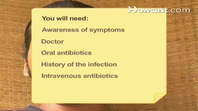
“People need to realize this is imminently preventable,” he said. Lyme disease develops following an infection with the bacteria Borrelia burgdorferi. It's transmitted to humans through the bite of infected blacklegged ticks. The tick must be attached to its host for 36 to 48 hours to transmit the bacteria.
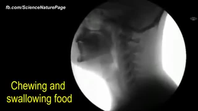
The human body as seen with MRI and X-RAY

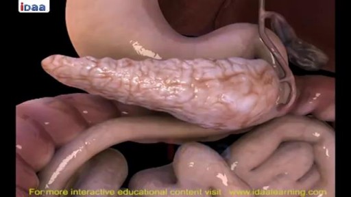
The cat's stomach is a sac-like structure designed to store large volumes of food and continue the digestive process. The esophagus carries food to the stomach, where it enters via a valve-like structure called the cardiac sphincter. On the interior surface of the stomach is a series of folds called gastric folds. These folds function to help grind and digest food. The inner stomach lining secretes acids and enzymes to break down food. Once the initial stomach digestive process is complete, the partially digested food exits the stomach through the pyloric sphincter area and then enters the duodenum (first segment of the small intestine). Once eaten, most food leaves the stomach within twelve hours after entering.
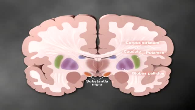
Parkinson's disease is a progressive disorder of the nervous system that affects movement. It develops gradually, sometimes starting with a barely noticeable tremor in just one hand. But while a tremor may be the most well-known sign of Parkinson's disease, the disorder also commonly causes stiffness or slowing of movement. In the early stages of Parkinson's disease, your face may show little or no expression, or your arms may not swing when you walk. Your speech may become soft or slurred. Parkinson's disease symptoms worsen as your condition progresses over time. Although Parkinson's disease can't be cured, medications may markedly improve your symptoms. In occasional cases, your doctor may suggest surgery to regulate certain regions of your brain and improve your symptoms.
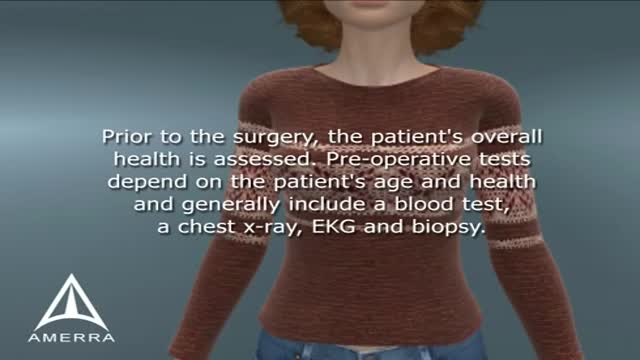
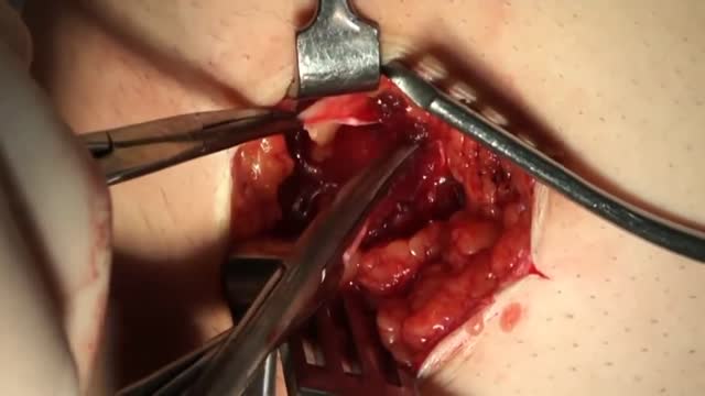
A modified radical mastectomy is a procedure in which the entire breast is removed, including the skin, areola, nipple, and most axillary lymph nodes; the pectoralis major muscle is spared. Historically, a modified radical mastectomy was the primary method of treatment of breast cancer. [1, 2] As the treatment of breast cancer evolved, breast conservation has become more widely used. [3, 4] However, mastectomy still remains a viable option for women with breast cancer. [5, 6]
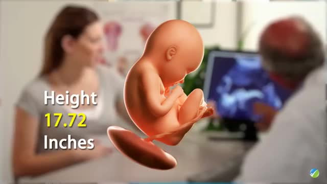
During 34 week of pregnancy, the baby is getting ready for delivery, you may feel less wriggling and kicking. Watch out this video to learn more about being 34 weeks pregnant.
The external jugular vein receives the greater part of the blood from the exterior of the cranium and the deep parts of the face, being formed by the junction of the posterior division of the retromandibular vein with the posterior auricular vein.
An appendectomy (sometimes called appendisectomy or appendicectomy) is the surgical removal of the vermiform appendix. This procedure is normally performed as an emergency procedure, when the patient is suffering from acute appendicitis.
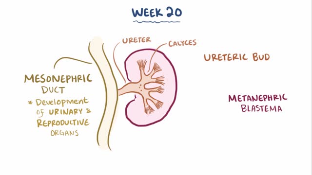
Multicystic dysplastic kidney (MCDK) is a condition that results from the malformation of the kidney during fetal development. The kidney consists of irregular cysts of varying sizes. Multicystic dysplastic kidney is a common type of renal cystic disease, and it is a cause of an abdominal mass in infants.
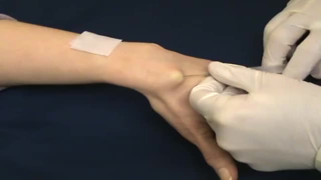
A nonsurgical method of treating a ganglion is to drain the fluid from (aspirate) the ganglion sac. Your doctor can do this in the office using the following procedure: The ganglion area is cleaned with an antiseptic solution. A local anesthetic is injected into the ganglion area to numb the area. When the area is numb, the ganglion sac is punctured with a sterile needle. The fluid is drawn out of the ganglion sac. The ganglion collapses. A bandage and, in some cases, a splint are used for a few days to limit movement and prevent the ganglion sac from filling again. Treating a ganglion by draining the fluid with a needle may not work because the ganglion sac remains intact and can fill again, causing the ganglion to return. For this reason, your doctor may puncture the sac with the needle 3 or 4 times so the sac will collapse completely. Even then, the ganglion is likely to come back.
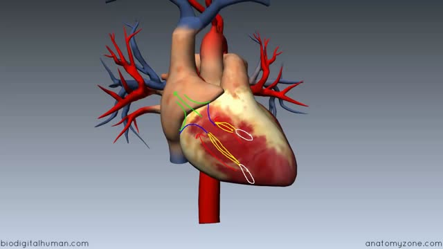
Heart Anatomy - Right Ventricle c
