- Physical Examination
- Surgical Examination
- Ophthalmology
- Clinical Skills
- Orthopedics
- Surgery Videos
- Laparoscopy
- Pediatrics
- Funny Videos
- Cardiothoracic Surgery
- Nursing Videos
- Plastic Surgery
- Otorhinolaryngology
- Histology and Histopathology
- Neurosurgery
- Dermatology
- Pediatric Surgery
- Urology
- Dentistry
- Oncology and Cancers
- Anatomy Videos
- Health and Fitness
- Radiology
- Anaesthesia
- Physical Therapy
- Pharmacology
- Interventional Radiology
- Cardiology
- Endocrinology
- Gynecology
- Emergency Medicine
- Psychiatry and Psychology
- Childbirth Videos
- General Medical Videos
- Nephrology
- Physiology
- Diet and Food Health
- Diabetes Mellitus
- Neurology
- Women Health
- Osteoporosis
- Gastroenterology
- Pulmonology
- Hematology
- Rheumatology
- Toxicology
- Nuclear Medicine
- Infectious Diseases
- Vascular Disease
- Reproductive Health
- Burns and Wound Healing
- Other
Top videos
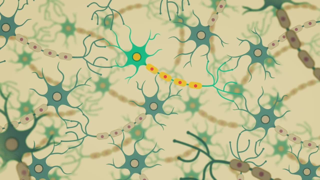
Dementia is the name for a group of symptoms that commonly include problems with memory, thinking, problem solving, language and perception. In a person with dementia, these symptoms are bad enough to affect daily life.
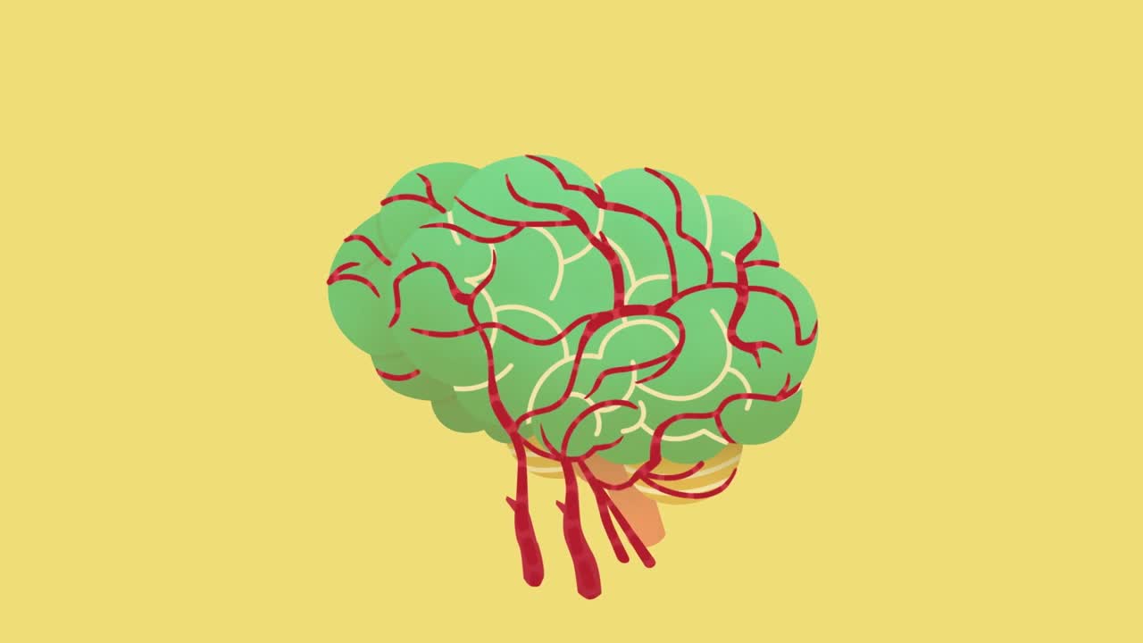
Vascular dementia is a general term describing problems with reasoning, planning, judgment, memory and other thought processes caused by brain damage from impaired blood flow to your brain. You can develop vascular dementia after a stroke blocks an artery in your brain, but strokes don't always cause vascular dementia. Whether a stroke affects your thinking and reasoning depends on your stroke's severity and location. Vascular dementia also can result from other conditions that damage blood vessels and reduce circulation, depriving your brain of vital oxygen and nutrients
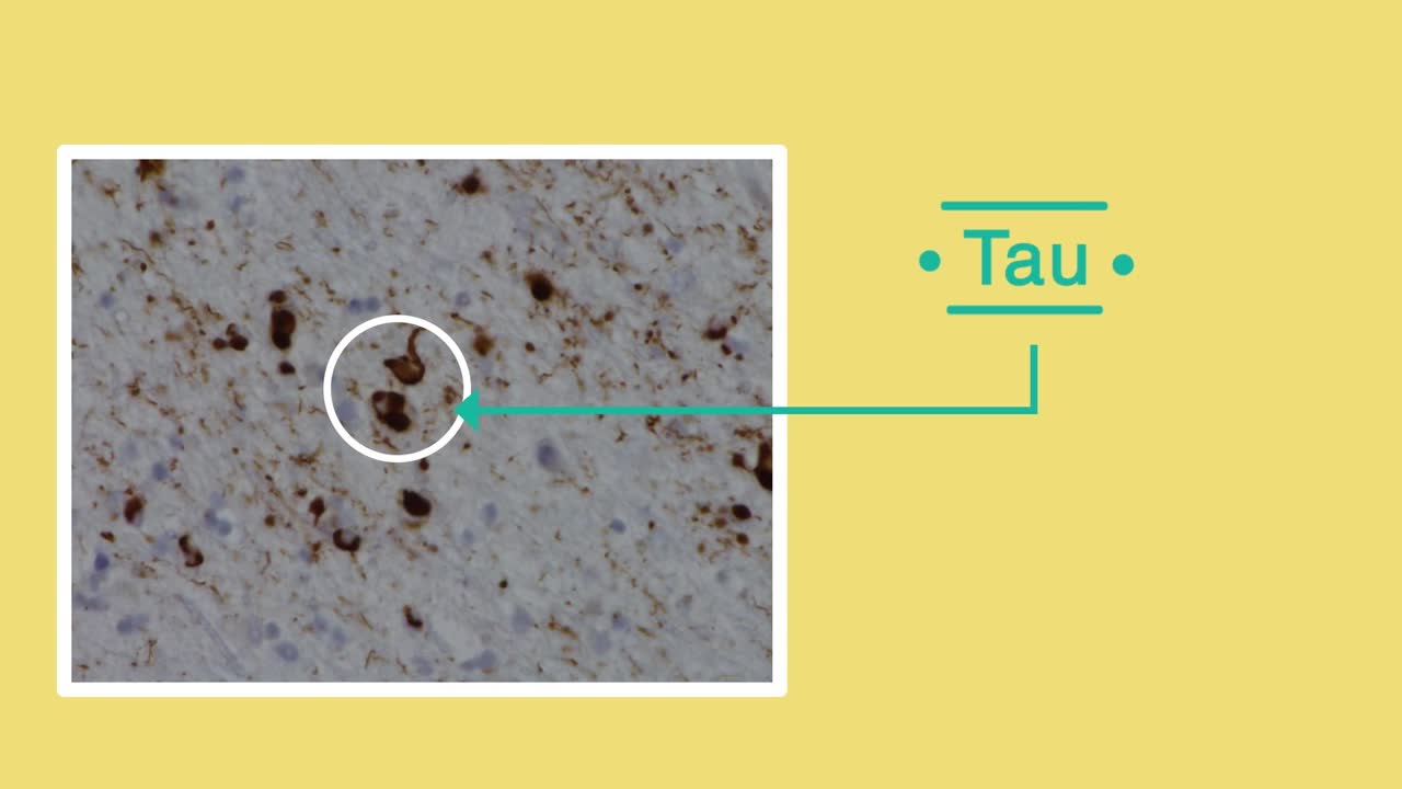
Frontotemporal dementia is the name for a range of conditions in which cells in the frontal and temporal lobes of the brain are damaged. These lobes control behaviour, emotional responses and language. This means that people will experience changes in personality and behaviour, or may struggle with language – for example, in finding the right word. Frontotemporal dementia is a less common form of dementia which is more likely to affect younger people – those under 65.
Furosemide is used to reduce extra fluid in the body (edema) caused by conditions such as heart failure, liver disease, and kidney disease. This can lessen symptoms such as shortness of breath and swelling in your arms, legs, and abdomen. This drug is also used to treat high blood pressure. Lowering high blood pressure helps prevent strokes, heart attacks, and kidney problems. Furosemide is a "water pill" (diuretic) that causes you to make more urine. This helps your body get rid of extra water and salt.

Oral sex can be an enjoyable, healthy part of an adult relationship. But there are some things that many people don't know about oral sex. Here are four facts that might surprise you. 1. Oral sex is linked to throat cancer. Cancer? Yes, you can get throat cancer from oral sex, says American Cancer Society Chief Medical Officer Otis Brawley, MD. It's not oral sex, per se, that causes cancer, but the human papillomavirus (HPV), which can be passed from person to person during sex, including oral sex.
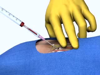
Arterial Cannulation

This is an introduction to ventilator settings like FIO2, PEEP, Flow rate,trigger,TV, and RR. I also discuss how these settings relate to CO2 and O2 control and to complications like oxygen toxicity and barotrauma with an emphasis on physiology.
Multiple studies demonstrate the safety of propofol in pediatric EDPS. Each has identified a drop in blood pressure and transient hypoxemia as the most frequent complications. In all of the studies in which hypotension was identified there was no evidence of poor perfusion. The hypoxemia in all of these studies quickly responded to minimal intervention with no apparent lasting complications. Although these were pediatric studies, the results were very similar to ours in complication rates and sedation times. Our study did not demonstrate the frequency of decreased blood pressure seen in these pediatric studies but had similar hypoxemia rates.
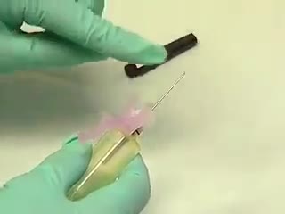
The venipuncture procedure is complex, requiring both knowledge and skill to perform. Each phlebotomist generally establishes a routine that is comfortable for her or him. Several essential steps are required for every successful collection procedure: Identify the patient. Assess the patient's physical disposition (i.e. diet, exercise, stress, basal state). Check the requisition form for requested tests, patient information, and any special requirements. Select a suitable site for venipuncture. Prepare the equipment, the patient and the puncture site. Perform the venipuncture. Collect the sample in the appropriate container. Recognize complications associated with the phlebotomy procedure. Assess the need for sample recollection and/or rejection. Label the collection tubes at the bedside or drawing area. Promptly send the specimens with the requisition to the laboratory.
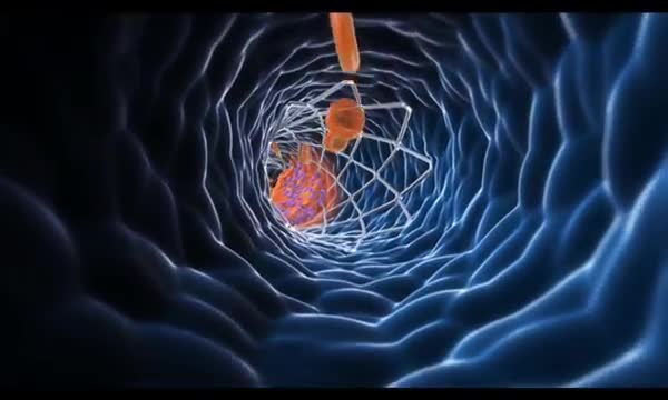
This video describes the effects of heart disease and explains how stents are used to treat damaged arteries.
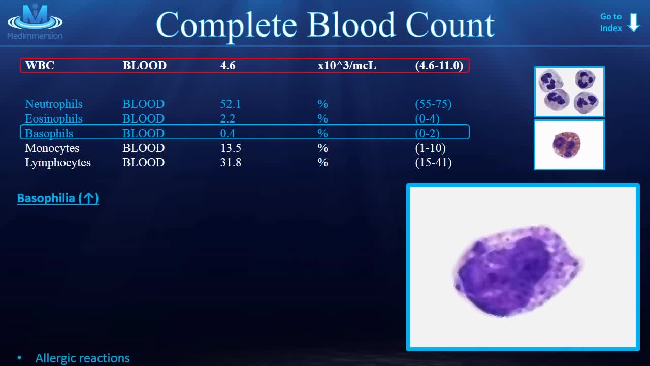
Learn the CBC once and for all!
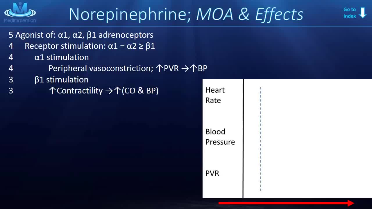
Norepinephrine is synthesized from dopamine by dopamine β-hydroxylase.[7] It is released from the adrenal medulla into the blood as a hormone, and is also a neurotransmitter in the central nervous system and sympathetic nervous system where it is released from noradrenergic neurons.
A good starting point for any scientist in any field is to recognize that there is much we do not know. We do not know, for example, why there is more matter than antimatter in the universe. We do not know very well how the evolution of the dinosaurs filtered out. And, perhaps most surprising of all is that we do not know very well how many organs the human body has or what all its functions are.
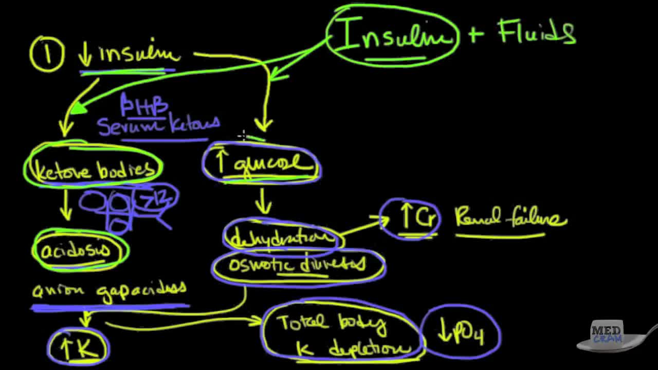
This is video 2 of 2 on diabetic ketoacidosis (DKA).
The MAKOplasty® procedure is an FDA-cleared treatment option for patients who suffer from osteoarthritis damage in the medial (inner) portion of the knee. ... Only the diseased portion of your knee is removed, leaving the healthy bone and tissue surrounding it untouched.
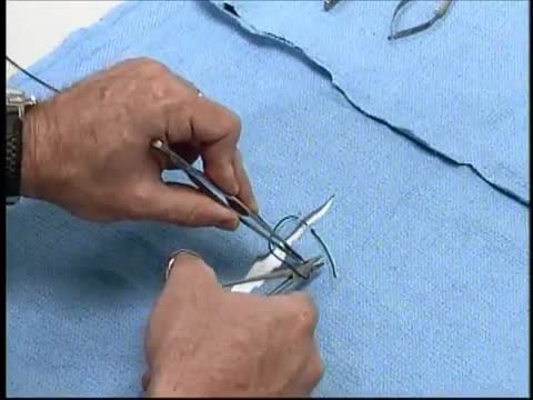
At first, grasping the needle is difficult because it will have a tendency to want to jump around. What can oftentimes help is to get hold of the thread with the left-hand forceps at a point 2 to 3 cm away from the needle. Dangle the needle until it just comes to rest on the surface. This will then allow you to use the angulated needle holder to grab the needle easily. Your needle is in a stable position if it is set up to 90 degrees to the axis of the tips of the forceps. You can make minor corrections by touching the needle with your left-hand forceps, or by partially relaxing your grip and nudging the needle tip against another firm object. You should hold the needle just behind its midpoint (If you hold it too near the tip, it will point downward. If you hold it too near the thread end, it will point upward.).
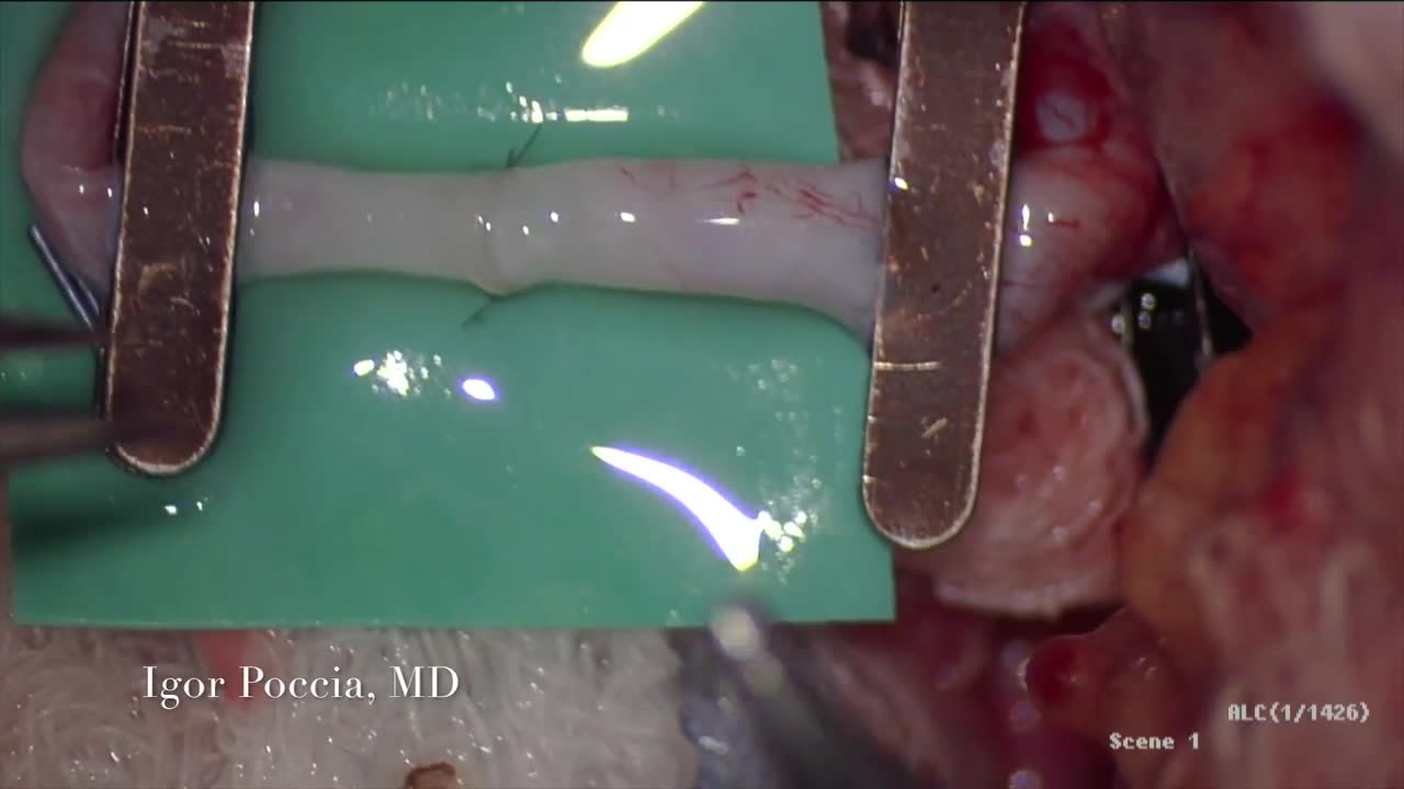
A circulatory anastomosis is a connection (an anastomosis) between two blood vessels, such as between arteries (arterio-arterial anastomosis), between veins (veno-venous anastomosis) or between an artery and a vein (arterio-venous anastomosis). An end artery (or terminal artery) is an artery that is the only supply of oxygenated blood to a portion of tissue. Examples of an end artery include the splenic artery that supplies the spleen and the renal artery that supplies the kidneys.
Throughout the body, there are several points at which blood vessels unite. The junctions are termed anastomoses. In the simplest sense, an anastomosis is any connection (made surgically or occurring naturally) between tube-like structures. Naturally occurring arterial anastomoses provide an alternative blood supply to target areas in cases where the primary arterial pathway is obstructed. They are most abundant in regions of the body where the blood supply may can be easily damaged or blocked (such as the joints or intestines). This article focuses on the arterial anastomotic networks of the upper limb.
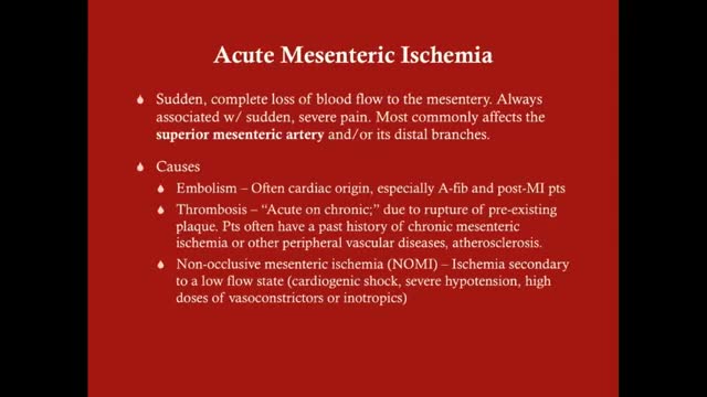
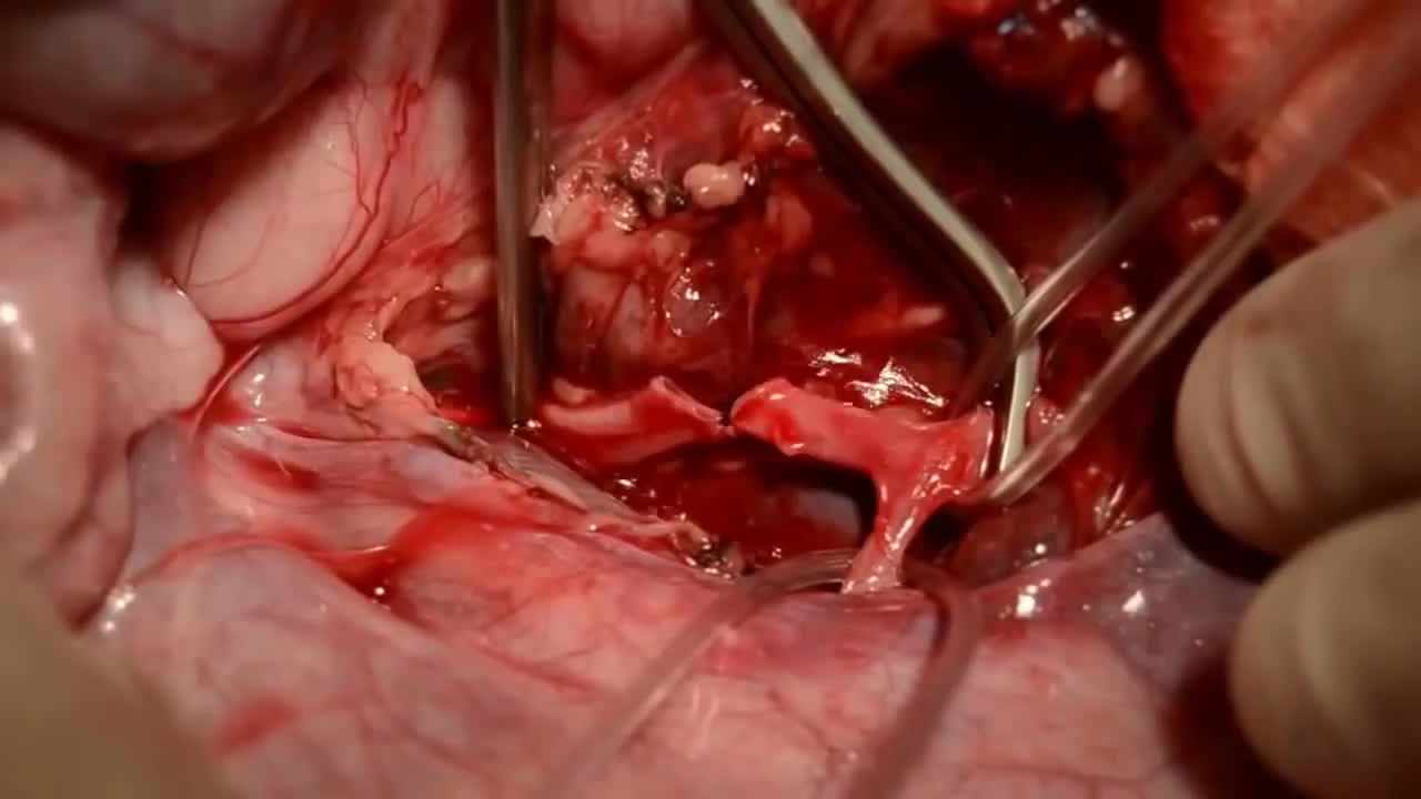
Acute mesenteric ischemia (AMI) is a syndrome caused by inadequate blood flow through the mesenteric vessels, resulting in ischemia and eventual gangrene of the bowel wall. Although relatively rare, it is a potentially life-threatening condition. Broadly, AMI may be classified as either arterial or venous. AMI as arterial disease may be subdivided into nonocclusive mesenteric ischemia (NOMI) and occlusive mesenteric arterial ischemia (OMAI); OMAI may be further subdivided into acute mesenteric arterial embolism (AMAE) and acute mesenteric arterial thrombosis (AMAT). AMI as venous disease takes the form of mesenteric venous thrombosis (MVT).
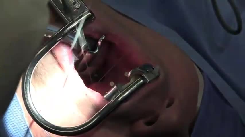
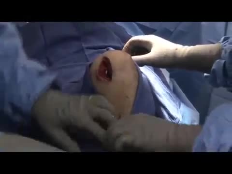
This procedure, and other types of soft palate surgery, targets the back of the roof of your mouth. It involves removing and repositioning excess tissue in the throat to make the airway wider. The surgeon can trim down your soft palate and uvula, remove your tonsils, and reposition some of the muscles of the soft palate. UPPP and other soft palate procedures are the most common type of surgery for sleep apnea. But UPPP alone is unlikely to cure moderate to severe sleep apnea. It may be combined with surgeries that target other sites in the upper airway.
