
- Physical Examination
- Surgical Examination
- Ophthalmology
- Clinical Skills
- Orthopedics
- Surgery Videos
- Laparoscopy
- Pediatrics
- Funny Videos
- Cardiothoracic Surgery
- Nursing Videos
- Plastic Surgery
- Otorhinolaryngology
- Histology and Histopathology
- Neurosurgery
- Dermatology
- Pediatric Surgery
- Urology
- Dentistry
- Oncology and Cancers
- Anatomy Videos
- Health and Fitness
- Radiology
- Anaesthesia
- Physical Therapy
- Pharmacology
- Interventional Radiology
- Cardiology
- Endocrinology
- Gynecology
- Emergency Medicine
- Psychiatry and Psychology
- Childbirth Videos
- General Medical Videos
- Nephrology
- Physiology
- Diet and Food Health
- Diabetes Mellitus
- Neurology
- Women Health
- Osteoporosis
- Gastroenterology
- Pulmonology
- Hematology
- Rheumatology
- Toxicology
- Nuclear Medicine
- Infectious Diseases
- Vascular Disease
- Reproductive Health
- Burns and Wound Healing
- Other
Top videos
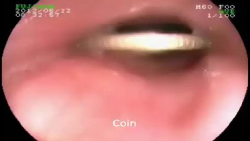

Endoscopic Removal of Coin from Esophagus
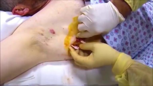
Armpit Abscess Drainage

What is hemodialysis and how does it work? Who needs it? How do you prepare for it? In the United States, over 30 million Americans have kidney disease, and sometimes, kidney disease progresses to kidney failure or end-stage renal disease. When this happens, you cannot survive unless you have a kidney transplant or some form of dialysis. So today we're going to talk about hemodialysis.
Your kidneys are the two kidney bean-shaped organs that are located in your lower back, or in your flanks. And the kidneys are responsible for filtering out or cleaning your blood. They get rid of excess waste, excess toxins, and excess fluids. If your kidneys stop functioning, then you develop renal failure or end-stage renal disease.
What is Hemodialysis?
Hemodialysis, or blood dialysis, is the filtering of your blood outside of your body. So, if your kidneys stop working properly, the hemodialysis acts as a substitute kidney. Now it's important to note that hemodialysis does not actually correct your own kidney function. It does not fix or treat your kidneys.
#hemodialysis #drfrita
What is The Dialyzer?
The dialyzer is actually the filter. It's the main powerhouse of the hemodialysis system, and it is what actually acts as the substitute kidney. In the dialyzer, you have these hollow fibers that run through it, and these fibers are bathed in something called dialysates, or dialysis fluid.
How Often Are Patients Treated With Hemodialysis?
Most patients who are on hemodialysis are on it between three and six hours, about three days a week, especially if they go to a center.
How Does Hemodialysis Work?
So when you are on dialysis, how does your blood get from your body to the hemodialysis machine and then back to your body? Well, it does so through tubes, and those tubes are connected to your access, and we'll talk about access in just a moment. But as far as the tubing, the tubing is connected to your body.
Types Of Hemodialysis Access
Arteriovenous Fistula or AV Fistula
The AV fistula is the gold standard as far as hemodialysis access is concerned because it gives you the most efficient hemodialysis and it is the least likely to be infected.
Arteriovenous Graft or AV Graft
The AV graft is very similar to the AV fistula in that you still have a surgically connected artery and a vein, usually in the arm, but in the case where if you have veins that are rather thin or arteries that are thin and maybe too weak in order to really give you a properly functioning, substantial AV fistula, then the vascular surgeon may opt to add an artificial material in order to make that shunt a little stronger, or little more durable. And so, an AV graft is another option for dialysis access.
Catheter
If you're in a situation where you need temporary dialysis, or if you have acute kidney injury, then you may have a temporary Vascath placed, and it's usually placed in a vein of the neck, the internal jugular vein, or it can be placed in the groin, or in the femoral vein.
Who Needs Hemodialysis Treatment?
How do you know if you need hemodialysis, and when is it time to prepare? Well, if you follow up with your kidney doctor (nephrologist) regularly, he or she will be watching your labs. They'll be able to see those signs of your kidneys not functioning properly.

Dr. Katherine Scovner from the Division of Nephrology at Massachusetts General Hospital discusses kidney dialysis.

"Axillary Artery to Vein AV Graft for Dialysis Access"
Houston Methodist DeBakey Heart & Vascular Center, presents a cardiovascular procedure featuring Maham Rahimi, MD, M. Mujeeb Zubair, MD, and Louis Gomez, MD, as they demonstrate “Axillary Artery to Vein AV Graft for Dialysis Access".
Surgery: Maham Rahimi, MD, M. Mujeeb Zubair, MD, and Louis Gomez, MD
Narration: M. Mujeeb Zubair, MD
__________________________
FOR MORE INFORMATION
DeBakey CV Education: https://www.houstonmethodist.o....rg/education/medical
Follow Us:
Facebook: https://www.facebook.com/debakeycvedu
Twitter: https://twitter.com/DeBakeyCVedu
Livestream: https://livestream.com/debakey
Want concise, relevant reviews of the hottest topics in CV medicine? Subscribe for FREE to the Methodist DeBakey Cardiovascular Journal for quarterly, peer-reviewed issues delivered to your door.
https://journal.houstonmethodist.org/

#dialysis #uvahealth
If your kidney function is declining and medications and other treatments aren’t working, dialysis can offer life-saving care. UVA has one of the largest dialysis programs in the country. Nephrologist Daphne Knicely, MD, explains the types of home dialysis and how they can work to fit your life.
Find out more at: https://uvahealth.com/services/dialysis
Transcript
Dialysis is just a way to replace the kidneys when they're not working anymore. So when the kidneys stop working, they stop getting rid of water, stop balancing the chemistry, stop getting rid of the toxins. Then dialysis does its job by balancing the chemistries, getting rid of the toxins, and help remove fluid. It doesn't fix the kidneys. It just replaces them.
I usually think of dialysis as two components. There's hemodialysis and peritoneal dialysis. So peritoneal dialysis can only be done at home. Hemodialysis can be done in a center, or it can be done at home.
Hemodialysis is where you have some sort of access to the blood. Either some sort of shunt in the arm that connects an artery and vein, or a catheter. And it allows for blood to leave you, go through a machine, get cleaned, chemistries balanced, and then comes back to you.
For home hemodialysis, the patient actually learns how to do that treatment. It's a very simple machine, very user-friendly. Training is usually about anywhere from four weeks up to eight weeks, and you work one-on-one with a nurse. You still see the physician. You come in about once a month, maybe twice a month, to get labs. You'll see a social worker, and a nutritionist at the same time.
Peritoneal dialysis takes place by putting a tube into your abdomen. And we take dialysis fluid that's chemically balanced. When we put it into the abdomen, it uses those little blood vessels to pull toxins out, to balance chemistries, kind of like little filters. Now, after it sits in there for several hours, we drain it out.
Anyone that needs dialysis is a candidate for home dialysis. There's not one type of dialysis that's going to make you live longer. They're all equal. The goal is to pick the type of dialysis that fits with your life.
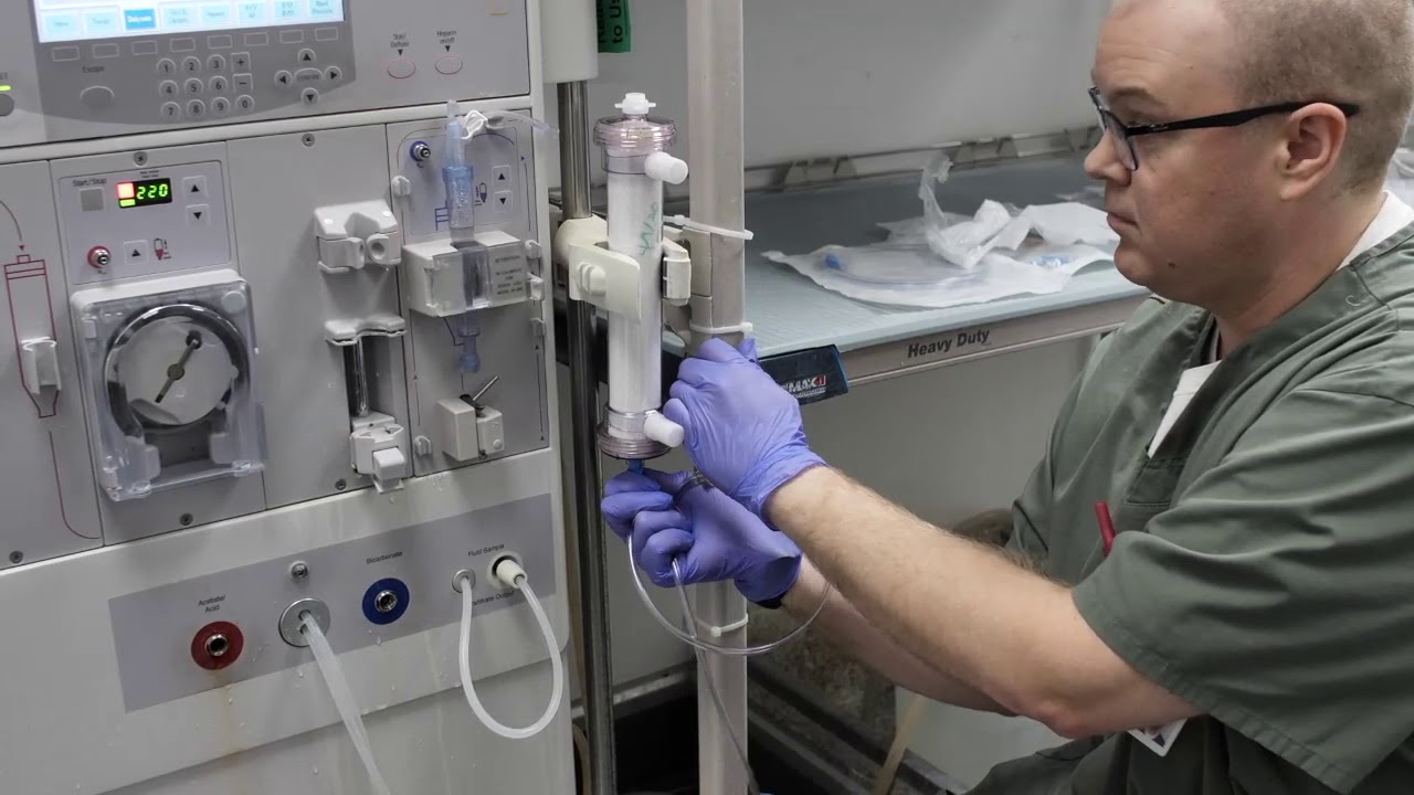
In this instructional video, Director of Critical Care Nephrology, Sevag Demirjian, MD goes over the steps for in-hospital production of ultra-pure continuous hemodialysis fluid.
By using the information in this video and/or any other materials made available by Cleveland Clinic related to the dialysate solution, you agree to comply with and be bound by the terms of the Permissive Use Agreement, a copy of which is available at https://bit.ly/3f9lN4j
Eric's Home Dialysis
For more information please visit: https://www.yalemedicine.org/c....onditions/acl-injury
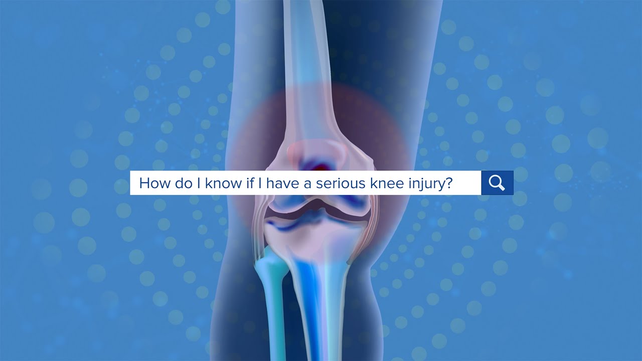
Serious injuries, by and large, cause a lot of swelling in the knee. Especially in younger patients. Now, someone could be arthritic and they overdo it going for a big long hike and they get some swelling the next day. But rapid onset of swelling, it's like hard to make out where your kneecap is, is a pretty big cardinal sign that there's something serious that's happened to your knee. Rapid onset swelling is usually due to blood in the joint. "A meniscus that really tears and flips in the front. You tear your quad or your patellar tendon, your kneecap dislocates, you tear a little blood vessel, your ACL tears, a piece of cartilage in bone gets knocked off and causes bleeding. So a lot of the really significant injuries, people get rapid onset swelling within three to four hours and they should seek attention There's always exceptions to rules, but if your knee looks like a grapefruit, you should go get it checked.

It depends upon which ligament is injured. If it is medial collateral ligament you feel pain when you walk ,sit and stand and you will be liming as well. If it is anterior cruciate ligament you feel pain when you walk on uneven ground.

We are looking for 5 patients with knee pain who want to get significantly better in the next 30 days. Click this link to let me know you're interested and learn more.
https://www.drdavidgeier.com/work-with-me/contact/
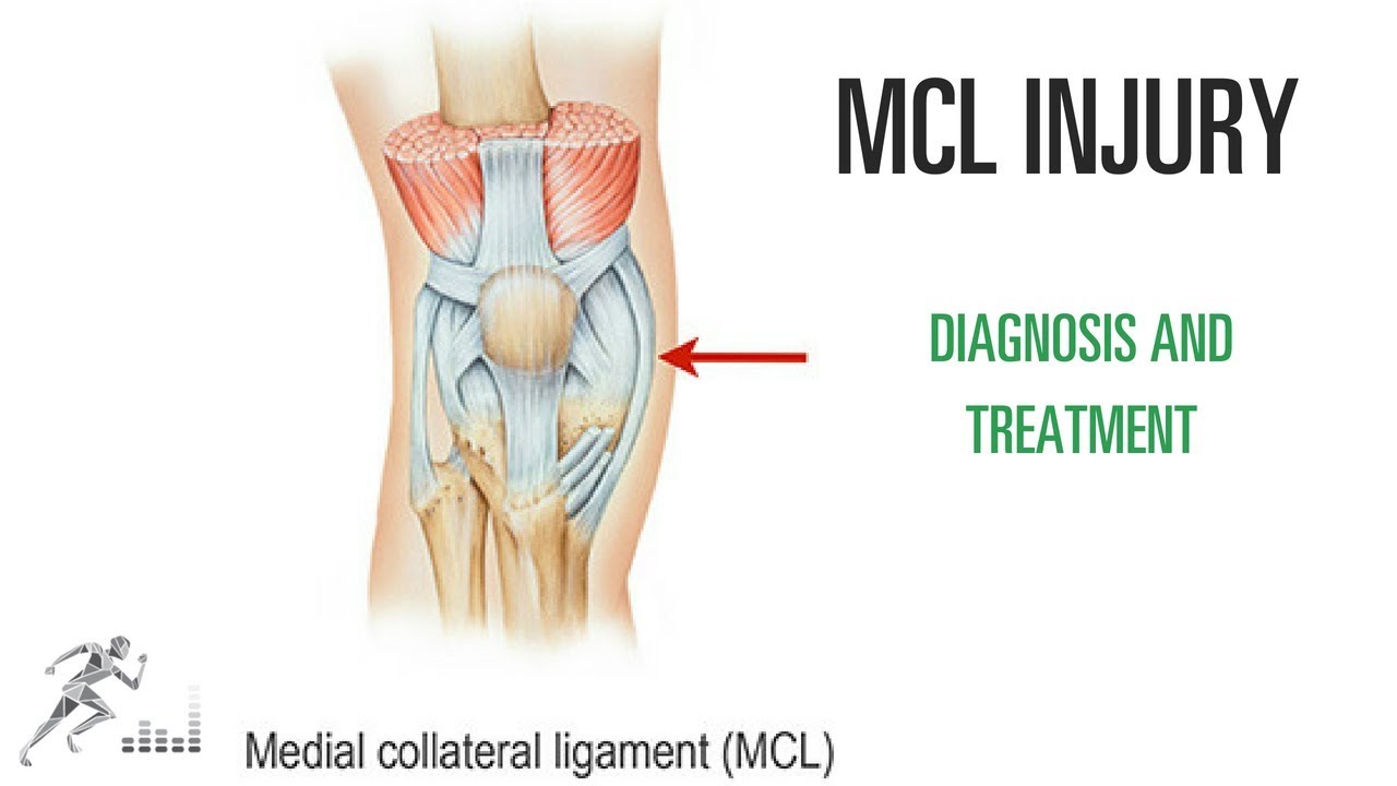
One of the most common knee injuries in contact and collision sports is a medial collateral ligament (MCL) injury. This is a ligament on the medial (side closest to the midline) side of your knee that provides stability against side-to-side stress to the knee. You might injure it by cutting maneuvers in sports like soccer or hockey. You can also suffer an MCL injury if another player hits you on the outside of your knee.
Please note: I don't respond to questions and requests for specific medical advice left in the comments to my videos. I receive too many to keep up (several hundred per week), and legally I can't offer specific medical advice to people who aren't my patients (see below). If you want to ask a question about a specific injury you have, leave it in the comments below, and I might answer it in an upcoming Ask Dr. Geier video. If you need more detailed information on your injury, go to my Resources page: https://www.drdavidgeier.com/resources/
The content of this YouTube Channel, https://www.youtube.com/user/drdavidgeier (“Channel”) is for INFORMATIONAL PURPOSES ONLY. The Channel may offer health, fitness, nutritional and other such information, but such information is intended for educational and informational purposes only. This content should not be used to self-diagnose or self-treat any health, medical, or physical condition. The content does not and is not intended to convey medical advice and does not constitute the practice of medicine. YOU SHOULD NOT RELY ON THIS INFORMATION AS A SUBSTITUTE FOR, NOR DOES IT REPLACE, PROFESSIONAL MEDICAL ADVICE, DIAGNOSIS, OR TREATMENT. You should consult with your healthcare professional before doing anything contained on this Channel. You agree that Dr. Geier is not responsible for any actions or inaction on your part based on the information that is presented on the Channel. Dr. David Geier Enterprises, LLC makes no representations about the accuracy or suitability of the content. USE OF THE CONTENT IS AT YOUR OWN RISK.
Unlike tears of the ACL, MCL injuries most often heal without surgery. You might need to wear a hinged knee brace for 2-6 weeks. The length of time you miss from sports or exercise varies depending on the location and severity of the injury.
In this video, I share my thoughts on the nature of an MCL injury, the diagnosis, the treatment options and return to sports.
Please remember, while I appreciate your questions, I cannot and will not offer specific medical advice by email, online, on my show, or in the comments at the end of these posts. My responses are meant to provide general medical information and education. Please consult your physician or health care provider for your specific medical concerns.

Welcome to the latest episode of HT Physio Quick Tips!
In this episode, Farnham's leading over-50's physiotherapist, Will Harlow, reveals the most common knee injuries that can be sustained from a fall. You'll learn the 5 most common knee injuries from falls, how to differentiate between them and the key signs to look for before getting help.
To register your interest for the upcoming Optimum Knee Health course and to be among the first to know when it is released, reach out to Will@ht-physio.co.uk
To get a copy of Will's new book, Thriving Beyond Fifty, you can find it on Amazon below:
UK link: https://amzn.to/3mAISFv
US link: https://amzn.to/43TE5Q8
(Amazon Affiliate links)
If you're suffering from nagging knee pain that hurts in the morning and stops you from walking as far as you'd like, you can take our free knee pain guide - which will give you 5 expert tips to put a stop to knee pain at home - by visiting here: https://ht-physio.co.uk/knee-pain-guide-download/
If you're over-50 with a painful problem in the Farnham, Surrey area, you can learn more about how Will Harlow and HT Physio can help you overcome a painful problem here: https://ht-physio.co.uk/
**Any information in this video should not be used as a substitute for individual medical advice. Please seek advice from your local healthcare professional before taking action on the information in this video.**

Subscribe to the Nucleus Biology channel to see new animations on biology and other science topics, plus short quizzes to ace your next exam: https://bit.ly/3lH1CzV
For Employees of Hospitals, Schools, Universities and Libraries: Download 8 FREE medical animations from Nucleus by signing up for a free trial: http://nmal.nucleusmedicalmedi....a.com/free-trial-mem
This animation by Nucleus shows you the function of plant and animal cells for middle school and high school biology, including organelles like the nucleus, nucleolus, DNA (chromosomes), ribosomes, mitochondria, etc. Also included are ATP molecules, cytoskeleton, cytoplasm, microtubules, proteins, chloroplasts, chlorophyll, cell walls, cell membrane, cilia, flagellae, etc.
0:07 What is a cell?
0:35 What are the 2 categories of cells?
1:22 What is an Organelle? DNA, Chromatin, Chromosomes
2:06 Organelles: Ribosomes, Endoplasmic Reticulum
2:59 Organelles: ER function, Vesicles, Golgi Body (Apparatus)
3:50 Organelles: Vacuole, Lysosome, Mitochondrion
4:45 Organelles: Cytoskeleton
5:04 Plant Cell Chloroplast, Cell Wall
5:43 Unique Cell Structures: Cilia
Watch another version of this video, narrated by biology teacher Joanne Jezequel here: https://youtu.be/cbiyKH9uPUw
#cell #nucleus #biology
----
Watch other Nucleus Biology videos:
- Controlled Experiments: https://youtu.be/D3ZB2RTylR4
- Independent vs. Dependent Variables: https://youtu.be/nqj0rJEf3Ew
- Active Transport: https://youtu.be/ufCiGz75DAk
----
Learn more about the company that created this video: http://www.nucleusmedicalmedia.com/
https://www.instagram.com/nucleusmedicalmedia
This animation won a Platinum Best of Show Aurora Award in 2016.

https://bit.ly/3HIStRc #shorts
Coloscopy | Colon Polyp Resection | Polypectomy
Colonoscopies are essential for detecting colorectal abnormalities, including colon polyps. Polypectomy, the surgical removal of these growths, can prevent them from becoming cancerous. This article offers a brief overview of colonoscopies, colon polyps, and polypectomy procedures.
A colonoscopy is an endoscopic examination allowing healthcare providers to visualize the colon and rectum using a colonoscope. The colonoscope, a flexible tube with a camera and light source, helps detect abnormalities, including polyps or tumors.
Colon polyps are abnormal growths arising from the colon's inner lining. While most polyps are benign, some can become malignant. Adenomatous polyps have a higher potential to become cancerous, whereas hyperplastic and inflammatory polyps pose a lower risk.
Polypectomy involves removing colon polyps during a colonoscopy. Two primary techniques include snare polypectomy, using a wire loop to cut the polyp, and cold forceps polypectomy, which employs forceps to grasp and remove smaller polyps.
Following a polypectomy, patients may experience mild discomfort or bleeding. Regular surveillance is crucial to minimize colorectal cancer risk. The frequency of surveillance colonoscopies depends on the number, size, and type of polyps found, as well as the patient's overall risk factors.
Colonoscopies and polypectomies play vital roles in detecting and removing colon polyps, reducing the risk of colorectal cancer, and maintaining optimal colon health.
Do you want to learn more about colon polyps and colonoscopy? check our:
Article @ https://bit.ly/41w5Ooq
For more free resources, find us on Pinterest & Facebook pages:
https://www.pinterest.ca/medicalartsofficial/
https://www.facebook.com/Medicalartsofficial
https://www.youtube.com/@medic....alarts?sub_confirmat
https://www.instagram.com/medicalartsofficial/
https://www.tiktok.com/@medicalarts
#endoscopicsurgery #digestivesystem
coloscopy
polyp
colon polyp
polypectomy
colonoscopy
colon polyp animation
gi endoscopy
@MedicalArts , 2023.

This particular video is intended as a demonstration of Neurologic Examination. This demonstration is intended as an example of a neurologic exam which may be used as part of the initial evaluation of patients with complaints that may have an underlying neurologic origin. This video is solely for educational purposes and intended for use to prepare for OSCEs incorporating standardized patient encounters. It is not intended as a demonstration of a comprehensive neurologic examination and is not intended as medical advice or medical guidelines.
It is not intended as a complete instructional video and should not be considered a source of complete physical examination instruction.
Instead, it should be treated as a supplement to independent learning using primary Osteopathic Clinical Skills instructional resources. Clinical skills are best learned and developed with support from faculty in the context of a complete Osteopathic Medical School Curriculum.
Osteopathic Clinical Skills is a channel dedicated to discussing and exploring Osteopathic Clinical Skills concepts for medical students, residents, and clinicians and presenting them in an easy to understand manner.
Attributions:
Many thanks to the University of North Texas Health Science Center Texas College of Osteopathic Medicine (UNTHSC - TCOM) for permitting use of the Simulation facilities and equipment during the production of this video.
Additional thanks to the UNTHSC-TCOM standardized patient and faculty volunteers who participated in this production and provided permission for the use of their image in this video.

General Examination - Clinical Skills OSCE - Dr Gill
The general examination is one of those early exams, which is essentially used to start medical students off with their clinical skills studies.
In the real world, it's mainly used with regard to gaining an overview of a patient, such as for a medical check up, or a baseline examination, for example, a health report.
They have been a couple of comments about the pulse monitor used in the video. For those who are interested. I’ve reached out to the manufacturer, and they’ve requested that the following code is provided to viewers, in order to get 20% off, if they decide on themselves.
Product model number: Vibeat SP20
Official Website: https://vibeatstore.com/produc....ts/sp20-handheld-pul
Special 20% OFF code: JAMES
--------------
Different medical schools, nursing colleges and other health professional courses will have their own preferred approach to a clinical assessment - you should concentrate on THEIR marks schemes for your assessments.
Some people watching this video may experience an ASMR effect
#DrGill #Asmr #Clinicalskills
#drgill #clinicalskills #asmr

This particular video is intended as a demonstration of a physical exam that may be useful in evaluating a patient with shoulder pain.
It is not intended as a complete instructional video and should not be considered a source of complete physical examination instruction. It is also intended not as a perfect example of a physical exam that would be performed for a patient in clinical practice, but is designed to optimize function and efficiency for a OSCE testing setting.
Instead, it should be treated as a supplement to independent learning using primary Osteopathic Physical Examination instructional resources. Clinical skills are best learned and developed with support from faculty in the context of a complete Osteopathic Medical School Curriculum.
Osteopathic Clinical Skills is a channel dedicated to discussing and exploring Osteopathic Clinical Skills concepts for medical students, residents, and clinicians and presenting them in an easy to understand manner.
Attributions:
Many thanks to the University of North Texas Health Science Center Texas College of Osteopathic Medicine (UNTHSC - TCOM) for permitting use of the Medical Education Training (MET) facilities and equipment during the production of this video.
Additional thanks to the UNTHSC-TCOM learner and faculty volunteers who participated in this production and provided permission for the use of their image in this video.

In this video, the viewer will learn the key aspects of the newborn physical exam, and how to distinguish between normal and abnormal findings.
Direct Links to chapters:
0:00-Intro
1:30-Head
3:49-Face
8:05-Neck
8:30-Chest
10:13-Abdomen
11:01-Groin
13:17-Extremities
14:05-Back
14:47-Neurologic
Please visit: www.openpediatrics.org
OPENPediatrics™ is an interactive digital learning platform for healthcare clinicians sponsored by Boston Children's Hospital and in collaboration with the World Federation of Pediatric Intensive and Critical Care Societies. It is designed to promote the exchange of knowledge between healthcare providers around the world caring for critically ill children in all resource settings. The content includes internationally recognized experts teaching the full range of topics on the care of critically ill children. All content is peer-reviewed and open access-and thus at no expense to the user.
For further information on how to enroll, please email: openpediatrics@childrens.harvard.edu
Please note: OPENPediatrics does not support nor control any related videos in the sidebar, these are placed by Youtube. We apologize for any inconvenience this may cause.
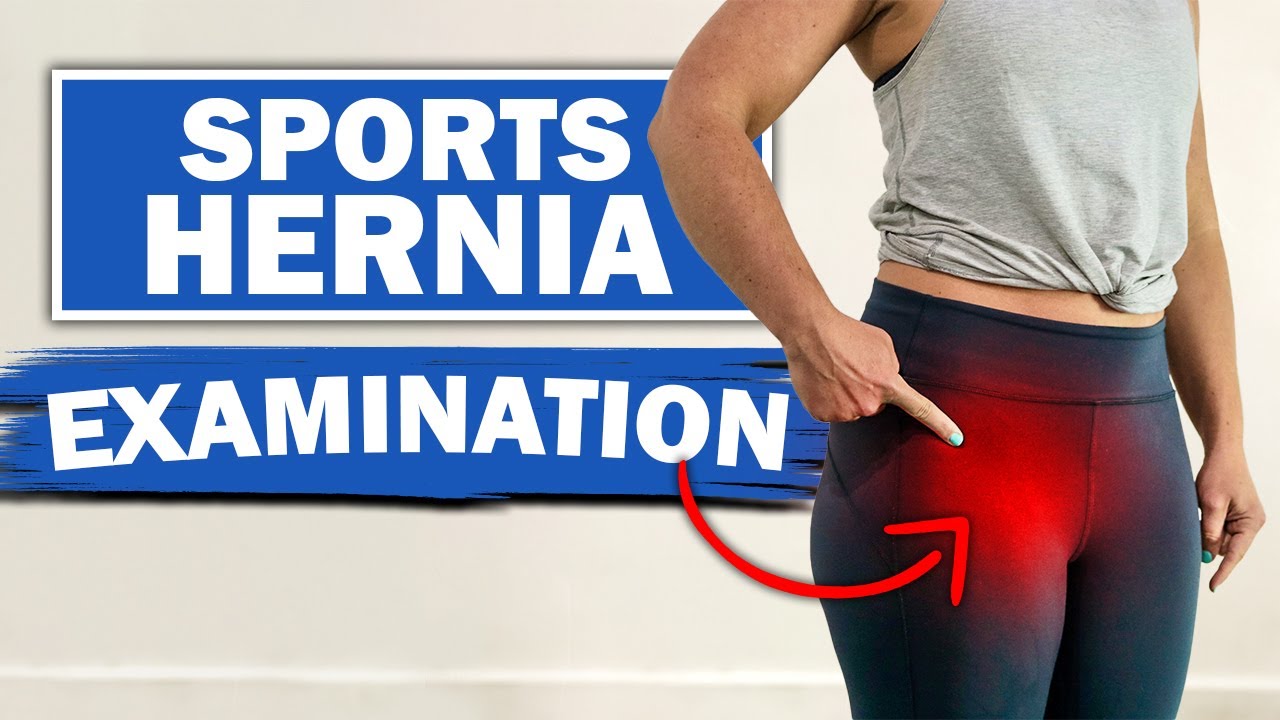
We will show you what a sports hernia examination (aka athletic pubalgia, gilmore's groin, lower abdominal pain) and rule out a diagnosis of hip impingement. Rehab exercises are suggested based on the results.
If you're experiencing any of these symptoms, don't hesitate to schedule a sports hernia examination. I can help you determine the best treatment plan to promote your recovery and avoid future injury. Subscribe to my channel to stay updated on the latest medical news and tips!
If you would like to know more about sports hernias and other diagnoses for front of hip, groin, adductor and lower abdominal strain, watch our detailed webinar here: https://bit.ly/37thtNF
For treatment, come visit us or schedule a virtual session. www.p2sportscare.com
Costa Mesa CA 715-502-4243
#sportshernia #abdominal #hippain
Sports Hernia Diagnosis
What Is A Sports Hernia?
A sports hernia is tearing of the transversalis fascia of the lower abdominal or groin region. A common misconception is that a sports hernia is the same as a traditional hernia. The mechanism of injury is rapid twisting and change of direction within sports, such as football, basketball, soccer and hockey.
The term “sports hernia” is becoming mainstream with more professional athletes being diagnosed. The following are just to name a few:
Torii Hunter
Tom Brady
Ryan Getzlaf
Julio Jones
Jeremy Shockey
If you follow any of these professional athletes, they all seem to have the same thing in common: Lingering groin pain. If you play fantasy sports, this is a major headache since it seems so minor, but it can land a player on Injury Reserve on a moments notice. In real life, it is a very frustrating condition to say the least. It is hard to pin point, goes away with rest and comes back after activity, but is hardly painful enough to make you want to stop. It lingers and is always on your mind. And if you’re looking for my step-by-step sports hernia rehab video course here it is.
One the best definitions of Sport hernias is the following by Harmon:
The phenomena of chronic activity–related groin pain that it is unresponsive to conservative therapy and significantly improves with surgical repair.”
This is truly how sports hernias behave in a clinical setting. It is not uncommon for a sports hernia to be unrecognized for months and even years. Unlike your typical sports injury, most sports medicine offices have only seen a handful of cases. It’s just not on most doctors’ radar. The purpose of this article is not only to bring awareness about sports hernias, but also to educate.
Will you find quick fixes in this article for sports hernia rehab?
Nope. There is no quick fix for this condition, and if someone is trying to sell you one, they are blowing smoke up your you-know-what.
Is there a way to decrease the pain related to sports hernias?
Yes. Proper rehab and avoidance of activity for a certain period of time will assist greatly, but this will not always stop it from coming back. Pain is the first thing to go and last thing to come. Do not be fooled when you become pain-free by resting it. Pain is only one measure of improvement in your rehab. Strength, change of direction, balance and power (just to name a few) are important, since you obviously desire to play your sport again. If you wanted to be a couch potato, you would be feeling better in no time. Watching Sports Center doesn’t require any movement.
Why is this article so long?
There is a lot of information on sports hernias available to you on the web. However, much of the information is spread out all over the internet and hard for athletes to digest due to complicated terminology. This article lays out the foundational terminology you will need to understand what options you have with your injury. We will go over anatomy, biomechanics, rehab, surgery, and even the fun facts. The information I am using is from the last ten years of medical research, up until 2016. We will be making updates overtime when something new is found as well. So link to this page and share with friends. This is the best source for information on sports hernias you will find.
Common Names (or Aliases?) for Sports Hernias
Sportsman’s Hernia
Athletic Pubalgia
Gilmore’s Groin
How Do You Know If You Have A Sports Hernia?
Typical athlete characteristics:
Male, age mid-20s
Common sports: soccer, hockey, tennis, football, field hockey
Motions involved: cutting, pivoting, kicking and sharp turns
Gradual onset
How A Sports Hernia Develops
Chronic groin pain typically happens over time, which is why with sports hernias, we do not hear many stories of feeling a “pop” or a specific moment of injury. It is the result of “overuse” mechanics stemming from a combination of inadequate strength and endurance, lack of dynamic control, movement pattern abnormalities, and discoordination of motion in the groin area.
#SPORTSHERNIAEXAM #california
