
- Physical Examination
- Surgical Examination
- Ophthalmology
- Clinical Skills
- Orthopedics
- Surgery Videos
- Laparoscopy
- Pediatrics
- Funny Videos
- Cardiothoracic Surgery
- Nursing Videos
- Plastic Surgery
- Otorhinolaryngology
- Histology and Histopathology
- Neurosurgery
- Dermatology
- Pediatric Surgery
- Urology
- Dentistry
- Oncology and Cancers
- Anatomy Videos
- Health and Fitness
- Radiology
- Anaesthesia
- Physical Therapy
- Pharmacology
- Interventional Radiology
- Cardiology
- Endocrinology
- Gynecology
- Emergency Medicine
- Psychiatry and Psychology
- Childbirth Videos
- General Medical Videos
- Nephrology
- Physiology
- Diet and Food Health
- Diabetes Mellitus
- Neurology
- Women Health
- Osteoporosis
- Gastroenterology
- Pulmonology
- Hematology
- Rheumatology
- Toxicology
- Nuclear Medicine
- Infectious Diseases
- Vascular Disease
- Reproductive Health
- Burns and Wound Healing
- Other
Top videos


#clean #leg #surgery #knee
✍️Dr. Matthew Harb talk about knee replacement surgery
https://www.MatthewHarbMD.com/links
👨⚕️Orthopedic Hip and Knee Surgeon
📍Located in Washington DC, and Maryland
📚Education and Insight
🛠Minimally invasive, outpatient, hip and knee replacement surgery
👉Visit me Online: https://www.MatthewHarbMD.com
☎️Schedule a virtual or in office appointment: https://www.MatthewHarbMD.com/links
📲Follow me on Social Media:
💥TIKTOK: https://www.tiktok.com/@matthewharbmd
💥INSTAGRAM: https://www.instagram.com/thebonesurgeon
📲Please SUBSCRIBE for new #shorts #doctor videos!
https://www.youtube.com/channel/UCbte-7tl7kaAm1xfWgz6xvQ?app=desktop&sub_confirmation=1
✍️QUESTION — Have a question or comment about medicine, orthopedic surgery, or musculoskeletal conditions. Post in the comment sections and let me know!
Dr. Matthew Harb specializes in minimally invasive, muscle sparing, hip and knee replacement surgery. Minimally invasive surgery allows patients to recover faster and have less pain post operatively. Implants are tailored and custom fit to each patient to allow for improved performance. Dr. Harb’s expertise in rapid recovery protocols allow for quick recovery after surgery and excellent outcomes in patients with hip and knee arthritis. With minimally invasive, muscle sparing surgery patients can return to their lifestyles and get back to doing the things they love sooner. Dr. Harb performs outpatient joint replacement surgery with many of his patients walking independently and going home the day of surgery.
“My focus is excellence in patient care, expedited recovery after surgery, and getting people back to the normal activities they love. Our team focused approach is committed to superb outcomes, improving lives, and returning patients to living pain free.”

Mini-Laparoscopic Cholecystectomy with Intraoperative Cholangiogram for Symptomatic Cholelithiasis (Gallstones) - Standard
Authors: Brunt LM1, Singh R1, Yee A2
Published: September 26, 2017
AUTHOR INFORMATION
1 Department of Surgery, Washington University, St. Louis, Missouri
2 Division of Plastic and Reconstructive Surgery, Washington University, St. Louis, Missouri
DISCLOSURE
No authors have a financial interest in any of the products, devices, or drugs mentioned in this production or publication.
ABSTRACT
Minimal invasive laparoscopic cholecystectomy is the typical surgical treatment for cholelithiasis (gallstones), where patients present with a history of upper abdominal pain and episodes of biliary colic. The classic technique for minimal invasive laparoscopic cholecystectomy involves four ports: one umbilicus port, two subcostal ports, and a single epigastric port. The Society of American Gastrointestinal and Endoscopic Surgeons (SAGES) has instituted a six-step strategy to foster a universal culture of safety for cholecystectomy and minimize risk of bile duct injury. The technical steps are documented within the context of the surgical video for (1) achieving a critical view of safety for identification of the cystic duct and artery, (2) intraoperative time-out prior to management of the ductal structures, (3) recognizing the zone of significant risk of injury, and (4) routine intraoperative cholangiography for imaging of the biliary tree. In this case, the patient presented with symptomatic biliary colic due to a gallstone seen on the ultrasound in the gallbladder. The patient was managed a mini-laparoscopic cholecystectomy using 3mm ports for the epigastric and subcostal port sites with intraoperative fluoroscopic cholangiogram. Specifically, the senior author encountered a tight cystic duct preventing the insertion of the cholangiocatheter and the surgical video describes how the author managed the cystic duct for achieving a cholangiogram, in addition to the entire technical details of laparoscopic cholecystectomy.

Michigan Medicine’s Cardiac Surgery Simplified series highlights a multitude of surgical procedures in order to educate patients, healthcare providers, and trainees interested in learning about cardiac surgery performed at the Frankel Cardiovascular Center.
Like and subscribe to our channel to learn more about our pioneering procedures including minimally invasive valve surgery and safer methods to repair aortic aneurysms and dissections.
To learn more about cardiac surgery at Michigan Medicine, visit: https://medicine.umich.edu/dept/cardiac-surgery
To learn more about Frankel Cardiovascular Center, visit: https://www.umcvc.org/
To watch the full playlist, visit: https://www.youtube.com/playli....st?list=PLNxqP-XbH8B
-------------------------------------------------------
Subscribe to Michigan Medicine’s YouTube channel for upcoming videos and future live streams featuring our experts answering your questions.
-------------------------------------------------------
Follow Michigan Medicine on Social:
Twitter: https://twitter.com/umichmedicine
Instagram: https://www.instagram.com/umichmedicine/
Facebook: https://www.facebook.com/MichiganMedi...
Follow the U-M Frankel Cardiovascular Center on Social:
Twitter: https://twitter.com/umichcvc
Facebook: https://www.facebook.com/Universityof...
#MichiganMedicine #MedEd #CardiacSurgery #UniversityOfMichiganHealth #FrankelCardiovascularCenter #Cardiology

This request usually follows orbit examination of a patient with thyroid eye disease. The aim is to look for signs of hyperthyroidism and less commonly hypothyroidism.
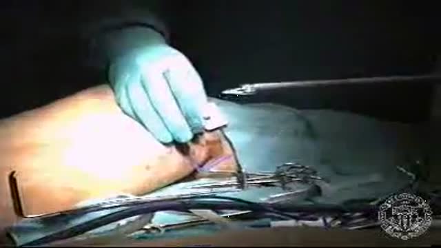
Endoscopic Vein Harvest
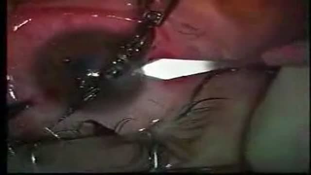
Removal of a foreign body from the eye (fish hook)

Cannula are often introduced into blood vessels in 80% of patients in the hospital for treatment. This can be a daunting experience to patients and stressful to doctors as multiple attempts are used. This may result in introducing spreading MRSA, E Coli & Chlostredium living on your skin into blood and results in Invasive MRSA infection.
Skin is often not adequatly cleaned during subsequent atempts as doctors/nurses do not wait for 1 min after applying cleaning solution on the skin before they puncture your skin.
Multiple punctured sites allow CA-MRSA to enter blood stream resulting in bacteremia and death.
Our mission is to reduce spreading invasive CA-MRSA in the hospitals by developing alternative technique to introduce cannulae.
Medifix was created by doctors with a mission to reduce the threat of spreading antibiotic resustant bacteria to mankind.
Michael Marcus, MD Pediatric Pulmonary www.DrMDK.com Maimonides Medical Center Fellowship:Children’s Hospital of Philadelphia www.DrMDK.com
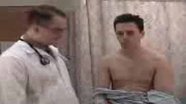
Examination of the heart from the USMLE collection
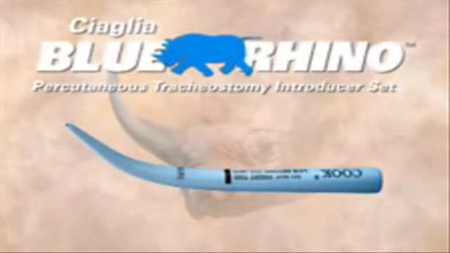
a video showing how to perform Percutaneous tracheostomy
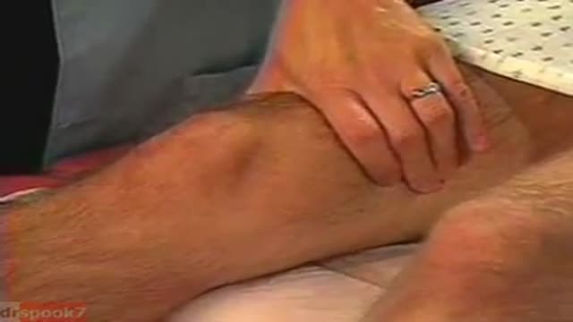
The Knee Exam
Observation:
1. Make sure that both knees are fully exposed. The patient should be in either a gown or shorts. Rolled up pant legs do not provide good exposure!
2. Watch the patient walk. Do they limp or appear to be in pain? When standing, is there evidence of bowing (varus) or knock-kneed (valgus) deformity? There is a predilection for degenerative joint disease to affect the medical aspect of the knee, a common cause of bowing. Varus Knee Deformity, more marked on the left leg. 3. Make note of any scars or asymmetry. Chronic/progressive damage, as in degenerative joint disease, may lead to abnormal contours and appearance. Is there obvious swelling as would occur in an effusion? Redness suggesting inflammation? 4. Is there evidence of atrophy of the quadriceps, hamstring, or calf muscle groups? Knee problems/pain can limit the use of the affected leg, leading to wasting of the muscles.
While both legs have well developed musculature,
the left calf and hamstring are bulkier than the right. 5. Look at the external anatomy, noting structures above and below the knee itself: 1. Patella 2. Patellar tendon 3. Quadriceps/Hamstring/Calf muscles 4. Medial and lateral joint lines. 5. Femur and Tibia 6. Tibial tuberosity
Ballotment (helpful if the effusion is large) 1. Slightly flex the knee which is to be examined.
2. Place one hand on the supra-pateallar pouch, which is above the patella and communicates with the joint space. Gently push down and towards the patella, forcing any fluid to accumulate in the central part of the joint.
3. Gently push down on the patella with your thumb.
4. If there is a sizable effusion, the patella will feel as if it's floating and "bounce" back up when pushed down.
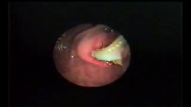
A 30 YEAR WOMEN WITH INTRACTABLE BILIARY COLIC CASE REPORT: This 30 year women developed severe pain right upper quadrant for last 10 days. She sought many consultations and was given intravenous analgesics both (nonnarcortic and narcotic). Pain did not subside and she sought my consultation. Examination revealed her to be in agony with severe upper abdominal pain. General physical examination was otherwise unremarkable. Abdominal examination revealed mild tenderness in right hypochondrium with doubtful Murphy's sign. Urgent abdominal ultrasound showed a linear structure in bile ducts making slow writhing movements. The structure had an anechoic tube (alimentary canal) inside suggestive of a large Ascarid. Urgent ERCP was performed and bile duct and pancreatic duct cannulated selectively. Pancreatic duct was normal. Bile ducts contained a long linear filling defect extending from lower end of common bile duct to right intrahepatic duct (see image gallery for ERCP plate). A basket was introduced in the duct (see video clip) and the linear structure was engaged with soft closure and extracted out of the bile duct. Accompanying the basket was a 25 cm thick highly motile Ascarid. To recover the worm, endoscope was withdrawn along with the basket and the friendly catch. While the endoscope was being withdrawn and the basket was in the duodenum with the worm out of bile duct, patient indicated of relief of abdominal pain. A relook cholangiogram showed no more structures in the duct. She was given antihelmintic therapy and passed hundreds of worms with the feces. The worms recovered form stools were both male and female population and varied in length and size. However the lone worm recovered form bile ducts was the longest and the thickest male worm. The phenomenal behavior of this ubiquitous infection remains unexplained. (Source Records from Dr. Khuroo's Medical Clinic. Review prepared by Mehnaaz Sultan Khuroo Host website www.drkhuroo.org , E-mail: mkhuroo@yahoo.com ).
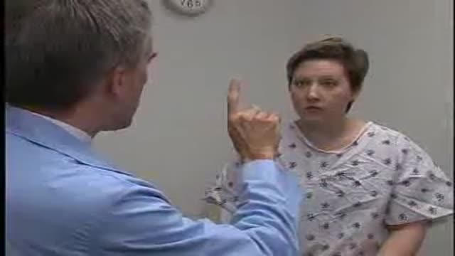
Part 3: from Loyola Medical School, Chicago showing clinical examination of the neurological system.
Deep Tie
Ileostomy Closure
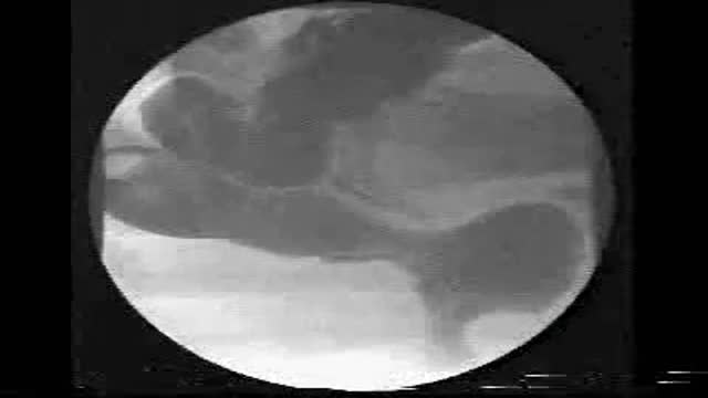
Defecography showing Enterocele
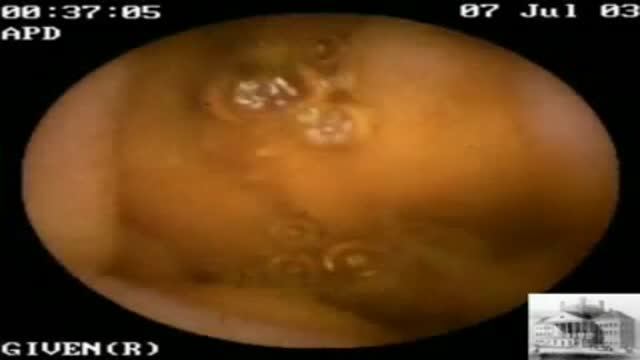
This 81 year old man with severe CAD and CHF was referred for VCE following a negative endoscopic workup for chronic guaiac positive stools. Seen on only three frames, this sequence reveals a single mid small bowel telangectasia, a possible source for his chronic GI blood loss. He has been managed c...onservatively and continues to require intermittent transfusions despite oral iron therapy.
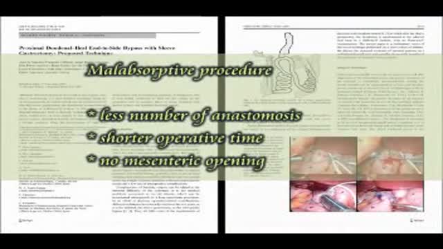
Loop duodenal switch is an end-to-side proximal duodeno-ileal bypass with a sleeve gastrectomy. The proximal duodenal stump is anastomosed to an ileal loop, 200 cm from the ileocecal valve. The procedure is a malabsorptive operation with some theoretical advantages: only one anastomosis is performed..., and so the operative time is shorter, and there is no mesenteric opening. It is not a mini-gastric bypass, as the gastric antrum, the pylorus and the first centimeters of the duodenum are preserved. The short term outcome shows a very good weight loss curve with no metabolic disturbances.
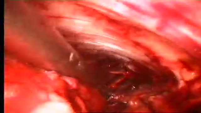
Purpose The complication rate in patients treated with the Linton procedure was unacceptably high. SEPS is minimal invasive treatment modality for chronic venous insufficiency and venous ulcers. Materials and Methods252 limbs of 229 patients who underwent SEPS procedure and/or safenous vein ablati...on from May 2003 to January 2008. Tourniquet was not used and two-port technique was preferred for operation. Skin graft was not used. Honeysoft (medical honey) was used for wound care in selected cases. Results According to CEAP clinical Classification 112 limbs were class 6, 70 limbs (class 5), 70 limbs (Class4) respectively. Greater saphenous vein stripping and/or high ligation, and varicose vein excision accompanied SEPS in 241limbs who had combined Sapheno-femoral junction and perforator vein insufficiencyand SEPS was performed alone 23 limbs who had recanalised deep venous thrombosis (19) and PVI alone(4). Mean patient follow-up was 35 months. No early deaths or thromboembolism occurred. Complications included severe subcutaneous emphysema(1), neuralgia (7), 1 year later cellulites (1). Ulcers healed in 124 limbs in two months and 58 limbs in 3 months. ulcer recurrence was seen on 12(%6.6) limbs. Clinical severity and disability scores improved significantly after surgery. Conclusion All venous ulcers healed with SEPS combined or not ablation of superficial venous reflux and remain healed 5 year period and symptom-free except recurrent ulcers during the long-term follow-up. SEPS is an effective and safety treatment modality.
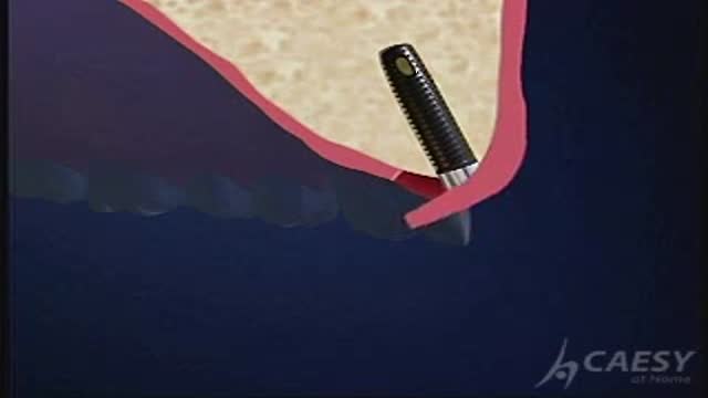
Single tooth implant
