- Physical Examination
- Surgical Examination
- Ophthalmology
- Clinical Skills
- Orthopedics
- Surgery Videos
- Laparoscopy
- Pediatrics
- Funny Videos
- Cardiothoracic Surgery
- Nursing Videos
- Plastic Surgery
- Otorhinolaryngology
- Histology and Histopathology
- Neurosurgery
- Dermatology
- Pediatric Surgery
- Urology
- Dentistry
- Oncology and Cancers
- Anatomy Videos
- Health and Fitness
- Radiology
- Anaesthesia
- Physical Therapy
- Pharmacology
- Interventional Radiology
- Cardiology
- Endocrinology
- Gynecology
- Emergency Medicine
- Psychiatry and Psychology
- Childbirth Videos
- General Medical Videos
- Nephrology
- Physiology
- Diet and Food Health
- Diabetes Mellitus
- Neurology
- Women Health
- Osteoporosis
- Gastroenterology
- Pulmonology
- Hematology
- Rheumatology
- Toxicology
- Nuclear Medicine
- Infectious Diseases
- Vascular Disease
- Reproductive Health
- Burns and Wound Healing
- Other
Top videos
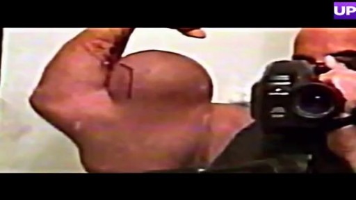
Bodybuilder Drains Synthol Hematoma From Bicep
Dont worry sister!
Focal seizures (also called partial seizures [citation needed] and localized seizures) are seizures which affect initially only one hemisphere of the brain. [citation needed] The brain is divided into two hemispheres, each consisting of four lobes – the frontal, temporal, parietal and occipital lobes.
A video showing the examination of femoral hernia.
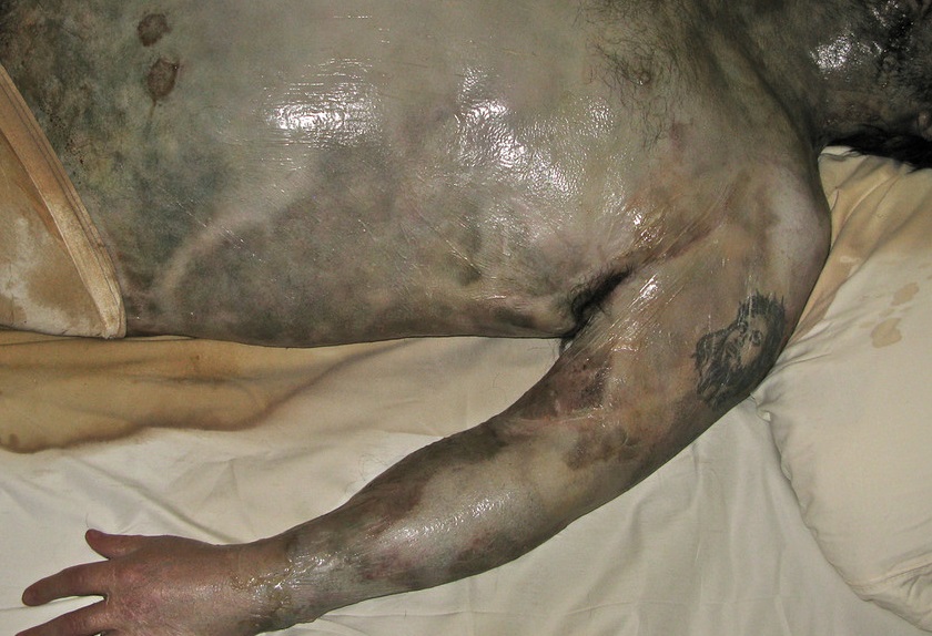
Watch that Full Human Body Decay Process Video
Tension pneumothorax describes the progressive accumulation of air in the pleural cavity (normally a potential space) through a defect in the visceral pleura. This leads to positive pressure being maintained and increasing throughout the respiratory cycle causing vessels within the mediastinum to be compressed with catastrophic consequences if left untreated. Clinical signs include hypoxia, hypotension, tachycardia, reduced breath sounds and hyper resonance ipsilaterally, with tracheal deviation (away from the affected side) and distended neck veins being late clinical signs.
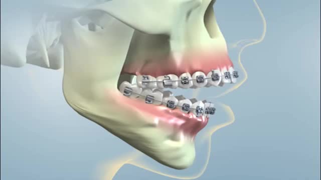
Jaw surgery, also known as orthognathic (or-thog-NATH-ik) surgery, corrects irregularities of the jaw bones and realigns the jaws and teeth to improve the way they work. Making these corrections may also improve your facial appearance. Jaw surgery may be a corrective option if you have jaw problems that can't be resolved with orthodontics alone. In most cases, you also have braces on your teeth before surgery and during recovery after surgery until healing and alignment are complete. Your orthodontist can work with your oral and jaw and face (maxillofacial) surgeon to determine your treatment plan.
A very funny video
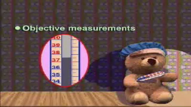
Pediatric measurements: length, body weight...etc.
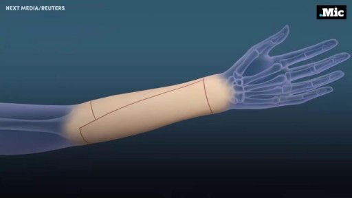
Here's how female-to-male gender reassignment surgery works.
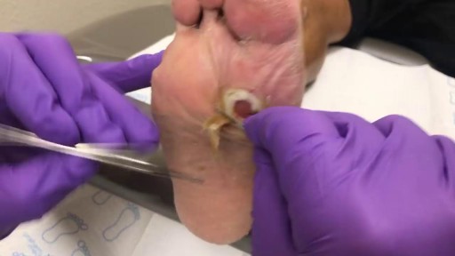
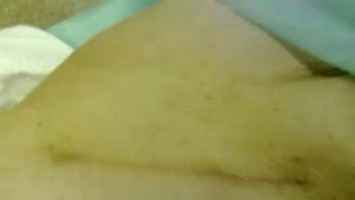
This is a diabetic foot ulcer. The patient reportedly went on vacation and noticed this ulcer upon their return. Debridement (removal of damaged tissue) to the level of healthy bleeding tissue is medically necessary as damaged tissue acts an impediment to wound healing. Due to their diabetic neuropathy, they did not feel any pain or indication that a wound was forming. This ulcer appeared to have penetrated to the level of subcutaneous tissue or even fascia, but turned out to be much deeper than that. These are serious wounds and are the beginnings of what lead to foot and leg amputations if they are not treated promptly by your healthcare provider, AKA Podiatrist.
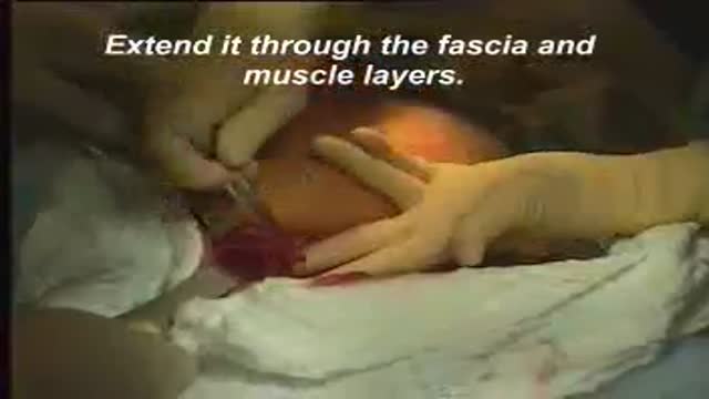
A video showing the steps of cesarean section surgery

Generic minoxidil is known to treat hair-fall issues in men and women, it is best for hair growth, hair re-development, etc. it is available in the strength of 5mg and easily available at online pharmacy store. For more information visit to http://www.medstorerx.com/generic-minoxidil.aspx
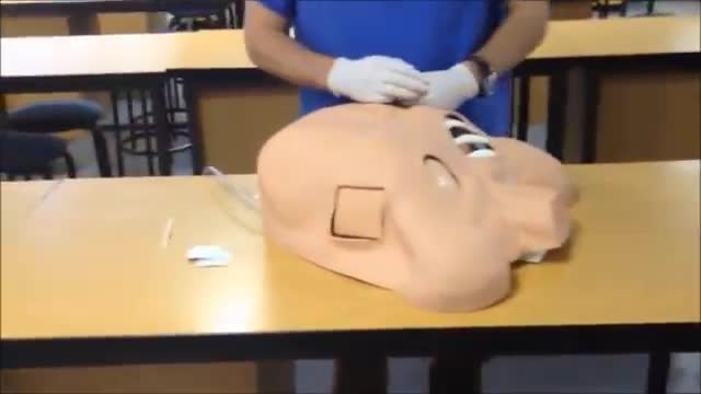
Ellis demonstrates how to insert and then remove an NG tube. This includes drawing gastric residual and checking the pH. After the demonstration, Ellis provides additional tips about clamping the NG tube and using the blue pigtail.
Our Critical Nursing Skills video tutorial series is taught by Ellis Parker MSN, RN-BC, CNE, CHS and intended to help RN and PN nursing students study for your nursing school exams, including the ATI, HESI and NCLEX.
#NCLEX #HESI #Kaplan #ATI #NursingSchool #NursingStudent #Nurse #RN #PN #Education #LVN #LPN #ClinicalSkills #NGTube #nurseeducator
00:00 What to expect
00:30 Preparing NG tube patient
00:56 Preparing NG tube equipment
1:29 Measuring the NG tube
2:02 Preparing for NG tube insertion
2:28 Inserting the NG tube
3:17 Checking placement with pH
4:23 Anchoring with split-tape
5:32 Connecting to suction
6:05 Disconnecting from suction
6:17 What to do before removal?
7:03 Removing NG tube
7:40 Additional tips on clamping
8:31 The blue pigtail
🚨 Reminder: shipping deadlines are looming 👀
🎁 Regular Shipping: Order by Friday, December 15
🚀 Expedited Shipping: Order by Monday, December 18
🔍 Still searching for last-minute gifts? Consider a Level Up RN Gift Card! 💌 It’s not only a thoughtful present but also the perfect way to share treasures like Pharmacology Flashcards OR digital treasures like Flashables Digital Nursing Flashcards & the Level Up RN membership. Give the gift of knowledge this holiday season! 🧠⚡️💖 bit.ly/LevelUpRNGC
🚪 Access our Cram Courses, Quizzes and Videos all in one ad free space with Level Up RN Membership https://bit.ly/LevelUpRNMembership
Want more ways to MASTER Clinical Skills? Check out our flashcards & videos!
👇👇👇👇👇👇👇👇👇👇
👉 https://bit.ly/clinicalnursingskills 👈
☝️👆☝️👆☝️👆☝️👆☝️👆
This is your one-stop-shop for materials to help you LEARN & REVIEW so you can PASS Nursing School.
🤔🤔🤔 DO YOU WANT TO PASS your classes, proctored exams and the NCLEX? 🤔🤔🤔 Our resources are the best you can buy. They are built with a single goal: help you pass with no fluff. Everything you need, and nothing you don’t. Don’t take our word for it, though! Check out our hundreds of ⭐️⭐️⭐️⭐️⭐️ reviews from nurses who passed their exams and the NCLEX with Level Up RN.
🗂️ Our Ultimate Nursing School Survival kit is your number 1 resource to get through nursing school and to pass the NCLEX. Whether you're just starting school or you’re already prepping for the NCLEX, this bundle of flashcards is the best you can buy. It covers all the information you need to know to pass all your exams and it has FREE shipping!
➡️ https://bit.ly/TUNSSK ⬅️
L👀king for EVEN MORE resources to survive Nursing School? Make your Nursing School experience your own! Life’s difficult enough—learning shouldn’t be.
🪅 Games https://nursesquad.com
💻 Digital resources https://bit.ly/NursingStudyCourses
📅 Organizational tools https://bit.ly/OrganizingSchool
✨Want perks? Join our channel!
https://youtube.com/leveluprn/join
🏷 Head to https://leveluprn.com/specials for all our latest deals!🥳️
📧 LOOKING FOR FREE RESOURCES TO HELP WITH YOUR EXAMS? Get exclusive tips, latest video releases and more delivered to your email!
➡️ https://leveluprn.com/signup ⬅️
⚕ 👩 LEVEL UP NURSE SQUAD 👩⚕️
All of the nurses at Level Up RN are here to help! Cathy Parkes started helping her fellow classmates back when she was in nursing school, tutoring so they could pass their exams and graduate. After she got her BSN and started working as an RN at Scripps Encinitas Hospital, she started this YouTube channel to help nursing students around the world. Since then she has built a team of top-notch dedicated nurses and nurse educators who are focused on improving nursing education and supporting career advancement for nurses everywhere. With flashcards, videos, courses, organizational tools and more, we are singularly focused on helping students and nurses Level Up on their exams and nursing careers.

Watch that video of people who Survived Deadly Snake Bites
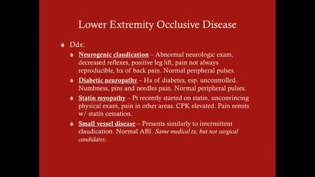
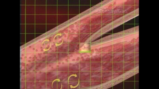
Claudication, which is defined as reproducible ischemic muscle pain, is one of the most common manifestations of peripheral arterial occlusive disease (PAOD) caused by atherosclerosis. Claudication occurs during physical activity and is relieved after a short rest. Pain develops because of inadequate blood flow. Examination of a patient with claudication should include a complete lower-extremity evaluation and pulse examination, including measuring segmental pressures. Attempt to palpate pulses from the abdominal aorta to the foot, with auscultation for bruits in the abdominal and pelvic regions. When palpable pulses are not present, a handheld Doppler device may be used to assess circulation.
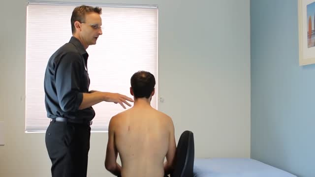
Rhomboid muscle strain and spasm causes upper back pain between the shoulder blades. Some patients describe the muscle spasms and discomfort as knots in the back. Early treatment is important to speed healing and recovery. Ice therapy for the first few days followed by moist heat can help relieve symptoms.Oct 12, 2015
When the hematocrit rises to 60 or 70%, which it often does in polycythemia, the blood viscosity can become as great as 10 times that of water, and its flow through blood vessels is greatly retarded because of increased resistance to flow. This will lead to decreased oxygen delivery.

LIS Closed done at 5 O clock position, using Scalpel blade 15. After feeling the groove between internal and external anal sphincter, the blade is passed in and the lower 1/2 of Internal anal sphincter is cut. Remain below dentate line. If anal mucosa is accidently cut suture with 4-0 rapid vicryl. In event of bleeding, pinchcock for 5 minutes.
