- Physical Examination
- Surgical Examination
- Ophthalmology
- Clinical Skills
- Orthopedics
- Surgery Videos
- Laparoscopy
- Pediatrics
- Funny Videos
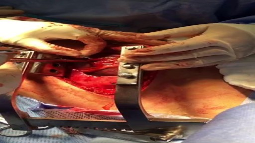
- Cardiothoracic Surgery
- Nursing Videos
- Plastic Surgery
- Otorhinolaryngology
- Histology and Histopathology
- Neurosurgery
- Dermatology
- Pediatric Surgery
- Urology
- Dentistry
- Oncology and Cancers
- Anatomy Videos
- Health and Fitness
- Radiology
- Anaesthesia
- Physical Therapy
- Pharmacology
- Interventional Radiology
- Cardiology
- Endocrinology
- Gynecology
- Emergency Medicine
- Psychiatry and Psychology
- Childbirth Videos
- General Medical Videos
- Nephrology
- Physiology
- Diet and Food Health
- Diabetes Mellitus
- Neurology
- Women Health
- Osteoporosis
- Gastroenterology
- Pulmonology
- Hematology
- Rheumatology
- Toxicology
- Nuclear Medicine
- Infectious Diseases
- Vascular Disease
- Reproductive Health
- Burns and Wound Healing
- Other
Top videos
Digoxin is derived from the leaves of a digitalis plant. Digoxin helps make the heart beat stronger and with a more regular rhythm. Digoxin is also used to treat atrial fibrillation, a heart rhythm disorder of the atria (the upper chambers of the heart that allow blood to flow into the heart).
Myelodysplastic syndromes are a group of cancers in which immature blood cells in the bone marrow do not mature or become healthy blood cells. In a healthy person, the bone marrow makes blood stem cells (immature cells) that become mature blood cells over time.Aug 12, 2015
Pompe disease is a rare multisystem genetic disorder that is characterized by absence or deficiency of the lysosomal enzyme alpha-glucosidase (GAA). This enzyme is required to breakdown (metabolize) the complex carbohydrate glycogen and convert it into the simple sugar glucose.
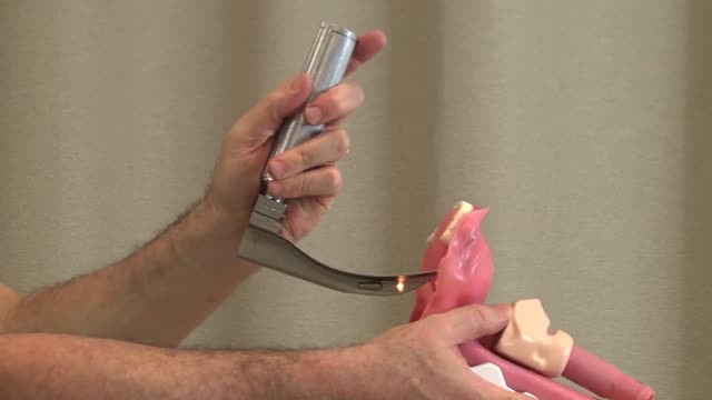
Direct Laryngoscopy: MICU Fellows Airway Course

How to place an NG tube in a baby,

Experts do not know the exact cause of Zollinger-Ellison syndrome. About 25 to 30 percent of gastrinomas are caused by an inherited genetic disorder called multiple endocrine neoplasia type 1 (MEN1). MEN1 causes hormone-releasing tumors in the endocrine glands and the duodenum.

See the effects of cannabis first hand, unedited, on Parkinson's tremor dyskinesia, and voice.
A blood transfusion is a routine medical procedure that can be lifesaving. During a blood transfusion, donated blood is added to your own blood. A blood transfusion may also be done to supplement various components of your blood with donated blood products. In some cases, a blood transfusion is done with blood that you've donated ahead of time before you undergo elective surgery. During a typical blood transfusion, certain parts of blood are delivered through an intravenous (IV) line that's placed in one of the veins in your arm. A blood transfusion usually takes one to four hours, though in an emergency it can be done much faster.
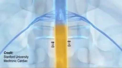
Wireless charger for medical devices that are implanted deep inside the body.

Vaccination is now mandatory in Italy.
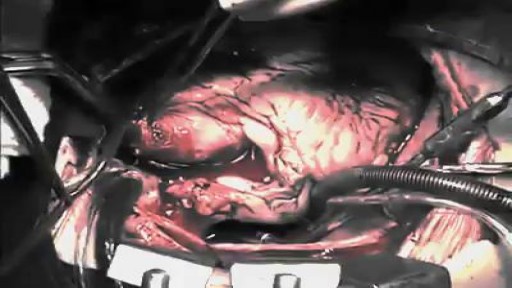
The first operation is harvesting the heart from the donor. The donor is usually an unfortunate person who has suffered irreversible brain injury, called "brain death". Very often these are patients who have had major trauma to the head, for example, in an automobile accident. The victim's organs, other than the brain, are working well with the help of medications and other "life support" that may include a respirator or other devices. A team of physicians, nurses, and technicians goes to the hospital of the donor to remove donated organs once brain death of the donor has been determined. The removed organs are transported on ice to keep them alive until they can be implanted. For the heart, this is optimally less than six hours. So, the organs are often flown by airplane or helicopter to the recipient's hospital.

Woman suffers allergic reaction to nut protein in boyfriend's sperm.A British woman has become the first recorded case of someone suffering an allergic...
The first week after birth: What to expect
30 yr old man presented to ER after Motor Vehicle Crash..blunt chest trauma...

http://plantar-fasciitis-solution.info-pro.co Foot Arch Pain, Sharp Pain In Heel, Pain In Foot, Achilles Heel Pain, Chronic Plantar Fasciitis What is Plantar Fasciitis? Plantar fasciitis is a common injury that affects the heel of a person’s foot. The arches of the feet are supported by a tough and fibrous tissue known as the plantar fascia and when this tissue is used repetitively, injury may occur. It can be easy to overuse the feet, especially when participating in activities such as sporting events. Hence, plantar fasciitis is more commonly found in athletes or others who are constantly using their feet for long durations. With excessive use, the planar fascia will eventually give in and this condition may also be progressive. Runners and those who are known to participate in similar activities need to ensure that they do not damage this important band of tissue. In addition, body weight could be a factor that leads to the occurrence of plantar fasciitis. If a person is overweight, the feet and subsequently the plantar fascia tissue could become overwhelmed. Improper footwear could also cause a strain on the plantar fascia tissue and this could gradually become severe over time. plantar fasciitis relief in 7 days click here. http://plantar-fasciitis-solution.info-pro.co

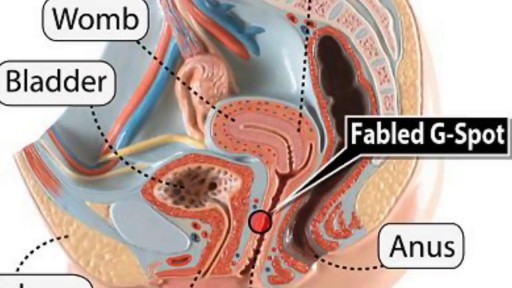
Watch that video to know What is G Spot?
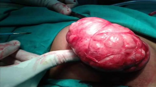
Watch that Big Size Fibrodenoma Removal Under Local Anesthesia
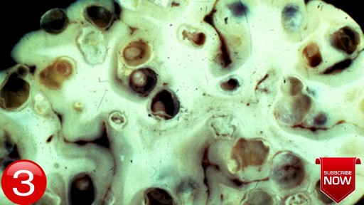
Watch that video of Terrible Horrifying Creatures Found Living Inside a Human Body

one of the best videos I've ever seen..
