Najbolji videi
The Irish Thoracic Society explain the Active Cycle of Breathing Technique for patients with acute and chronic respiratory illnesses and diseases and respiratory distress
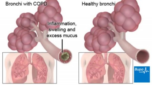
COPD (chronic obstructive pulmonary disease) makes it hard for you to breathe. The two main types are chronic bronchitis and emphysema. The main cause of COPD is long-term exposure to substances that irritate and damage the lungs. This is usually cigarette smoke. Air pollution, chemical fumes, or dust can also cause it. At first, COPD may cause no symptoms or only mild symptoms. As the disease gets worse, symptoms usually become more severe. They include A cough that produces a lot of mucus Shortness of breath, especially with physical activity Wheezing Chest tightness Doctors use lung function tests, imaging tests, and blood tests to diagnose COPD. There is no cure. Treatments may relieve symptoms. They include medicines, oxygen therapy, surgery, or a lung transplant. Quitting smoking is the most important step you can take to treat COPD.
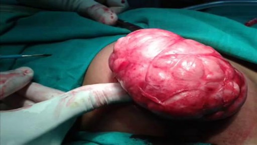
Splenectomy for massive splenomegaly (>1500 g) provides palliation but is associated with a high rate of perioperative complications in a population of patients with advanced hematological malignancies. Predictive factors for survival and whether the palliative goals are achieved in the long-term are not well defined.
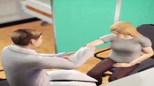
If you’re considering an epidural to help manage the pain of childbirth, you’re not alone. More than 60 percent of women delivering at hospitals elect for an epidural during labor. And with good reason: An epidural is considered one of the safest methods of pain control, with just one in 3,000 pregnancies experiencing serious complications. It’s also good for you, since you’ll remain awake and alert during the birth, as well as for your baby, since the drugs will barely reach your bloodstream (so they can’t get into hers).
Is Air Travel During Pregnancy Safe? Traveling by air is considered safe for women while they are pregnant; however, the following ideas might make your trip safer and more comfortable. Most airlines allow pregnant women to travel through their eighth month.
This video may contain images of a medical doctor providing emergency care for a patient.
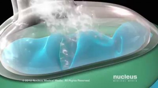
Hysterectomy is the surgical removal of the uterus. It ends menstruation and the ability to become pregnant. Depending on the reason for the surgery, a hysterectomy may also involve the removal of other organs and tissues such as the ovaries and/or fallopian tubes.
For strong lungs, chew 3 to 5 peppermint leaves each day. To treat congestion, add a few drops of peppermint oil to a pot of hot water and inhale the steam. You can also drink 2 cups of peppermint tea daily. To make the tea, add 1 teaspoon of dried peppermint leaves to a cup of hot water.
Skin Graft? Skin grafting is a surgical procedure that involves removing the skin from one area of the body and moving it, or transplanting it, to a different area of the body. This surgery may be done if a part of your body has lost its protective covering of skin due to burns, injury, or illness

Vaccination is now mandatory in Italy.
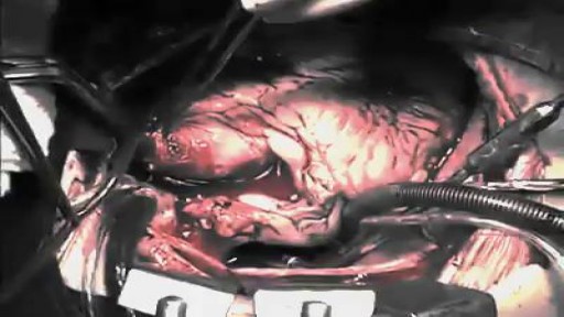
The first operation is harvesting the heart from the donor. The donor is usually an unfortunate person who has suffered irreversible brain injury, called "brain death". Very often these are patients who have had major trauma to the head, for example, in an automobile accident. The victim's organs, other than the brain, are working well with the help of medications and other "life support" that may include a respirator or other devices. A team of physicians, nurses, and technicians goes to the hospital of the donor to remove donated organs once brain death of the donor has been determined. The removed organs are transported on ice to keep them alive until they can be implanted. For the heart, this is optimally less than six hours. So, the organs are often flown by airplane or helicopter to the recipient's hospital.
Woman suffers allergic reaction to nut protein in boyfriend's sperm.A British woman has become the first recorded case of someone suffering an allergic...
The first week after birth: What to expect
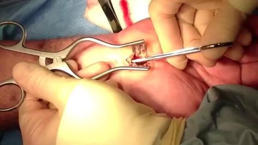
Carpal tunnel release is a surgery used to treat and potentially heal the painful condition known as carpal tunnel syndrome. Doctors used to think that carpal tunnel syndrome was caused by an overuse injury or a repetitive motion performed by the wrist or hand, often at work. They now know that it’s most likely a congenital predisposition (something that runs in families) – some people simply have smaller carpal tunnels than others. Carpal tunnel syndrome can also be caused by injury, such as a sprain or fracture, or repetitive use of a vibrating tool. It's also been linked to pregnancy, diabetes, thyroid disease, and rheumatoid arthritis.
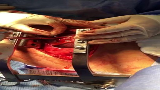
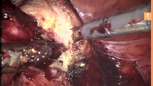
30 yr old man presented to ER after Motor Vehicle Crash..blunt chest trauma...
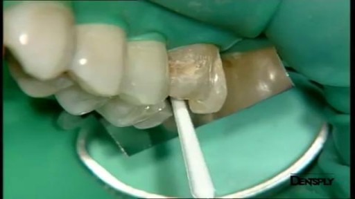
Art restoration of anterior teeth

http://plantar-fasciitis-solution.info-pro.co Foot Arch Pain, Sharp Pain In Heel, Pain In Foot, Achilles Heel Pain, Chronic Plantar Fasciitis What is Plantar Fasciitis? Plantar fasciitis is a common injury that affects the heel of a person’s foot. The arches of the feet are supported by a tough and fibrous tissue known as the plantar fascia and when this tissue is used repetitively, injury may occur. It can be easy to overuse the feet, especially when participating in activities such as sporting events. Hence, plantar fasciitis is more commonly found in athletes or others who are constantly using their feet for long durations. With excessive use, the planar fascia will eventually give in and this condition may also be progressive. Runners and those who are known to participate in similar activities need to ensure that they do not damage this important band of tissue. In addition, body weight could be a factor that leads to the occurrence of plantar fasciitis. If a person is overweight, the feet and subsequently the plantar fascia tissue could become overwhelmed. Improper footwear could also cause a strain on the plantar fascia tissue and this could gradually become severe over time. plantar fasciitis relief in 7 days click here. http://plantar-fasciitis-solution.info-pro.co

In this video, learn how to suture like a plastic surgeon! We'll go over the different types of sutures, appropriate needle sizes, and the correct technique for suturing different types of wounds. Whether you're a medical student or just interested in improving your suturing skills, this video is for you! Join us and start mastering this essential surgical skill.
Check out what we offer to medical students and residents!
Our Research Courses: Take the chance now to start your research career!
Research course🔬📄: https://cutt.ly/z7Xp9u5
Systematic Reviews course📄: https://cutt.ly/67XpVSr
Statistical analysis 📊: https://cutt.ly/o7Xaf2W
How to find 👀 Research 🔬 Positions in the U.S: https://cutt.ly/g7XakpF
~~~~~~
Our services:
One-on-one Residency advising👩🏫👨🏫: https://cutt.ly/47XiCl1
USMLE tutoring: https://cutt.ly/67XoZYl
Interview preparation💼: https://cutt.ly/d7XiFPP
Personal statement editing ✍🏻📃: https://cutt.ly/17Xi4NG
ERAS CV, supplemental ERAS CV, or your regular CV🔥 editing: https://cutt.ly/j7XogUO
MATCH application package: https://cutt.ly/B7XoR06
#suturing #stitch #surgery #plasticsurgery
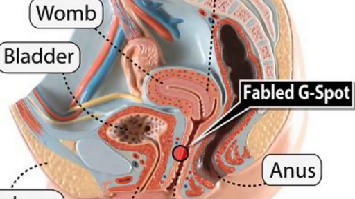
Watch that video to know What is G Spot?
Watch that Big Size Fibrodenoma Removal Under Local Anesthesia
