- Physical Examination
- Surgical Examination
- Ophthalmology
- Clinical Skills
- Orthopedics
- Surgery Videos
- Laparoscopy
- Pediatrics
- Funny Videos
- Cardiothoracic Surgery
- Nursing Videos
- Plastic Surgery
- Otorhinolaryngology
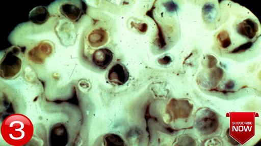
- Histology and Histopathology
- Neurosurgery
- Dermatology
- Pediatric Surgery
- Urology
- Dentistry
- Oncology and Cancers
- Anatomy Videos
- Health and Fitness
- Radiology
- Anaesthesia
- Physical Therapy
- Pharmacology
- Interventional Radiology
- Cardiology
- Endocrinology
- Gynecology
- Emergency Medicine
- Psychiatry and Psychology
- Childbirth Videos
- General Medical Videos
- Nephrology
- Physiology
- Diet and Food Health
- Diabetes Mellitus
- Neurology
- Women Health
- Osteoporosis
- Gastroenterology
- Pulmonology
- Hematology
- Rheumatology
- Toxicology
- Nuclear Medicine
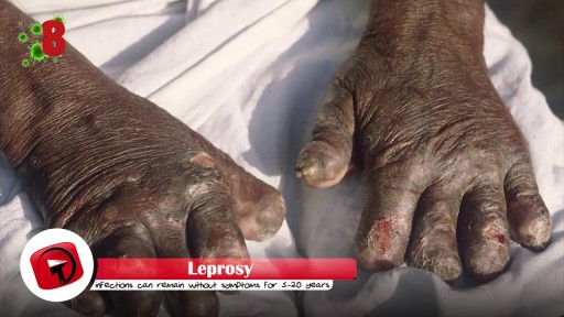
- Infectious Diseases
- Vascular Disease
- Reproductive Health
- Burns and Wound Healing
- Other
Top videos
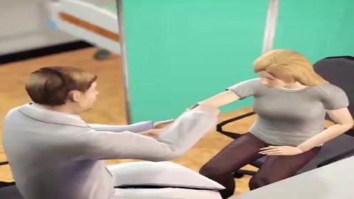
Hysterectomy is the surgical removal of the uterus. It ends menstruation and the ability to become pregnant. Depending on the reason for the surgery, a hysterectomy may also involve the removal of other organs and tissues such as the ovaries and/or fallopian tubes.

For strong lungs, chew 3 to 5 peppermint leaves each day. To treat congestion, add a few drops of peppermint oil to a pot of hot water and inhale the steam. You can also drink 2 cups of peppermint tea daily. To make the tea, add 1 teaspoon of dried peppermint leaves to a cup of hot water.
Exam- COPD Patient
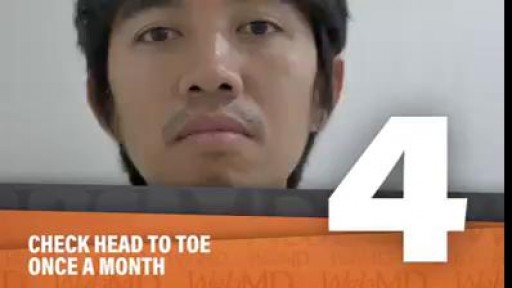
Skin isn't just your body's biggest organ-- it's also the fastest growing.
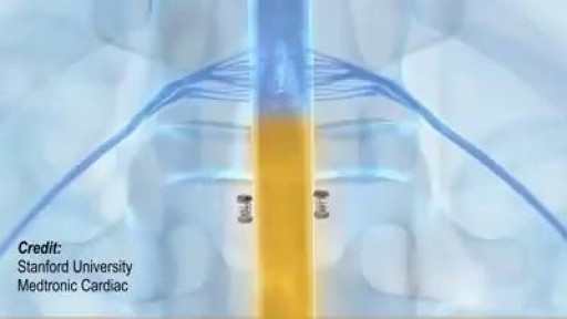
Wireless charger for medical devices that are implanted deep inside the body.

Vaccination is now mandatory in Italy.
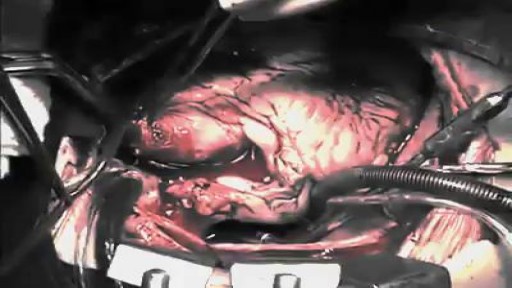
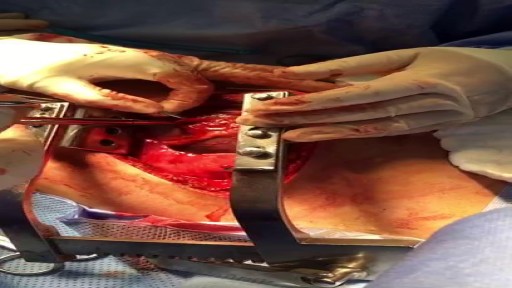
The first operation is harvesting the heart from the donor. The donor is usually an unfortunate person who has suffered irreversible brain injury, called "brain death". Very often these are patients who have had major trauma to the head, for example, in an automobile accident. The victim's organs, other than the brain, are working well with the help of medications and other "life support" that may include a respirator or other devices. A team of physicians, nurses, and technicians goes to the hospital of the donor to remove donated organs once brain death of the donor has been determined. The removed organs are transported on ice to keep them alive until they can be implanted. For the heart, this is optimally less than six hours. So, the organs are often flown by airplane or helicopter to the recipient's hospital.
Woman suffers allergic reaction to nut protein in boyfriend's sperm.A British woman has become the first recorded case of someone suffering an allergic...
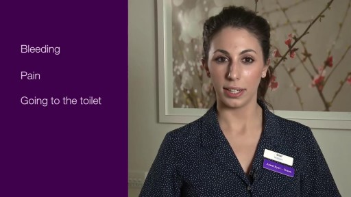
The first week after birth: What to expect
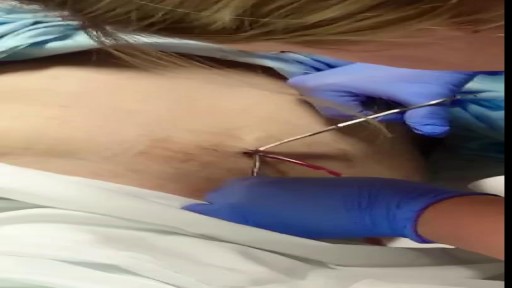
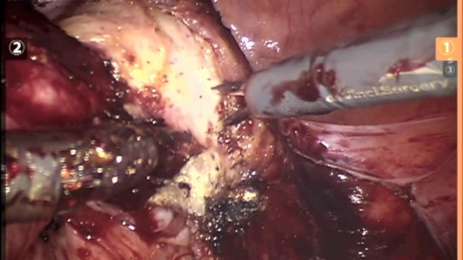
Removal of drain tube after spleen surgery
30 yr old man presented to ER after Motor Vehicle Crash..blunt chest trauma...
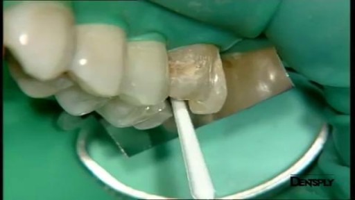
Art restoration of anterior teeth

http://plantar-fasciitis-solution.info-pro.co Foot Arch Pain, Sharp Pain In Heel, Pain In Foot, Achilles Heel Pain, Chronic Plantar Fasciitis What is Plantar Fasciitis? Plantar fasciitis is a common injury that affects the heel of a person’s foot. The arches of the feet are supported by a tough and fibrous tissue known as the plantar fascia and when this tissue is used repetitively, injury may occur. It can be easy to overuse the feet, especially when participating in activities such as sporting events. Hence, plantar fasciitis is more commonly found in athletes or others who are constantly using their feet for long durations. With excessive use, the planar fascia will eventually give in and this condition may also be progressive. Runners and those who are known to participate in similar activities need to ensure that they do not damage this important band of tissue. In addition, body weight could be a factor that leads to the occurrence of plantar fasciitis. If a person is overweight, the feet and subsequently the plantar fascia tissue could become overwhelmed. Improper footwear could also cause a strain on the plantar fascia tissue and this could gradually become severe over time. plantar fasciitis relief in 7 days click here. http://plantar-fasciitis-solution.info-pro.co
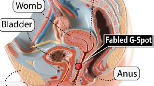
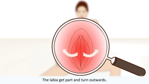
Watch that video to know What is G Spot?
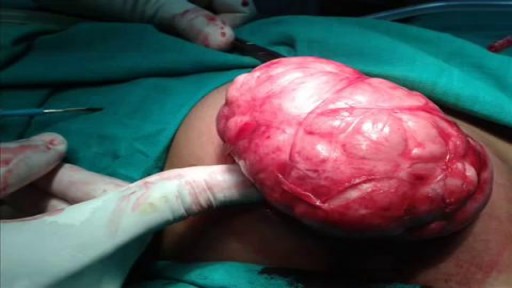
Watch that Big Size Fibrodenoma Removal Under Local Anesthesia
Watch that video of Terrible Horrifying Creatures Found Living Inside a Human Body

Aumento De Gluteos, Metacrilato En Gluteos, Aumento De Gluteos Natural, Operacion De Nalgas.--- http://aumente-gluteos.plus101.com/ --- Con una combinación de dieta, ejercicio y mejoras artificiales, puedes cambiar la forma de los glúteos rápidamente, sin importar tu tipo de cuerpo. Aunque no verás un cambio significativo en una semana, si dedicas un tiempo y haces ejercicios enfocados en los tres músculos principales de los glúteos: el glúteo mayor, el glúteo medio y el glúteo menor, tendrás unos glúteos más grandes. Enfócate en consumir muchas proteínas. Las proteínas son esenciales para el crecimiento y el desarrollo de los músculos, por lo que es importante comer el tipo correcto de proteínas. La proteína combinada con el ejercicio correcto aumentará definitivamente el tamaño de los glúteos. Algunas fuentes saludables de proteínas incluyen los huevos, las pechugas de pollo sin piel, el salmón, el atún, el queso cottage, el pavo, los frijoles, las legumbres, la carne de res magra y las nueces de soya. En cuanto a la carne, busca una que sea magra y sin procesar. Cuando compres el pescado, trata de hornearlo en lugar de freírlo. Elige el tipo correcto de carbohidratos y grasas. Existen muchas dietas que dicen que eliminan por completo los carbohidratos y las grasas, pero lo mejor no es eliminar los alimentos de la dieta, sino sustituirlos por opciones más saludables. Evita el exceso de calorías y la mala alimentación, alejándote de los carbohidratos procesados, como las papas fritas y la pasta. Los carbohidratos saludables incluyen la quinua, el camote, el arroz integral, los granos de avena enteros y los panes integrales. Las fuentes de grasas saludables que pueden ayudarte a perder peso y a tonificar los glúteos son los aceites de pescado, el aceite de oliva extra virgen, la mantequilla de almendras y las nueces. Abastécete de vegetales. Los vegetales suelen ser una parte olvidada de la dieta para agrandar los músculos. Al agregar vegetales a cada comida te darás cuenta de que tus niveles de energía serán más constantes y por lo tanto, podrás hacer un entrenamiento más fuerte ya que no sentirás demasiado cansancio. Descubre por qué las cirugías y los implantes no son la solución más efectiva. Olvídate del quirófano y ahorra tu dinero, porque con mi método resolverás el problema de “Síndrome de los glúteos planos” rápidamente. ingresa ahora a: http://aumente-gluteos.plus101.com/
Watch that video of The Most Unbelievable Medical Condition
Watch that video to learn everything about the female orgasm
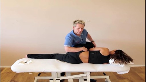
This video is demonstrating how to correct the most common sacroiliac dysfunction and that is an anterior innominate rotation using a muscle energy technique
