- Physical Examination
- Surgical Examination
- Ophthalmology
- Clinical Skills
- Orthopedics
- Surgery Videos
- Laparoscopy
- Pediatrics
- Funny Videos
- Cardiothoracic Surgery
- Nursing Videos
- Plastic Surgery
- Otorhinolaryngology
- Histology and Histopathology
- Neurosurgery
- Dermatology
- Pediatric Surgery
- Urology
- Dentistry
- Oncology and Cancers
- Anatomy Videos
- Health and Fitness
- Radiology
- Anaesthesia
- Physical Therapy
- Pharmacology
- Interventional Radiology
- Cardiology
- Endocrinology
- Gynecology
- Emergency Medicine
- Psychiatry and Psychology
- Childbirth Videos
- General Medical Videos
- Nephrology
- Physiology
- Diet and Food Health
- Diabetes Mellitus
- Neurology
- Women Health
- Osteoporosis
- Gastroenterology
- Pulmonology
- Hematology
- Rheumatology
- Toxicology
- Nuclear Medicine
- Infectious Diseases
- Vascular Disease
- Reproductive Health
- Burns and Wound Healing
- Other
Top videos
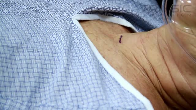
Surgery is the only way to treat parathyroid disease (hyperparathyroidism). There are no medications or pills that work to cure or treat parathyroid problems or high calcium. The parathyroid tumor must be removed by a surgeon. As soon as the parathyroid tumor has been removed, you are cured! It is very likely this will change your life. If you have hyperparathyroidism you need to have parathyroid surgery. If you have an expert surgeon this operation should be very easy.
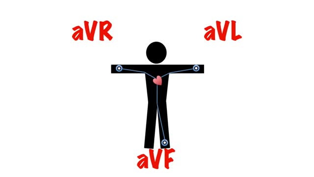
It then spreads down the bundle of his and then purkinje fibres to cause ventricular contraction. So when viewing the heart from the front, the direction of depolarisation is 11 o'clock to 5 o'clock. The general direction of depolarisation is known as the cardiac axis.
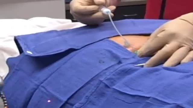
A facet joint injection is a relatively simple, straightforward procedure, and is usually performed in an office based procedure suite or in an ambulatory surgical center. As with many spinal injections, facet joint injections are best performed using fluoroscopy (live X-ray) for guidance to properly target and place the needle (and to help avoid nerve injury or other injury).

An ICD is a battery-powered device placed under the skin that keeps track of your heart rate. Thin wires connect the ICD to your heart. If an abnormal heart rhythm is detected the device will deliver an electric shock to restore a normal heartbeat if your heart is beating chaotically and much too fast.

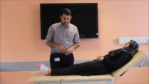
Shoulder pain and exercises Milwaukee WI

Blood Transfusion-Transmitted Diseases
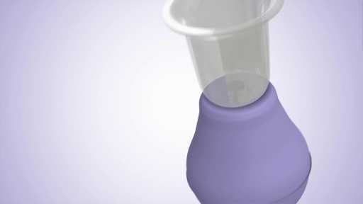
The Lansinoh Latch Assist has been designed to extend inverted nipples - watch this video to see how.?

Anemia is a condition in which the body does not have enough healthy red blood cells. Red blood cells provide oxygen to body tissues. There are many types of anemia. Pernicious anemia is a decrease in red blood cells that occurs when the intestines cannot properly absorb vitamin B12.

The Watchman can be inserted in less than an hour and could save your life.
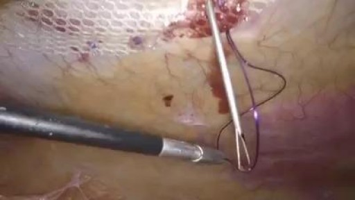
Laparoscopic postoperative ventral hernial (POVT) repair Laparoscopic surgery, also called minimally invasive surgery (MIS), bandaid surgery,

As a curious child, you might remember staring at an older relative's thick stockings at the blue, gnarled veins lying under the skin like bumpy snakes. Known as varicose veins, these blood vessels, which return blood from the legs to the heart, are actually a more superficial system. The real, working venous system for the legs lies deeper, says to Robert A. Weiss, MD, assistant professor of dermatology at the Johns Hopkins School of Medicine in Baltimore. This is good news, because it means that if the surface veins begin to clump up and bulge, they can be removed or destroyed without ruining circulation to the leg. The National Institutes of Health estimates that 60% of all men and women suffer from some form of vein disorder. A quarter of varicose vein sufferers are men, although Weiss notes that it is almost always women who seek help for spider veins.

Watch that video to know How To Whiten Your Yellow Teeth Naturally

Watch that video of Unbelievable Mutations and Medical Conditions

Watch that video of People should have gone to the dentist a lot sooner

Dieta Alcalina Recetas, Listado De Alimentos Alcalinos, Que Es El Agua Alcalina, Menu Para Adelgazar-- http://dieta-alcalina-alimentos.good-info.co -- Entendiendo los Efectos del nivel de pH en el cuerpo El nivel de pH en el cuerpo tiene la habilidad de afectar cada célula del cuerpo. Cuando la sangre tiene un pH alcalino en vez de un pH ácido, ocurre un efecto positivo en cada función corporal del sistema. El cerebro, el sistema circulatorio, los nervios, los músculos, el sistema respiratorio, el sistema digestivo y reproductivo se pueden beneficiar de un nivel adecuado de pH. Por otro lado, cuando el pH del cuerpo es muy ácido, es susceptible a muchas enfermedades y problemas. Ganancia de peso, enfermedades del corazón, envejecimiento prematuro, fatiga, problemas nerviosos, alergias, enfermedades musculares y cáncer son las más probables a ocurrir cuando el pH del cuerpo no está al nivel óptimo. Ya que todos estos problemas son más probables a ocurrir cuando el pH del cuerpo está muy ácido, tiene sentido consumir una dieta rica en alimentos alcalinos. El objetivo principal es usualmente comer aproximadamente entre 75-80% de alimentos alcalinos junto con solamente entre 20-25% de alimentos ácidos. Si se mantiene este nivel en la dieta, el resultado final es un nivel de pH bajo en el cuerpo, el que se requiere para una salud óptima. Descubre como la dieta alcalina funciona & por qué los alimentos alcalinos son altamente recomendados para tu salud. Haz clic aquí http://dieta-alcalina-alimentos.good-info.co

Watch that video of Huge Foot blister Freezing With Liquid Nitrogen

Heart failure can occur if the heart cannot pump (systolic) or fill (diastolic) adequately. Symptoms include shortness of bronicreath, fatigue, swollen legs, and rapid heartbeat. Treatments can include eating less salt, limiting fluid intake, and taking prescription medications. In some cases a defibrillator or pacemaker may be implanted.

Pregnancy occurs when an egg is fertilized by a sperm, grows inside a woman's uterus (womb), and develops into a baby. In humans, this process takes about 264 days from the date of fertilization of the egg, but the obstetrician will date the pregnancy from the first day of the last menstrual period (280 days 40 weeks).

Eye Color Change Surgery with Implant
