- Physical Examination
- Surgical Examination
- Ophthalmology
- Clinical Skills
- Orthopedics
- Surgery Videos
- Laparoscopy
- Pediatrics
- Funny Videos
- Cardiothoracic Surgery
- Nursing Videos
- Plastic Surgery
- Otorhinolaryngology
- Histology and Histopathology
- Neurosurgery
- Dermatology
- Pediatric Surgery
- Urology
- Dentistry
- Oncology and Cancers
- Anatomy Videos
- Health and Fitness
- Radiology
- Anaesthesia
- Physical Therapy
- Pharmacology
- Interventional Radiology
- Cardiology
- Endocrinology
- Gynecology
- Emergency Medicine
- Psychiatry and Psychology
- Childbirth Videos
- General Medical Videos
- Nephrology
- Physiology
- Diet and Food Health
- Diabetes Mellitus
- Neurology
- Women Health
- Osteoporosis
- Gastroenterology
- Pulmonology
- Hematology
- Rheumatology
- Toxicology
- Nuclear Medicine
- Infectious Diseases
- Vascular Disease
- Reproductive Health
- Burns and Wound Healing
- Other
Top videos

How to approach histology for Human Anatomy students. Using a key will help get you through it! Add some penguin fairy dust will help too!
Please note: I mis-spoke and said "striated" instead of "stratified epithelium" a couple of times... apologies!
There are lots of histology keys out there, but the one I showed in the video is here: http://www.penguinprof.com/upl....oads/8/4/3/1/8431323
Want more?
Subscribe: http://www.youtube.com/user/ThePenguinProf
FB Page: https://www.facebook.com/ThePenguinProf
Twitter: https://twitter.com/penguinprof
Web: http://www.penguinprof.com/
---------------------------------------------------------------------------------------------------
Details:
Tissue in the human body:
Epithelial: Is made of cells arranged in a continuous sheet with one or more layers, has apical & basal surfaces.
A basement membrane is the attachment between the basal surface of the cell & the underlying connective tissue.
Two types of epithelial tissues: (1) Covering & lining epithelia and (2) Glandular Epithelium.
The number of cell layers & the shape of the cells in the top layer can classify epithelium.
Simple Epithelium - one cell layer
Stratified epithelium - two or more cell layers
Pseudostratified Columnar Epithelium - When cells of an epithelial tissue are all anchored to the basement Membrane but not all cells reach the apical surface.
Glandular Epithelium -- (1) Endocrine: Release hormones directly into the blood stream and (2) Exocrine - Secrete into ducts.
Connective: contains many different cell types including: fibroblasts, macrophages, mast cells, and adipocytes. Connective Tissue Matrix is made of two materials: ground substance - proteins and polysaccharides, fiber -- reticular, collagen and elastic.
Classification of Connective Tissue:
Loose Connective - fibers & many cell types in gelatinous matrix, found in skin, & surrounding blood vessels, nerves, and organs.
Dense Connective - Bundles of parallel collagen fibers& fibroblasts, found in tendons& ligaments.
Cartilage - Cartilage is made of collagen & elastin fibers embedded in a matrix glycoprotein & cells called chondrocytes, which was found in small spaces.
Cartilage has three subtypes:
Hyaline cartilage -- Weakest, most abundant type, Found at end of long bones, & structures like the ear and nose,
Elastic cartilage- maintains shape, branching elastic fibers distinguish it from hyaline and
Fibrous Cartilage - Strongest type, has dense collagen & little matrix, found in pelvis, skull & vertebral discs.
Muscle: is divided into 3 categories, skeletal, cardiac and smooth.
Skeletal Muscle -- voluntary, striated, striations perpendicular to the muscle fibers and it is mainly found attached to bones.
Cardiac Muscle -- involuntary, striated, branched and has intercalated discs
Smooth Muscle -- involuntary, nonstriated, spindle shaped and is found in blood vessels & the GI tract.
Nervous: Consists of only two cell types in the central nervous system (CNS) & peripheral nervous system (PNS):
Neurons - Cells that convert stimuli into electrical impulses to the brain, and Neuroglia -- supportive cells.
Neurons -- are made up of cell body, axon and dendrites. There are 3 types of neurons:
Motor Neuron -- carry impulses from CNS to muscles and glands,
Interneuron - interpret input from sensory neurons and end responses to motor neurons
Sensory Neuron -- receive information from environment and transmit to CNS.
Neuroglia -- is made up of astrocytes, oligodendrocytes, ependymal cells and microglia in the CNS, and schwann cells and satellite cells in the PNS.
Purchase a license to download a non-watermarked copy of this video here: https://www.alilamedicalmedia.....com/-/galleries/all-
Voice by: Sue Stern.
©Alila Medical Media. All rights reserved.
Support us on Patreon and get FREE downloads and other great rewards: patreon.com/AlilaMedicalMedia
Perfect for patient education purposes.
All images/videos by Alila Medical Media are for information purposes ONLY and are NOT intended to replace professional medical advice, diagnosis or treatment. Always seek the advice of a qualified healthcare provider with any questions you may have regarding a medical condition.
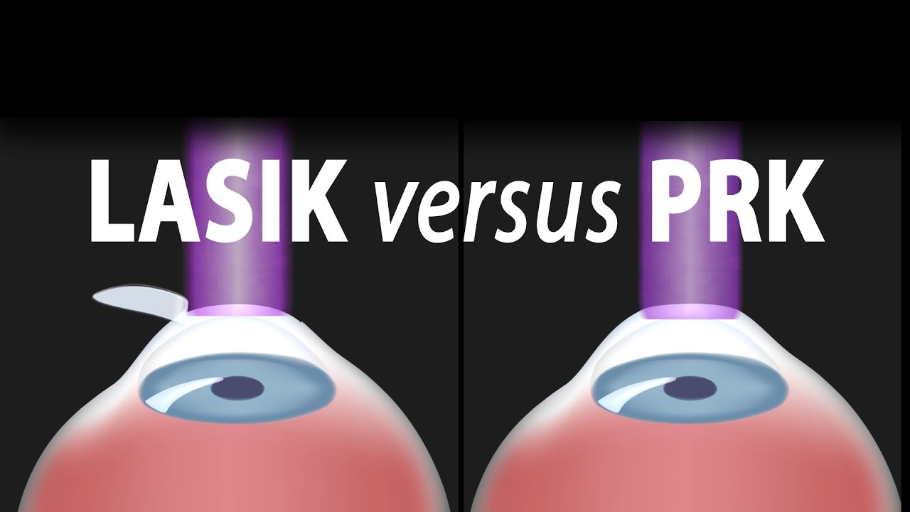
LASIK, or "laser-assisted in situ keratomileusis," is the most commonly performed laser eye surgery to treat myopia, hyperopia and astigmatism. The goal of the treatment is to reshape the cornea to correct the refractive error of the eye.
The cornea is the transparent dome-shaped structure in front of the eye. The cornea refracts light and accounts for about two-thirds of the eye's total optical power. Altering the curvature of the cornea changes the way light rays enter the eye. As a result, the light rays can be focused properly onto the retina for clearer vision.
For nearsighted people, the laser is used to flatten the cornea. For farsighted people, the cornea is made steeper. For patients with astigmatism, the laser is used to smooth the irregularly-shaped cornea into a more regular shape.
The outer layer of the cornea - the epithelium – is capable of replacing itself within a few days after being damaged or removed. The deeper layer of the cornea – the stroma, on the contrary, is a permanent corneal tissue with very limited regenerative capacity. The stroma, if reshaped by a laser, will remain that way permanently.
In this procedure, a thin, circular "FLAP" is created in the surface of the cornea to gain access to the permanent corneal tissue. This can be done with a mechanical cutting tool called a microkeratome, OR, for a blade-free experience, by a femtosecond laser. An excimer laser is then used to remove some corneal tissue to reshape the cornea. Excimer laser uses cool ultraviolet light beams to vaporize microscopic amounts of tissue in a precise manner to accurately reshape the cornea. The excimer laser is computer-controlled and is programmed based on the patient’s refractive error. The flap is then laid back in place and is allowed to heal.
LASIK eye surgery is mostly painless and can be completed within minutes. Improved vision can usually be seen overnight.
PRK, or photorefractive keratectomy, was the first type of laser eye surgery for vision correction and is the predecessor to the popular LASIK procedure. In PRK, NO flap is created. Rather, the epithelial cells on the eye surface are simply removed. An excimer laser is then used to reshape the cornea just like it does in LASIK.
The vision correction outcomes of PRK surgery are comparable to those of LASIK, but the recovery period is longer. This is because the epithelium is completely removed in PRK and it takes a few days to regenerate. PRK patients also have more discomfort and haziness of vision in the first few days after the surgery. Improved vision also takes longer to achieve.
PRK does, however, offer certain advantages. Because PRK does not involve creation of a flap, which contains both epithelial and deeper stromal tissue, the entire thickness of the stroma is available for treatment. The treatment range is therefore higher. This is particularly useful for patients with high levels of myopia or for those whose cornea is too thin for LASIK. PRK is also free of flap-related complication risks.

See what it’s like to get LASIK eye surgery from Lisa Homsy’s perspective. Keep watching until the end to see the final results!

The video is about the evolution of the anatomic UCLA laparoscopic technique over 1325 cases and demonstrates the key steps of our operation to improve patient safety and outcomes.
Learn more at http://urology.ucla.edu

This video contains five segments with best practices on how to prevent infection in patients with catheters, fistulas or grafts. It also includes segments on hand hygiene and glove use and dialysis station disinfection. The video is intended to be used by outpatient hemodialysis facilities as an educational tool to help remind their frontline staff, including technicians and nurses, about infection prevention measures. It can be used as an orientation video for new staff and as an annual in-service training tool to remind staff of proper protocols.
See the Spanish captioned version at: http://youtu.be/L5ypnOvOFMQ
Comments on this video are allowed in accordance with our comment policy: http://www.cdc.gov/SocialMedia..../Tools/CommentPolicy
This video can also be viewed at http://streaming.cdc.gov/vod.p....hp?id=dc66d96228817d

Dr. Erica Hodgman discusses pediatric surgery at the Johns Hopkins Children's Center Pediatric General Surgery program, what common surgeries the program specializes in, what makes the program unique and her work as a pediatric surgeon. #PediatricSurgery #JohnsHopkinsChildrenCenter
Questions Answered:
0:03 Describe the pediatric general surgery division at Johns Hopkins Children's Center.
1:00 What makes this program unique?
1:31 What are some common pediatric surgery cases?
2:23 Explain your work as a pediatric general surgeon?

Rafael Nadal missed seven months last year with a knee injury. That knee will face its toughest test when he plays in the French Open, his first Grand Slam event since his return.
Subscribe to the Times Video newsletter for free and get a handpicked selection of the best videos from The New York Times every week: http://bit.ly/timesvideonewsletter
Subscribe on YouTube: http://bit.ly/U8Ys7n
Watch more videos at: http://nytimes.com/video
---------------------------------------------------------------
Want more from The New York Times?
Twitter: https://twitter.com/nytvideo
Facebook: https://www.facebook.com/nytimes
Google+: https://plus.google.com/+nytimes/
Whether it's reporting on conflicts abroad and political divisions at home, or covering the latest style trends and scientific developments, New York Times video journalists provide a revealing and unforgettable view of the world. It's all the news that's fit to watch. On YouTube.
Analysis of Rafael Nadal's Knee Injury (Computer Animation)
http://www.youtube.com/user/TheNewYorkTimes

Originally broadcast November 21, 2014.
They advertise low, low prices. But does anyone actually pay that rate? Erica Johnson investigates.
More from CBC Marketplace, Canada's top consumer affairs show:
Watch episodes online at http://cbc.ca/marketplace
Like us on Facebook: http://facebook.com/cbcmarketplace
Talk to us on Twitter: http://twitter.com/cbcmarketplace
Follow our hosts @cbctom and @cbcerica

An estimated 20 million LASIK procedures have been performed since 1998. The FDA website is filled with stories of complications, including pain, dizziness and detached retinas. CBS2's Chris Wragge reports.
Patient information from Sunnybrook's Holland Musculoskeletal Program. For more, visit: http://sunnybrook.ca/holland

We will show how to know if you have a sports hernia. These are a few tests you can do on your own. Lower abdominal pain and tightness that increases with twisting and kicking. Stretching and exercises tend to make the discomfort increase.
Want more info? We have a free webinar that covers hip, groin, adductor, lower abdominal strains and sports hernia diagnosis in detail. Use this link to get access. https://bit.ly/37thtNF
#sportshernia #hernia #hippain
To work with us, contact us using this link https://bit.ly/3zCBnzZ or call us 714-502-4243. We have online programs, virtual and in-person options.
Costa Mesa, CA www.p2sportscare.com
Option 1: Groin On-Demand Webinar https://bit.ly/37thtNF
Option 2: Video Guide https://bit.ly/33aLIqC
Option 3 (the best): Work With Us https://www.p2sportscare.com/
Sports Hernia Diagnosis
What Is A Sports Hernia?
A sports hernia is tearing of the transversalis fascia of the lower abdominal or groin region. A common misconception is that a sports hernia is the same as a traditional hernia. The mechanism of injury is rapid twisting and change of direction within sports, such as football, basketball, soccer and hockey.
The term “sports hernia” is becoming mainstream with more professional athletes being diagnosed. The following are just to name a few:
Torii Hunter
Tom Brady
Ryan Getzlaf
Julio Jones
Jeremy Shockey
If you follow any of these professional athletes, they all seem to have the same thing in common: Lingering groin pain. If you play fantasy sports, this is a major headache since it seems so minor, but it can land a player on Injury Reserve on a moments notice. In real life, it is a very frustrating condition to say the least. It is hard to pin point, goes away with rest and comes back after activity, but is hardly painful enough to make you want to stop. It lingers and is always on your mind. And if you’re looking for my step-by-step sports hernia rehab video course here it is.
One the best definitions of Sport hernias is the following by Harmon:
The phenomena of chronic activity–related groin pain that it is unresponsive to conservative therapy and significantly improves with surgical repair.”
This is truly how sports hernias behave in a clinical setting. It is not uncommon for a sports hernia to be unrecognized for months and even years. Unlike your typical sports injury, most sports medicine offices have only seen a handful of cases. It’s just not on most doctors’ radar. The purpose of this article is not only to bring awareness about sports hernias, but also to educate.
Will you find quick fixes in this article for sports hernia rehab?
Nope. There is no quick fix for this condition, and if someone is trying to sell you one, they are blowing smoke up your you-know-what.
Is there a way to decrease the pain related to sports hernias?
Yes. Proper rehab and avoidance of activity for a certain period of time will assist greatly, but this will not always stop it from coming back. Pain is the first thing to go and last thing to come. Do not be fooled when you become pain-free by resting it. Pain is only one measure of improvement in your rehab. Strength, change of direction, balance and power (just to name a few) are important, since you obviously desire to play your sport again. If you wanted to be a couch potato, you would be feeling better in no time. Watching Sports Center doesn’t require any movement.
Why is this article so long?
There is a lot of information on sports hernias available to you on the web. However, much of the information is spread out all over the internet and hard for athletes to digest due to complicated terminology. This article lays out the foundational terminology you will need to understand what options you have with your injury. We will go over anatomy, biomechanics, rehab, surgery, and even the fun facts. The information I am using is from the last ten years of medical research, up until 2016. We will be making updates overtime when something new is found as well. So link to this page and share with friends. This is the best source for information on sports hernias you will find.
Common Names (or Aliases?) for Sports Hernias
Sportsman’s Hernia
Athletic Pubalgia
Gilmore’s Groin
How Do You Know If You Have A Sports Hernia?
Typical athlete characteristics:
Male, age mid-20s
Common sports: soccer, hockey, tennis, football, field hockey
Motions involved: cutting, pivoting, kicking and sharp turns
Gradual onset
How A Sports Hernia Develops
Chronic groin pain typically happens over time, which is why with sports hernias, we do not hear many stories of feeling a “pop” or a specific moment of injury. It is the result of “overuse” mechanics stemming from a combination of inadequate strength and endurance, lack of dynamic control, movement pattern abnormalities, and discoordination of motion in the groin area.

Marcus Greatens, M.D., an orthopedic surgeon at Mayo Clinic Health System provides insight into a few of the things patients can expect to experience during knee replacement surgery.
LIKE, SUBSCRIBE & HIT THE 🔔 http://bit.ly/2VW2yV1
Mayo Clinic Health System offers outstanding care close to home. Part of Mayo Clinic, we meet most of your health care needs locally.
#mayoclinichealthcare #mayoclinicexperience #mayoclinichealthsystem #mayoclinic
Follow Mayo Clinic Health System on
Facebook:
http://bit.ly/2VXSYRv
On Instagram at:
http://bit.ly/30obB03
And also on Twitter:
http://bit.ly/2JgsHI5

Laparoscopic surgery is now commonly used as a type of minimally invasive surgery, but what is it and why is it used?
Interested in learning more about minimally invasive techniques, or having surgery planned? Visit https://www.topdoctors.co.uk/doctor/charles-imber
✔ Follow us on Instagram: https://bit.ly/3fSrqXb
✔ Follow us on Facebook: https://bit.ly/3t5kGsW
✔ Follow us on Twitter: https://bit.ly/39TidKh


Is that knee pain just a sprain or a more serious ACL injury? Orthopedic surgeon Paul Fadale, M.D., offers tips on how to tell the difference. http://www.orthopedicsri.org/

What is hemodiafiltration? Hemodiafiltration, or HDF, is a renal replacement modality that combines diffusion and convection to improve removal of molecules in the middle molecular weight range versus hemodialysis.
Find our full video library only on Osmosis Prime: http://osms.it/more.
Join over 3 million current & future clinicians who learn by Osmosis, and over 130 universities around the world who partner with us to make medical and health education more engaging and efficient. We have unparalleled tools and materials to prepare you to succeed in school, on board exams, and as a future clinician. Sign up for a free trial at http://osms.it/more. If you're interested in exploring an institutional partnership, visit osmosis.org/educators to request a personalized demo.
Follow us on social:
Facebook: http://osms.it/facebook
Twitter: http://osms.it/twitter
Instagram for med: http://osms.it/instagram
Instagram for nursing: https://osms.it/ignursing
Linkedin: https://osms.it/linkedin
Our Vision: Everyone who cares for someone will learn by Osmosis.
Our Mission: To empower the world’s clinicians and caregivers with the best learning experience possible. Learn more here: http://osms.it/mission
Medical disclaimer: Knowledge Diffusion Inc (DBA Osmosis) does not provide medical advice. Osmosis and the content available on Osmosis's properties (Osmosis.org, YouTube, and other channels) do not provide a diagnosis or other recommendation for treatment and are not a substitute for the professional judgment of a healthcare professional in diagnosis and treatment of any person or animal. The determination of the need for medical services and the types of healthcare to be provided to a patient are decisions that should be made only by a physician or other licensed health care provider. Always seek the advice of a physician or other qualified healthcare provider with any questions you have regarding a medical condition. © 2023 Elsevier. All rights reserved.

Welcome to the latest episode of HT Physio Quick Tips!
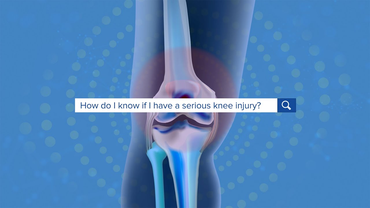
In this episode, Farnham's leading over-50's physiotherapist, Will Harlow, reveals the THREE most serious knee injuries and gives the signs and symptoms that indicate a serious knee injury has occurred.
To get a copy of Will's new book, Thriving Beyond Fifty, you can visit here: https://www.amazon.co.uk/Thriv....ing-Beyond-Fifty-Str
If you're suffering from nagging knee pain that hurts in the morning and stops you from walking as far as you'd like, you can take our free knee pain guide - which will give you 5 expert tips to put a stop to knee pain at home - by visiting here: https://ht-physio.co.uk/knee-pain-guide-download/
If you're over-50 with a painful problem in the Farnham, Surrey area, you can learn more about how Will Harlow and HT Physio can help you overcome a painful problem here: https://ht-physio.co.uk/
**Any information in this video should not be used as a substitute for individual medical advice. Please seek advice from your local healthcare professional before taking action on the information in this video.**

Dialysis patients need to choose their heart medicine carefully, as Canadian researchers say that some beta blockers are easily removed from the blood during treatment. Also, people who eat a Mediterranean diet may decrease their risk of developing kidney problems. Eboni Williams reports on the day's top health news.

Discover how hemodialysis works and the different options available for this dialysis treatment.
Related articles on DaVita.com:
What Is Hemodialysis? (http://www.davita.com/treatmen....t-options/hemodialys
How Does a Dialysis Machine Work? (http://www.davita.com/treatmen....t-options/hemodialys
For more information please visit: https://www.yalemedicine.org/c....onditions/acl-injury
Serious injuries, by and large, cause a lot of swelling in the knee. Especially in younger patients. Now, someone could be arthritic and they overdo it going for a big long hike and they get some swelling the next day. But rapid onset of swelling, it's like hard to make out where your kneecap is, is a pretty big cardinal sign that there's something serious that's happened to your knee. Rapid onset swelling is usually due to blood in the joint. "A meniscus that really tears and flips in the front. You tear your quad or your patellar tendon, your kneecap dislocates, you tear a little blood vessel, your ACL tears, a piece of cartilage in bone gets knocked off and causes bleeding. So a lot of the really significant injuries, people get rapid onset swelling within three to four hours and they should seek attention There's always exceptions to rules, but if your knee looks like a grapefruit, you should go get it checked.
