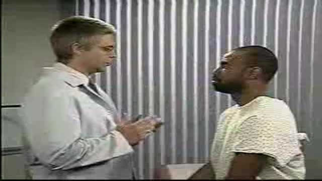
- Physical Examination
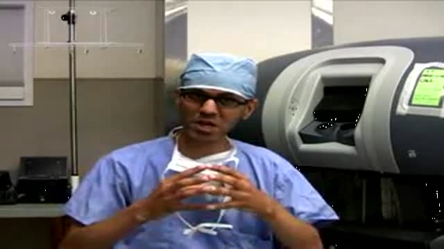
- Surgical Examination
- Ophthalmology
- Clinical Skills
- Orthopedics
- Surgery Videos
- Laparoscopy
- Pediatrics
- Funny Videos
- Cardiothoracic Surgery
- Nursing Videos
- Plastic Surgery
- Otorhinolaryngology
- Histology and Histopathology
- Neurosurgery
- Dermatology
- Pediatric Surgery
- Urology
- Dentistry
- Oncology and Cancers
- Anatomy Videos
- Health and Fitness
- Radiology
- Anaesthesia
- Physical Therapy
- Pharmacology
- Interventional Radiology
- Cardiology
- Endocrinology
- Gynecology
- Emergency Medicine
- Psychiatry and Psychology
- Childbirth Videos
- General Medical Videos
- Nephrology
- Physiology
- Diet and Food Health
- Diabetes Mellitus
- Neurology
- Women Health
- Osteoporosis
- Gastroenterology
- Pulmonology
- Hematology
- Rheumatology
- Toxicology
- Nuclear Medicine
- Infectious Diseases
- Vascular Disease
- Reproductive Health
- Burns and Wound Healing
- Other
Top videos
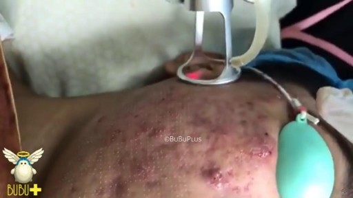
Laser Cystic Acne and Pimples Extraction

Una revision unica de fertilizacion, desarrollo embrionario y de los procedimientos llevados acabo durante un ciclo de fertilizacion invitro. Tome un tour virtual exclusivo de unos de los laboratorios de fertilizacion invitro mas avanzados del mundo y con tecnologia de punta en reproduccion asistida para que conozca con mas detalle como RMA de NY realiza estos procedimientos bajo control estricto de calidad.
Este video proporciona documentacion acerca de la aspiracion de ovulos, inseminacion de ovulos, desarrollo embrionario desde etapa de clivaje (2-3 dias) hasta etapa de blastocisto (5-6 dias), inyeccion intracitplasmatica de esperma (ICSI)), eclosion asistida, transferencia embrionaria y congelacion de embriones.
Mexico City
Dr. Benjamin Sandler
Reproductive Medicine Associates International
http://www.rmany.com/mexicointernatio...
Prolongacion Paseo de la Reforma 1232, Oficina 1213
Colonia Lomas de Bezares
Delegacion Miguel Hidalgo
Mexico, Distrito Federal 11910
Tel: 011-52-55-2167-2515
Fax: 011-52-55-2167-6434
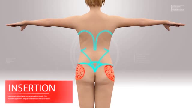
Liposuction & Facelift

the technique of retrograde intubation to maintain the patient's airway.
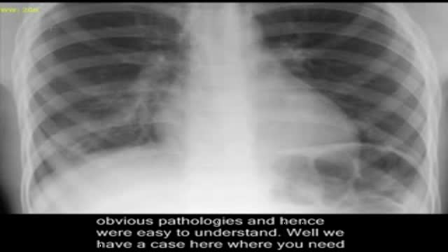
Subtle pneumonia. How to diagnose pneumonia on chest x-ray. Please visit my website for disclaimer. www.academyofprofessionals.com. Multiple choice questions are also available for those who might want to enhance their knowlege or test themselves.
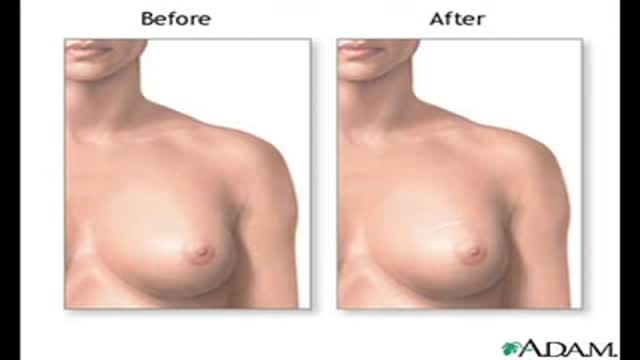
Symptoms of carcinoma of the breast

Adenocarcinoma of the Transverse Colon taken by Dr. Julio Murra Saca This is the case of a 42 year-old male, with no significant past medical history presented with abdominal pain and no weight loss was reported. Adenocarcinoma of the colon is a primary cause of mortality and
morbidity in North America and Western Europe. Colonic cancers are the most common GI carcinomas and have the best prognosis. The 5-year survival rate is approximately 50%.
Survival rates may be improved by screening and removal of adenomatous polyps. Almost all colonic cancers are primary adenocarcinomas.
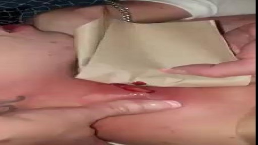
Abscess drainage in neck
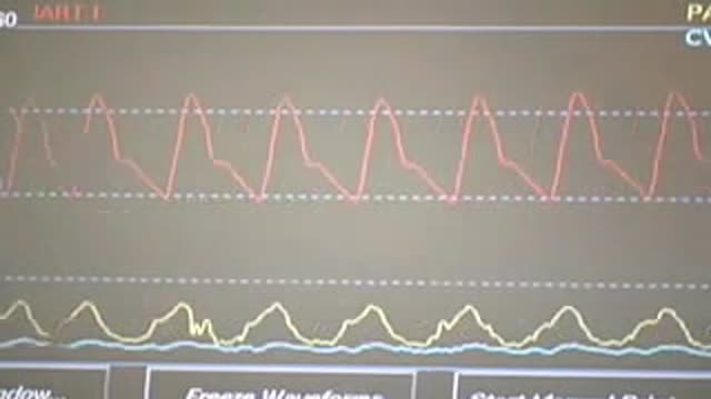
Central Venous Catheter Placement & Pulmonary Artery Catheter Video
Watch that video of The World's Worst Spider Bites

Superficial Palpation of the Abdomen
McMaster University technique of Laparoscopic Radical Nephrectomy
examination of Cranial nerves VI and VII: abducent and facial nerves

The complex circuitry interconnecting different areas in the brain, known collectively as white matter, is composed of millions of axons organized into fascicles and bundles. Upon macroscopic examination of sections of the brain, it is difficult to discern the orientation of the fibers. The same is true for conventional imaging modalities. However, recent advancements in magnetic resonance imaging (MRI) make such task possible in a live subject. By sensitizing an otherwise typical MRI sequence to the diffusion of water molecules it is possible to measure their diffusion coefficient in a given direction1. Normally, the axonal membrane and myelin sheaths pose barriers to the movement of water molecules and, thus, they diffuse preferentially along the axon2. Therefore, the direction of white matter bundles can be elucidated by determining the principal diffusivity of water. The three-dimensional representation of the diffusion coefficient can be given by a tensor and its mathematical decomposition provides the direction of the tracts3; this MRI technique is known as diffusion tensor imaging (DTI). By connecting the information acquired with DTI, three-dimensional depictions of white matter fascicles are obtained4. The virtual dissection of white matter bundles is rapidly becoming a valuable tool in clinical research.
Our journey begins with a transverse section of tightly packed axons as seen through light microscopy. Although represented as a two-dimensional "slice", we see that these axons in fact resemble tubes. A simulation of water molecules diffusing randomly inside the axons demonstrates how the membranes and myelin hinder their movement across them and shows the preferred diffusion direction --along the axons. The tracts depicted through DTI slowly blend in and we ride along with them. As we zoom out even more, we realize that it is a portion of the corpus callosum connecting the two sides of the brain we were traveling on and the great difference in relative scale of the individual axons becomes evident. The surface of the brain is then shown, as well as the rest of the white matter bundles--a big, apparently chaotic tangle of wires. Finally, the skin covers the brain.
With the exception of the simulated water molecules, all the data presented in the animation is obtained through microscopy and MRI. Computer algorithms for the extraction of the cerebral structures and a custom-built graphics engine make our journey through the brain's anatomy possible in a living person.
Micrograph courtesy of Dr. Christian Beaulieu, University of Alberta.
Music by Mario Mattioli.
References:
1. Stejskal, E.O., et al., J. Chem. Phys., 1965. 42:
2. Beaulieu, C., NMR Biomed., 2002. 15:435-55.
3. Basser, P.J., et al., J. Magn. Reson. B, 1994. 103:247-54.
4. Mori, S., et al., NMR Biomed., 2002. 15:468-80.
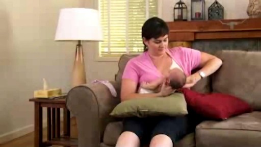
In breastfeeding, the latch is the moment everything comes together: Your baby takes a big mouthful of your nipple and areola (or "latches on"), begins to suck, and draws out your milk. When your baby has established a good latch, your nipple soreness is minimized and your little one gets the nourishment he needs. How do you pull all that off? First and most important, have faith in yourself and your baby. "Babies are designed to breastfeed," says Emily Pease, R.N., international board certified lactation consultant (IBCLC), of Swedish Hospital's Breastfeeding Center in Seattle. "They are born with instincts that help them find Mom's breast and latch on often with very little assistance. And if problems do come up, there are lots of ways to troubleshoot." Here are more steps to get a good latch right from the start.
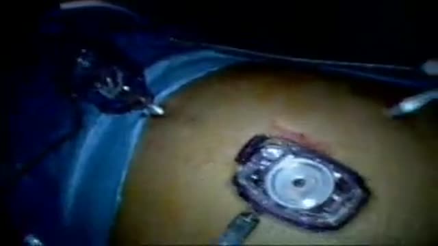
New robotic surgery procedure pioneered at Washington University School of Medicine in St. Louis to remove tumors from kidneys in a minimally invasive way

A local doctor says that the new pap smear guidelines makes sense for many women

Prostate anatomy
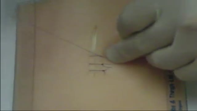
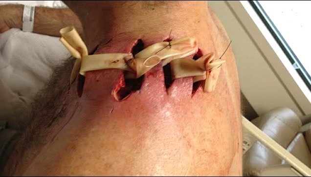
A video showing simple skin suture

Ultrasound Guided Sclerotherapy for Varicose Veins
