- Physical Examination
- Surgical Examination
- Ophthalmology
- Clinical Skills
- Orthopedics
- Surgery Videos
- Laparoscopy
- Pediatrics
- Funny Videos
- Cardiothoracic Surgery
- Nursing Videos
- Plastic Surgery
- Otorhinolaryngology
- Histology and Histopathology
- Neurosurgery
- Dermatology
- Pediatric Surgery
- Urology
- Dentistry
- Oncology and Cancers
- Anatomy Videos
- Health and Fitness
- Radiology
- Anaesthesia
- Physical Therapy
- Pharmacology
- Interventional Radiology
- Cardiology
- Endocrinology
- Gynecology
- Emergency Medicine
- Psychiatry and Psychology
- Childbirth Videos
- General Medical Videos
- Nephrology
- Physiology
- Diet and Food Health
- Diabetes Mellitus
- Neurology
- Women Health
- Osteoporosis
- Gastroenterology
- Pulmonology
- Hematology
- Rheumatology
- Toxicology
- Nuclear Medicine
- Infectious Diseases
- Vascular Disease
- Reproductive Health
- Burns and Wound Healing
- Other
Top videos

New device claims to stimulate brain for depression treatment
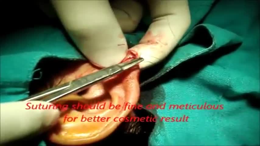
Earlobe surgery or repair is a reconstructive plastic surgery procedure performed to restore the appearance of earlobes that have been torn or stretched due to ear tearing, trauma, or piercing, including the insertion of ear gauges. This can also be called earlobe reconstructive surgery or split earlobe repair .

Generalized Anxiety Disorder, Symptoms Of Anxiety Attack, Shortness Of Breath Anxiety --- http://panic-attacks-anxiety.good-info.co --- Newly Discovered Panic "Off Switch" Gives You Anxiety Relief Without Pills or Therapy Here's an interesting fact about anxiety and panic attacks: Did you know that just like the hiccups, doctors still can't agree exactly why they happen to you? And did you also know there's a 60-second solution to panic and anxiety that you can do anywhere? Yes, it takes you just one minute and I'm going to share it with you today. Until one day about a year ago, I thought I might be doomed to let panic attacks rule my life. And I made this free online presentation to tell you about the one discovery about panic and general anxiety that finally cut through the confusion and changed everything. Pay very close attention, because whether you've only had one or two "attacks" so far… or even if you've been having them for years and it seems like a life sentence you'll never escape from… You're about to discover one weird thing that panic, anxiety and the hiccups – yes, the hiccups – have in common that goes right back to the stone age. Discover How To Begin Eliminating Panic And Anxiety From Your Life Forever Click Here: http://panic-attacks-anxiety.good-info.co

Epilepsy is a chronic disorder, the hallmark of which is recurrent, unprovoked seizures. Many people with epilepsy have more than one type of seizure and may have other symptoms of neurological problems as well. Sometimes EEG testing, clinical history, family history and outlook are similar among a group of people with epilepsy. In these situations, their condition can be defined as a specific epilepsy syndrome. The human brain is the source of human epilepsy. Although the symptoms of a seizure may affect any part of the body, the electrical events that produce the symptoms occur in the brain. The location of that event, how it spreads and how much of the brain is affected, and how long it lasts all have profound effects. These factors determine the character of a seizure and its impact on the individual. Esssentially, anything the brain can do, it can do in the form of a seizure. Having seizures and epilepsy can affect one's safety, relationships, work, driving and so much more. Public perception and treatment of people with epilepsy are often bigger problems than actual seizures.

wide resection of giant cell tumor ,then strut grafting using free fibula graft,knowles pinning of the graft.
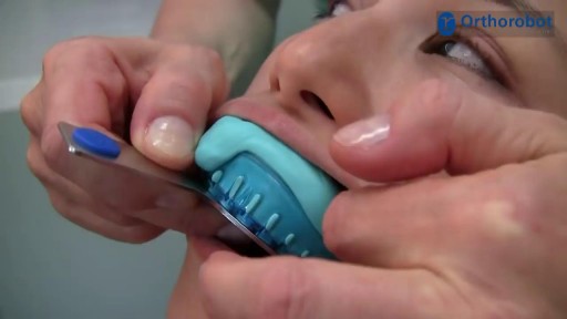
A short introduction on how to take a correction impression. The shown materials are recommended by Orthorobot and have proven to be fully compatible with the Orthorobot lab procedure.

There are 3 major parts of the respiratory system: the airway, the lungs, and the muscles of respiration. The airway, which includes the nose, mouth, pharynx, larynx, trachea, bronchi, and bronchioles, carries air between the lungs and the body's exterior.
Female Catheter Insertion
Open heart (coronary artery bypass, or CABG) surgery is performed in order to reroute, or "bypass," blood around blocked arteries, thereby improving the supply of oxygen-rich blood to the heart. Surgeons usually use an artery from the chest wall to construct the "detour" around the blocked part of the artery. Veins from the legs are also used.
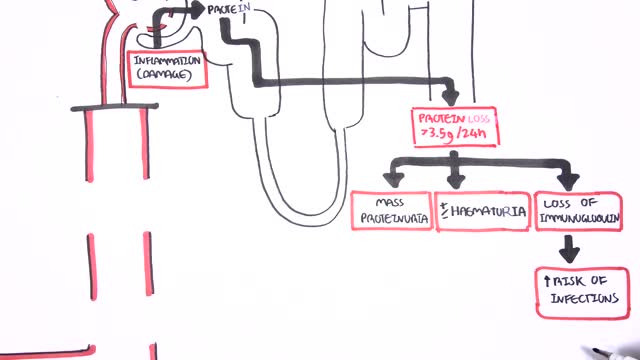
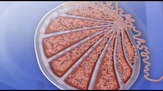
Nephrotic syndrome is a kidney disorder that causes your body to excrete too much protein in your urine. Nephrotic syndrome is usually caused by damage to the clusters of small blood vessels in your kidneys that filter waste and excess water from your blood. Nephrotic syndrome causes swelling (edema), particularly in your feet and ankles, and increases the risk of other health problems. Treatment for nephrotic syndrome includes treating the underlying condition that's causing it and taking medications. Nephrotic syndrome can increase your risk of infections and blood clots. Your doctor may recommend medications and dietary changes to prevent these and other complications of nephrotic syndrome.

Vital signs help us assess patients in the nursing profession, and there are six common vital signs that we assess as nurses:
1. Heart Rate (Pulse)
2. Respiration Rate
3. Temperature
4. Blood Pressure
5. Pain Rating
6. Oxygen Saturation
This video will demonstrate how to check vital signs (live) on a patient, along with normal rates for each assessment. I also give you a few tips for taking vital signs as a nurse, CNA, or other healthcare profession.
🟣ABG eBook: https://registerednursern.creator-spring.com/
🟣ABG physical book: https://amzn.to/3EsF0Mc (affiliate link)
More nursing skills: https://www.youtube.com/watch?v=G5-Rp-6FMCQ&list=PLQrdx7rRsKfUhd_qQYEbp0Eab3uUKhgKb
Website: https://www.registerednursern.com/
More Videos: https://www.youtube.com/watch?v=R2XMro13dD0&list=UUPyMN8DzkFl2__xnTEiGZ1w
Nursing Gear: https://teespring.com/stores/registerednursern
Instagram: https://www.instagram.com/registerednursern_com/
Facebook: https://www.facebook.com/RegisteredNurseRNs
Twitter: https://twitter.com/NursesRN
Popular Playlists:
NCLEX Reviews: https://www.youtube.com/playli....st?list=PLQrdx7rRsKf
Fluid & Electrolytes: https://www.youtube.com/playli....st?list=PLQrdx7rRsKf
Nursing Skills: https://www.youtube.com/playli....st?list=PLQrdx7rRsKf
Spermatogenesis is the process in which spermatozoa are produced from spermatogonial stem cells by way of mitosis and meiosis. The initial cells in this pathway are called spermatogonia, which yield primary spermatocytes by mitosis.
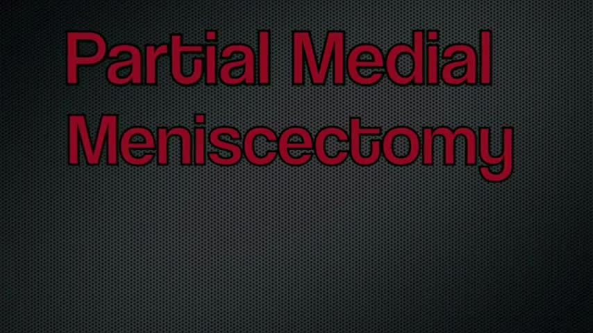
An arthroscopic meniscectomy is a procedure to remove some or all of a meniscus from the tibio-femoral joint of the knee using arthroscopic (aka 'keyhole') surgery. In a complete meniscectomy the meniscus including the meniscal rim is removed. A partial meniscectomy involves partial removal of the meniscus. This may vary from minor trimming of a frayed edge to anything short of removing the rim. This is a minimally invasive procedure often done as day suas an outpatient in a one-day clinic [1] This procedure is performed when a meniscal tear is too large to be corrected by a surgical meniscal repair.[1] When non-operative therapy provides some degree of symptom relief over the long-term, these benefits may wane with continued meniscal degeneration. In such patients, arthroscopic partial meniscectomy can be effective in improving patient quality of life.
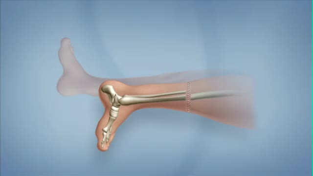
Rotationplasty is a type of autograft wherein a portion of a limb is removed, while the remaining limb below the involved portion is rotated and reattached. This procedure is used when a portion of an extremity is injured or involved with a disease, such as cancer. Typically, the ankle joint becomes the knee joint.
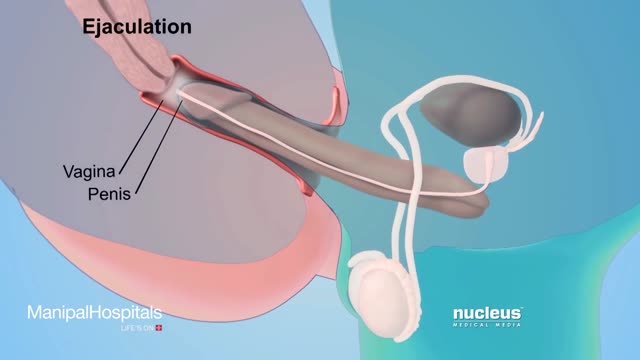
How To Use Male Condom Correctly - Manipal Hospitals

Must Watch Very Special New Funny Video 2023 Doctor Funny Video Injection Wala Funny Video | Comedy Video Episode 124 By Fun Comedy Ltd
@funcomedyltd
#funcomedyltd
#doctor
#comedy
#wala
Hello Dear Viewers,
If We have any mistake. please comment and tell us, what is our mistake? We will try to solve this mistake next. please watch our videos and give us confidence to trying best. Thank you for watching this video.
IMPORTANT NOTE:-
This video are no any kind of risk. This video are totally acting no risk no Dangerous act no Physical Harm or Death its ok for viewers.
injection wala comedy video injection wala video injection funny video injection injection wala injection injection doctor doctor doctor sui wala wala suji wala suji wala cartoon doctor cartoon funny video tui tui injection cartoon 22 cartoon video injection video cartoon cartoon comedy video doctor video wala cartoon busy fun ltd my family our fun tv fun tv 24 fun tv 420 funny day funny family ding dong bidik fun tv roma fun tv
#cartoon
#comedyvideo
#doctor_doctor
#busyfunltd
#newfunnyvideo2022
#newfunniestcomedy
#injectionfunnyvideo
#sui_wala
#myfamily
#busyfunltd
#funnyday
#bidikfuntv
#mohafuntv
#dingdong
Always consult your doctor and seek help early enough to prevent complications

Ultrasound Guided Sclerotherapy for Varicose Veins
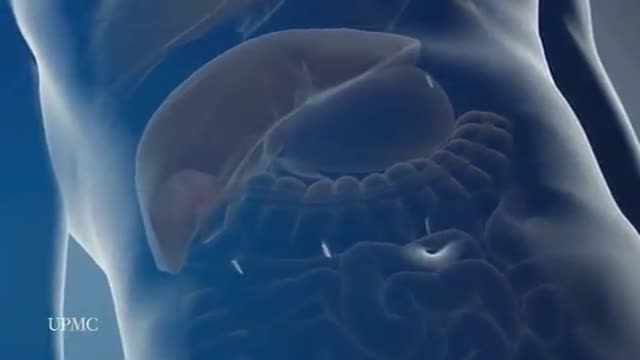
The most frequent incision utilized to open the abdomen for liver surgery is called a chevron incision. In this incision a cut is made on the abdomen below the rib cage. The cut starts under the armpit below the ribs on the right side of the abdomen and continues all the way across the abdomen to the opposite arm pit thereby the whole width of the abdomen is cut to provide access to the liver. The average length of the incision is approximately 24 to 30 inches. This is one of the longest incisions is utilized in abdominal surgery. The incision is frequently associated with significant discomfort after the surgery and in some patients the discomfort can continue for many months, particularly when some of the nerves in the abdominal wall have been cut during the surgery. Laparoscopic surgery provides advantages over open surgery for the liver since the chevron incision is completely avoided and the surgery is performed through tiny incisions. As a consequence the duration of stay in hospital, the amount and duration of post operative discomfort, and the length of recovery is much shorter after the laparoscopic procedure compared to open surgery
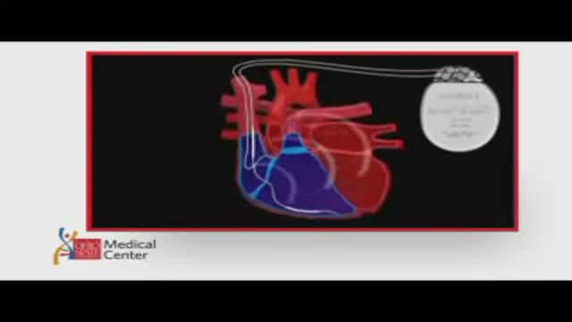
ICDs are useful in preventing sudden death in patients with known, sustained ventricular tachycardia or fibrillation. Studies have shown ICDs to have a role in preventing cardiac arrest in high-risk patients who haven't had, but are at risk for, life-threatening ventricular arrhythmias. View an animation of an ICD. Newer-generation ICDs may have a dual function which includes the ability to serve as a pacemaker. The pacemaker feature would stimulate the heart to beat if the heart rate is detected to be too slow. What is an Implantable Cardioverter Defibrillator (ICD)? An ICD is a battery-powered device placed under the skin that keeps track of your heart rate. Thin wires connect the ICD to your heart. If an abnormal heart rhythm is detected the device will deliver an electric shock to restore a normal heartbeat if your heart is beating chaotically and much too fast. ICDs have been very useful in preventing sudden death in patients with known, sustained ventricular tachycardia or fibrillation. Studies have shown that they may have a role in preventing cardiac arrest in high-risk patients who haven't had, but are at risk for, life-threatening ventricular arrhythmias.
