
- Physical Examination
- Surgical Examination
- Ophthalmology
- Clinical Skills
- Orthopedics
- Surgery Videos
- Laparoscopy
- Pediatrics
- Funny Videos
- Cardiothoracic Surgery
- Nursing Videos
- Plastic Surgery
- Otorhinolaryngology
- Histology and Histopathology
- Neurosurgery
- Dermatology
- Pediatric Surgery
- Urology
- Dentistry
- Oncology and Cancers
- Anatomy Videos
- Health and Fitness
- Radiology
- Anaesthesia
- Physical Therapy
- Pharmacology
- Interventional Radiology
- Cardiology
- Endocrinology
- Gynecology
- Emergency Medicine
- Psychiatry and Psychology
- Childbirth Videos
- General Medical Videos
- Nephrology
- Physiology
- Diet and Food Health
- Diabetes Mellitus
- Neurology
- Women Health
- Osteoporosis
- Gastroenterology
- Pulmonology
- Hematology
- Rheumatology
- Toxicology
- Nuclear Medicine
- Infectious Diseases
- Vascular Disease
- Reproductive Health
- Burns and Wound Healing
- Kidney Zone Podcast
- Other
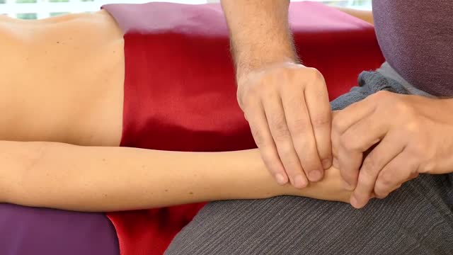
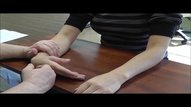
Carpal Tunnel Release Procedure

Educational only: videos on MedicalVideos.com are for general information and are not medical advice. Always consult your own doctor about your health.
Carpal tunnel surgery, also called carpal tunnel release (CTR) and carpal tunnel decompression surgery, is a surgery in which the transverse carpal ligament is divided. It is a treatment for carpal tunnel syndrome and recommended when there is static (constant, not just intermittent) numbness, muscle weakness, or atrophy, and when night-splinting no longer controls intermittent symptoms of pain in the carpal tunnel. In general, milder cases can be controlled for months to years, but severe cases are unrelenting symptomatically and are likely to result in surgical treatment.
This video in 30 seconds
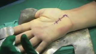
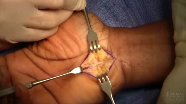
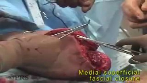
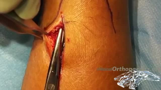
A surgeon narrates a short-form demonstration of open carpal tunnel release. He explains his ulnar incision placed away from a branch of the median nerve, how the junction of the thenar and hypothenar fascia marks the end of the ligament, and his slow division of the ligament and forearm fascia under direct vision.
Key points
- The incision is placed ulnar to the thenar crease, away from the median nerve
- The junction of thenar and hypothenar fascia marks the ligament end
- Guyon's canal is opened as part of the exposure
- The carpal ligament is divided slowly on its ulnar side
- The forearm fascia division completes the release under direct vision
Chapters
- 0:23 — Ulnar incision placement explained
- 1:13 — Locating the fascial junction landmark
- 2:58 — Starting division of the ligament
- 3:47 — Reaching the end of the release
- 4:24 — Proximal fascia division to finish
Video Transcript
A surgeon's narrated 'short form' of open carpal tunnel release. He explains his deliberately ulnar incision, placed away from the palmar cutaneous branch of the median nerve, how he finds the 'V' where the thenar and hypothenar fascia meet to locate the far end of the ligament, and his slow division of the carpal ligament and forearm fascia under direct vision.
Read the full transcript
This is the video short form for carpal tunnel release. And here's the table of contents for the various aspects of our how to do our carpal tunnel release. My incision is very ulnar to the thenar crease. It's long and it's ulnar to the thenar crease. And it's ulnar to the thenar crease for several reasons. One is I wanna be way away from the palmyrocutaneous branch of the median nerve.
I want any healing to be way away from the median nerve itself. And by coming ulnar, I achieve both of those goals. And also in many patients, if not most patients, there's a small cutaneous branch coming off the ulnar nerve and it's large enough to protect through this ulnar incision.
It's about two and a half to three and a half centimeters distal to the wrist crease. This incision also lets me see the V between the hypothenar and thenar muscles. And that cues me as to where the termination of the carpal ligament releases. This is a thin woman. I don't need to go above the wrist on her. But on some patients that are obese or have very thick hands knowing about the distal marker for the V and being able to extend the incision above the wrist is also very helpful.
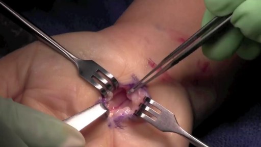
Now it's about in here where you would see that cutaneous nerve branch coming off the ulnar nerve. And I'm not seeing it in this patient, but I'm looking for it. I'll frequently open up Guyan's canal, not necessarily because I think I need to open up Guyan's canal, but because it lets me see the V for the hypothenar and thenar. Of course, when you do a carpal tunnel release by just doing that, you are also decompressing Guyan's canal. So now I'll start to look for the V. And I see the thenar muscles running in the direction
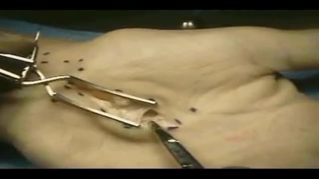
of the thenar muscles, the hypothenar muscles in the opposite direction. And that junction between the two is the V. So there's thenar fascia. And then over here, hypothenar fascia and muscle and right between the two of them is this one spot where the hypothenar and thenar fascia meets that's the termination of that carpal release. I've marked it there with that ink dot. So now I'll start proxily. It doesn't really matter if you start proxily or distly, but I usually start proxily and I go very slow. I slow down the speed of the surgery now
and just scratch through the carpal ligament, going very slowly. And I'm on the extreme ulnar side of the ligament. I have not had any problem with a subluxation or bow stringing of tendons. And I'm very ulnar to the median nerve.
Way away from the parmicutaneous branches, you can see. And this allows the healing relating to that division of the flexor retinaculum to be way away from the median nerve. Now I'm coming to the end, that V. And I get rid of my knife and I use my tenotomy so I can pick up and slowly just get fascia. You can see that bright yellow fat, which means you're at the end of the release. And the median nerve is way over there. It's interesting that even though I'm way ulnar
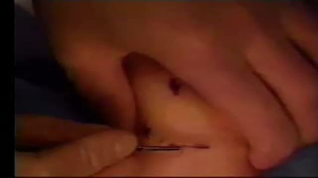
to the median nerve, it's very close to the median nerve. So if you're making your incision right on top of the median nerve, I believe you're gonna scar down to it. Now I then move to the very end of the table. And I'm releasing the antebrakeal fascia and or proximal portion of the ligament. I'm the only one that can see this. So I position those CEN retractors or small down curve retractors myself. And then I tug on that proximal divided end of the flexor retinaculum. I pull it towards myself.
So I pull it towards myself. I make sure that the wrist is slightly flexed, not dorsiflexed. And I'm cutting that ligament and antebrakeal fascia on the ulnar side. So I'm sort of veering away from the median nerve. And I'm going very slowly here. No pushing, no blind pushing. And when you get to the end of that ligament, and you can see I've got that wrist slightly flexed, so I'm moving the nerve down. I'm cutting on top of the tendon right now. And when I get to the very end,
everything is under direct vision. And I'm the only one that can see as I'm doing this. So I make sure I like where the retraction is. When you're done cutting that ligament, there's an abrupt drop off. So it's not a little slow drop off. You are done when you get to the end of that thick antebrakeal fascia. And everything is under complete observation. Median nerve is way over there, so way away from the overlying cut on the flexor retinoculum.
Transcript auto-generated from the video’s audio for accessibility and study. Educational content only — not medical advice.
Video Overview
From our Surgery Videos collection: Surgery videos: watch real operations step by step — general, laparoscopic and specialist surgery performed in the OR, explained for students and surgeons. Browse all Surgery Videos videos →
Medical Videos
Free medical and surgery videos for patients, students and professionals — trusted since 2007.
Founded by Dr. Mohamed Ibrahim, MD, PhD — physician-scientist.
Follow us on Facebook · Instagram
SORT BY-
Top Comments
-
Latest comments