Top videos

Epilepsy
Epilepsy samer kareem 2,273 Views • 3 years ago

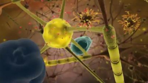
Epilepsy is a chronic disorder, the hallmark of which is recurrent, unprovoked seizures. Many people with epilepsy have more than one type of seizure and may have other symptoms of neurological problems as well. Sometimes EEG testing, clinical history, family history and outlook are similar among a group of people with epilepsy. In these situations, their condition can be defined as a specific epilepsy syndrome. The human brain is the source of human epilepsy. Although the symptoms of a seizure may affect any part of the body, the electrical events that produce the symptoms occur in the brain. The location of that event, how it spreads and how much of the brain is affected, and how long it lasts all have profound effects. These factors determine the character of a seizure and its impact on the individual. Esssentially, anything the brain can do, it can do in the form of a seizure. Having seizures and epilepsy can affect one's safety, relationships, work, driving and so much more. Public perception and treatment of people with epilepsy are often bigger problems than actual seizures.

End To End Arterial Arterial Anastomosis
End To End Arterial Arterial Anastomosis samer kareem 1,673 Views • 3 years ago

A circulatory anastomosis is a connection (an anastomosis) between two blood vessels, such as between arteries (arterio-arterial anastomosis), between veins (veno-venous anastomosis) or between an artery and a vein (arterio-venous anastomosis). An end artery (or terminal artery) is an artery that is the only supply of oxygenated blood to a portion of tissue. Examples of an end artery include the splenic artery that supplies the spleen and the renal artery that supplies the kidneys.

Revision of Mini Gastric ByPass
Revision of Mini Gastric ByPass Surgeon 9,572 Views • 3 years ago

Revision of Mini Gastric ByPass

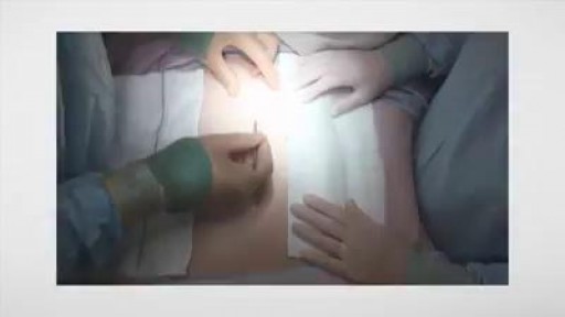
Laparoscopic Ovarian Drilling for PCOS - What is it? Can Ovarian Drilling cure PCOS?
Laparoscopic Ovarian Drilling for PCOS - What is it? Can Ovarian Drilling cure PCOS? Surgeon 139 Views • 3 years ago

***************************************************************************
MEDICAL ANIMATION TRANSCRIPT:

Laparoscopic Ovarian Drilling (LOD)

A surgical treatment for women with PCOS
Women with PCOS usually have ovaries with a thick outer layer.
Ovarian drilling works by breaking through the thick outer surface and lowering the amount of testosterone made by the ovaries

A small incision is made in the abdomen.
Carbon dioxide gas is used to inflate the abdomen.
Very small holes are made in the ovaries.
Ovarian drilling can help restore ovulation and improve the chances of becoming pregnant.
***************************************************************************
*TimeStamps*
0:00 Introduction
0:15 Procedure of Laparoscopic Ovarian Drilling (LOD)
***************************************************************************
Let us watch this 3D video to understand what is Laparoscopic Ovarian Drilling for PCOS, why it is done, how well it works, and what to expect.
***************************************************************************
Get credible information on various health topics follow us on:
* Facebook: https://www.facebook.com/eremedium
* Instagram: https://www.instagram.com/eremedium/
* LinkedIn: https://www.linkedin.com/company/13197441/
* Twitter: https://twitter.com/eremedium
***************************************************************************
Disclaimer: Eremedium blogs and videos are for informational purposes only and should not be construed as advice or as a substitute for consulting a physician. It is not a substitute for medical advice or treatment from a healthcare professional.

#pcos #pcostreatment #laparascopicovariandrilling

How to Know If You Are Pregnant
How to Know If You Are Pregnant Harvard_Student 100,624 Views • 3 years ago

Watch that video to learn How to Know If You Are Pregnant

What  Unborn Babies Can Do in the Womb
What Unborn Babies Can Do in the Womb samer kareem 3,174 Views • 3 years ago

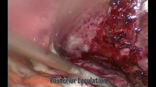
Laparoscopic Excision of Endometriosis - Brigham and Women's Hospital
Laparoscopic Excision of Endometriosis - Brigham and Women's Hospital Surgeon 151 Views • 3 years ago

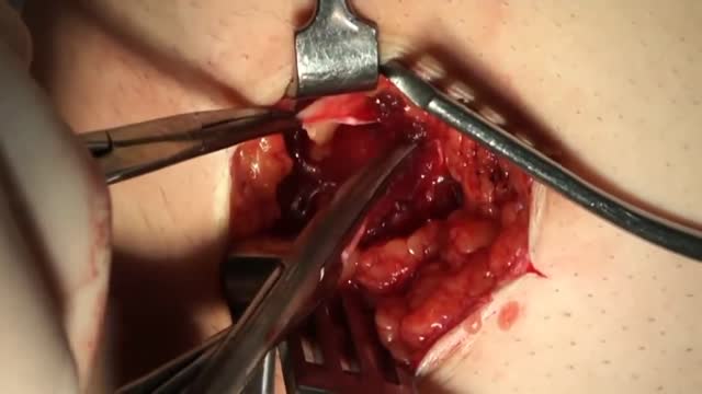
The video demonstrates complete excision of endometrosis in a variety of challenging situations.

Partial Medial Meniscectomy Arthroscopغ
Partial Medial Meniscectomy Arthroscopغ samer kareem 1,350 Views • 3 years ago

An arthroscopic meniscectomy is a procedure to remove some or all of a meniscus from the tibio-femoral joint of the knee using arthroscopic (aka 'keyhole') surgery. In a complete meniscectomy the meniscus including the meniscal rim is removed. A partial meniscectomy involves partial removal of the meniscus. This may vary from minor trimming of a frayed edge to anything short of removing the rim. This is a minimally invasive procedure often done as day suas an outpatient in a one-day clinic [1] This procedure is performed when a meniscal tear is too large to be corrected by a surgical meniscal repair.[1] When non-operative therapy provides some degree of symptom relief over the long-term, these benefits may wane with continued meniscal degeneration. In such patients, arthroscopic partial meniscectomy can be effective in improving patient quality of life.

What Is Cervicitis ?
What Is Cervicitis ? samer kareem 4,693 Views • 3 years ago

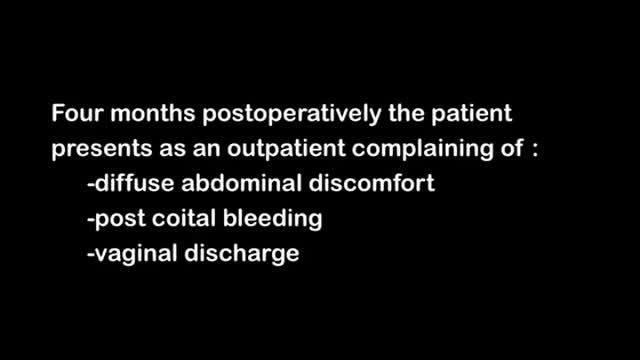
Cervicitis is an inflammation of the cervix, the lower, narrow end of the uterus that opens into the vagina. Possible symptoms of cervicitis include bleeding between menstrual periods, pain with intercourse or during a cervical exam, and abnormal vaginal discharge. However, it's also possible to have cervicitis and not experience any signs or symptoms. Often, cervicitis results from a sexually transmitted infection, such as chlamydia or gonorrhea. Cervicitis can develop from noninfectious causes, too. Successful treatment of cervicitis involves treating the underlying cause of the inflammation.

Robotic Knee Replacement Surgical Footage | Penn Orthopaedics
Robotic Knee Replacement Surgical Footage | Penn Orthopaedics Surgeon 110 Views • 3 years ago

Note: This video contains graphic surgical footage so viewer discretion is advised.

Director of the Penn Orthopaedics Robotics and Navigation Program, Dr. Christopher Travers, discusses robotic joint replacement surgery, which is one of the multiple options that Penn Orthopaedics offers for joint replacement surgery. He walks through a robotic knee replacement surgery, discussing what the procedure is, how it differs from traditional joint replacement surgery, and the benefits.

Refer a patient (physicians only):
https://www.pennmedicine.org/refer-your-patient

Learn more about the Penn Joint Replacement Program:
https://www.pennmedicine.org/f....or-patients-and-visi

Learn more about Dr. Travers:
https://www.pennmedicine.org/providers/profile/christopher-travers?fadf=pennmedicine&keyword=travers

#RoboticSurgery #JointReplacementSurgery #KneeReplacement #SurgicalFootage

Sarcoidosis Diagnosis
Sarcoidosis Diagnosis samer kareem 1,230 Views • 3 years ago

A diagnosis of sarcoidosis is established on the basis of compatible clinical and radiologic findings and histologic evidence of the presence of noncaseous epithelioid cell granulomas in one or more organs and the absence of causative organisms or particulates (16). Granulomas of known causes and local sarcoidlike reactions must be excluded. Granulomatous lesions may result from many conditions, including tuberculosis, berylliosis, leprosy, hypersensitivity pneumonitis, Crohn disease, primary biliary cirrhosis, and fungal disease. Moreover, local sarcoidlike reactions may be seen in lymph nodes that drain a neoplasm or a site of chronic inflammation (19). Such reactions also have been seen in patients who have undergone chemotherapy and radiation therapy (23). If biopsy of lymph nodes or pulmonary or pleural tissue is necessary for diagnosis, one of three techniques may be used: transbronchial biopsy, CT-guided biopsy, or surgical biopsy (24). The use of a surgical technique may be warranted when the results of biopsy with another procedure are not definitive and biopsy of mediastinal lymph nodes, lung, or both is required. This can generally be done with minimally invasive procedures, such as cervical mediastinoscopy, the Chamberlain procedure (a parasternal minithoracotomy for biopsy of the aortopulmonary window or para-aortic nodes), or video-assisted thoracoscopic surgical biopsy (25).

What is Nightmare Disorder?
What is Nightmare Disorder? samer kareem 1,699 Views • 3 years ago

There can be a number of psychological triggers that cause nightmares in adults. For example, anxiety and depression can cause adult nightmares. Post-traumatic stress disorder (PTSD) also commonly causes people to experience chronic, recurrent nightmares. Nightmares in adults can be caused by certain sleep disorders

Spermatogenesis
Spermatogenesis samer kareem 12,626 Views • 3 years ago

Spermatogenesis is the process in which spermatozoa are produced from spermatogonial stem cells by way of mitosis and meiosis. The initial cells in this pathway are called spermatogonia, which yield primary spermatocytes by mitosis.

Nephrotic Syndrome
Nephrotic Syndrome samer kareem 3,623 Views • 3 years ago

Nephrotic syndrome is a kidney disorder that causes your body to excrete too much protein in your urine. Nephrotic syndrome is usually caused by damage to the clusters of small blood vessels in your kidneys that filter waste and excess water from your blood. Nephrotic syndrome causes swelling (edema), particularly in your feet and ankles, and increases the risk of other health problems. Treatment for nephrotic syndrome includes treating the underlying condition that's causing it and taking medications. Nephrotic syndrome can increase your risk of infections and blood clots. Your doctor may recommend medications and dietary changes to prevent these and other complications of nephrotic syndrome.

Arterial Pulse in health and disease
Arterial Pulse in health and disease samer kareem 2,891 Views • 3 years ago

A detailed description of the Arterial Pulse including its waveform and pathological subtypes. Also discussed are the abnormal rates (tachycardia and bradycardia) and their causes, abnormal rhythm (including regularly regular and irregularly irregular pulses) and abnormal character (including pulses bisferiens, pulses parvus et tarsus, pulsus alternans, pulses paradoxus and others.) Description of pulse in various pathological states including Aortic stenosis and aortic regurgitation is also included. Finally there is also a description of the peripheral signs of aortic regurgitation.

Lumbar Multifidis Muscle Rehabilitation
Lumbar Multifidis Muscle Rehabilitation Doctor 21,711 Views • 3 years ago

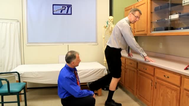
Video demonstrates the action of the isolated lumbar multifidis muscle

Ultrasound Guided Sclerotherapy for Varicose Veins
Ultrasound Guided Sclerotherapy for Varicose Veins Surgeon 21,032 Views • 3 years ago

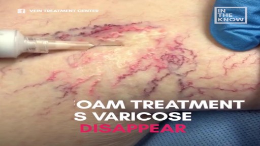
Ultrasound Guided Sclerotherapy for Varicose Veins

Could A Brain Implant Cure Depression?
Could A Brain Implant Cure Depression? samer kareem 1,832 Views • 3 years ago

Depression is a very serious mental illness that affects millions worldwide. Could a small brain implant cure it?

Cerebral Venous Sinus Thrombosis
Cerebral Venous Sinus Thrombosis samer kareem 5,840 Views • 3 years ago

Thrombosis of the venous channels in the brain is an uncommon cause of cerebral infarction relative to arterial disease, but it is an important consideration because of its potential morbidity. (See Prognosis.) Knowledge of the anatomy of the venous system is essential in evaluating patients with cerebral venous thrombosis (CVT), since symptoms associated with the condition are related to the area of thrombosis. For example, cerebral infarction may occur with cortical vein or sagittal sinus thrombosis secondary to tissue congestion with obstruction. (See Presentation.) Lateral sinus thrombosis may be associated with headache and a pseudotumor cerebri–like picture. Extension into the jugular bulb may cause jugular foramen syndrome, while cranial nerve palsies may be seen in cavernous sinus thrombosis as a compressive phenomenon. Cerebral hemorrhage also may be a presenting feature in patients with venous sinus thrombosis. (See Presentation.) Imaging procedures have led to easier recognition of venous sinus thrombosis (see the images below), offering the opportunity for early therapeutic measures. (See Workup.) Left lateral sinus thrombosis demonstrated on magn Left lateral sinus thrombosis demonstrated on magnetic resonance venography (MRV). This 42-year-old woman presented with sudden onset of headache. Physical examination revealed no neurologic abnormalities. View Media Gallery Axial view of magnetic resonance (MR) venogram dem Axial view of magnetic resonance (MR) venogram demonstrating lack of flow in transverse sinus. View Media Gallery The following guidelines for CVT have been provided by the American Heart Association and the American Stroke Association [1] : In patients with suspected CVT, routine blood studies consisting of a complete blood count, chemistry panel, prothrombin time, and activated partial thromboplastin time should be performed. Screening for potential prothrombotic conditions that may predispose a person to CVT (eg, use of contraceptives, underlying inflammatory disease, infectious process) is recommended in the initial clinical assessment. Testing for prothrombotic conditions (including protein C, protein S, or antithrombin deficiency), antiphospholipid syndrome, prothrombin G20210A mutation, and factor V Leiden can be beneficial for the management of patients with CVT. Testing for protein C, protein S, and antithrombin deficiency is generally indicated 2-4 weeks after completion of anticoagulation. There is a very limited value of testing in the acute setting or in patients taking warfarin. In patients with provoked CVT (associated with a transient risk factor), vitamin K antagonists may be continued for 3-6 months, with a target international normalized ratio of 2.0-3.0. In patients with unprovoked CVT, vitamin K antagonists may be continued for 6-12 months, with a target international normalized ratio of 2.0-3.0. For patients with recurrent CVT, venous thromboembolism (VTE) after CVT, or first CVT with severe thrombophilia (ie, homozygous prothrombin G20210A; homozygous factor V Leiden; deficiencies of protein C, protein S, or antithrombin; combined thrombophilia defects; or antiphospholipid syndrome), indefinite anticoagulation may be considered, with a target international normalized ratio of 2.0-3.0. For women with CVT during pregnancy, low-molecular-weight heparin (LMWH) in full anticoagulant doses should be continued throughout pregnancy, and LMWH or vitamin K antagonist with a target international normalized ratio of 2.0-3.0 should be continued for ≥6 weeks postpartum (for a total minimum duration of therapy of 6 months). It is reasonable to advise women with a history of CVT that future pregnancy is not contraindicated. Further investigations regarding the underlying cause and a formal consultation with a hematologist or maternal fetal medicine specialist are reasonable. It is reasonable to treat acute CVT during pregnancy with full-dose LMWH rather than unfractionated heparin. For women with a history of CVT, prophylaxis with LMWH during future pregnancies and the postpartum period is reasonable. Next: Etiology What to Read Next on Medscape Related Conditions and Diseases Quiz: Do You Know the Complications, Proper Workup, and Best Treatment Practices for Ischemic Stroke? Quiz: How Much Do You Know About Hypothyroidism? Quiz: Do You Know the Risk Factors, Symptoms, and Potential Treatments for Alzheimer Disease? Quiz: How Much Do You Know About Hypertension? Quiz: Test Your Knowledge of Epilepsy and Seizure-related Conditions A 25-Year-Old Man With Painless Diplopia NEWS & PERSPECTIVE Temporal Trends and Factors Associated With Diabetes Mellitus Among Patients Hospitalized With Heart Failure Watchful Waiting Tied to Worse Outcomes in LVAD Patients With Hemolysis Age of Transfused Blood Impacts Perioperative Outcomes Among Patients Who Undergo Major Gastrointestinal Surgery TOOLS Drug Interaction Checker Pill Identifier Calculators Formulary SLIDESHOW Chronic Alcohol Abuse: Complications and Consequences Most Popular Articles According to Neurologists DHA Supplements Linked to Less Progression to Alzheimer's in APOE4 Carriers Heading in Soccer Linked to CNS Symptoms 'Transient Smartphone Blindness' Misdiagnosed as Multiple Sclerosis? New Advances in Traumatic Brain Injury FDA Clears Deflazacort (Emflaza) for DMD View More Overview Background

What is Scoliosis surgery?
What is Scoliosis surgery? Scott 6,173 Views • 3 years ago

Most scoliosis surgeons agree that children who have very severe curves (45-50° and higher) will need surgery to lessen the curve and prevent it from getting worse. The operation for scoliosis is a spinal fusion. The basic idea is to realign and fuse together the curved vertebrae so that they heal into a single, solid bone. With the tools and technology available today, scoliosis surgeons are able to improve curves significantly.

Showing 63 out of 358