Video teratas
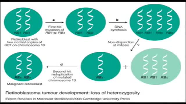
Retinoblastoma is an eye cancer that begins in the retina — the sensitive lining on the inside of your eye. Retinoblastoma most commonly affects young children, but can rarely occur in adults. Your retina is made up of nerve tissue that senses light as it comes through the front of your eye. The retina sends signals through your optic nerve to your brain, where these signals are interpreted as images. A rare form of eye cancer, retinoblastoma is the most common form of cancer affecting the eye in children. Retinoblastoma may occur in one or both eyes.
Obesity is the abnormal condition that causes a person to put on excessive amounts of weight due to accumulation of fat in their body. This extreme weight causes a variety of other disorders and diseases as complications associated with it. https://goo.gl/GgSAoY
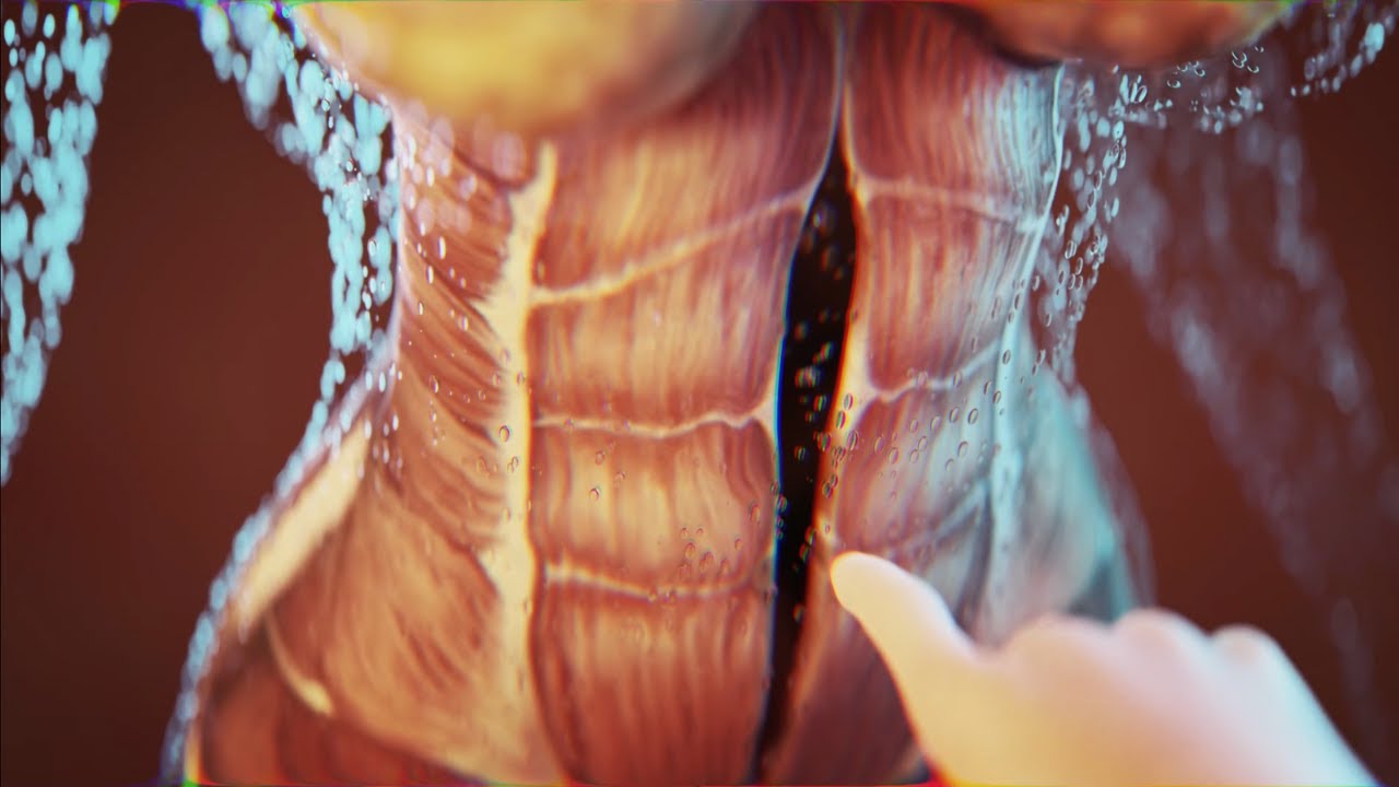
Today I'm using the best 3D animation to explain WHAT IS DIASTASIS RECTI and what you need to know about diastasis recti after pregnancy! Grab the Complete Diastasis Recti Healing Guide: https://landing.mailerlite.com..../webforms/landing/n0
If you are't sure what video to start with and you just want step-by-step daily instructions you can start my 30-day core healing program. You get a new 10-min core healing video daily for 30 days. https://pregnancyandpostpartum....tv.thinkific.com/cou
How I healed my 4-finger diastasis recti gap:
Jessica Pumple is a registered dietitian, and pre & postnatal fitness instructor and certified pregnancy and postpartum core exercise specialist (CPES). She helps pregnant women stay fit, have healthy babies, and easier labors. She helps new moms with postpartum recovery, to heal and strengthen their core and feel energized after pregnancy!
If you enjoy our content subscribe to our channel, hit the bell button, leave a comment and share with your friends so I can make you more of the videos you enjoy!
Disclaimer: This is general postnatal fitness only. Please check with your doctor or health care provider to see if this video is safe for you. Wait until you get clearance (usually 4-6 weeks or 6-8 weeks after a c-section).You are responsible for your own safety. Don’t do anything that feels unsafe for you or baby. Stop if you have any pain or discomfort, bleeding, chest pain or shortness of breath, dizziness or if you feel unwell. P&P Health Inc., Pregnancy and Postpartum TV and Jessica Pumple are not liable in any way for any injury, loss, damages, costs or expenses suffered by you in relation to this video or its content.
Copyright 2023 P&P Health Inc. All rights reserved
#diastasisrecti #whatisdiastasisrecti #3danimation
Music: Epidemic Sound
What combines research opportunities, intellectual challenge, and international collaboration in the study of a disease which affects many organs of the body and all sectors of society? And demands that specialists from many different backgrounds work together to crack sometimes intractable problems? It is, of course, oncology. As a career choice, it's demanding; it takes passion coupled with a willingness to put in the hours and to learn how to discuss death honestly and sensitively. But for the right person, it can be immensely rewarding.

The timing of the nausea or vomiting can indicate the cause. When appearing shortly after a meal, nausea or vomiting may be caused by food poisoning, gastritis (inflammation of the stomach lining), an ulcer, or bulimia. Nausea or vomiting one to eight hours after a meal may also indicate food poisoning.

It is a specialized bodily fluid that supplies essential substances and nutrients, such as sugar, oxygen, and hormones to our cells, and carries waste away from those cells, this waste is eventually flushed out of the body in urine, feces, sweat, and lungs (carbon dioxide). Blood also contains clotting agents.

An antecedent upper respiratory infection is present in 50% of patients. Abdominal pain is a presenting symptom in 1 0-15% of patients. The skin lesions are symmetric, involve dependent parts of the body, and classically progress from an erythematous, macular rash to papular purpura. The joints and kidneys are also commonly involved
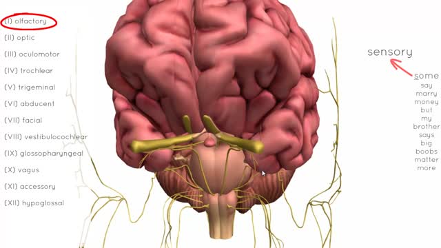
There are twelve cranial nerves in total. The olfactory nerve (CN I) and optic nerve (CN II) originate from the cerebrum. Cranial nerves III – XII arise from the brain stem (Figure 1). They can arise from a specific part of the brain stem (midbrain, pons or medulla), or from a junction between two parts: Midbrain – the trochlear nerve (IV) comes from the posterior side of the midbrain. It has the longest intracranial length of all the cranial nerves. Midbrain-pontine junction – oculomotor (III). Pons – trigeminal (V). Pontine-medulla junction – abducens, facial, vestibulocochlear (VI-VIII). Medulla Oblongata – posterior to the olive: glossopharyngeal, vagus, accessory (IX-XI). Anterior to the olive: hypoglossal (XII). The cranial nerves are numbered by their loca
Paradoxical movement is an obvious sign that the portion of the chest wall is not assisting with the breathing function. Other symptoms of flail chest can include: Bruises, grazes, and/or discoloration in the chest area. Telltale markings from a seat belt.
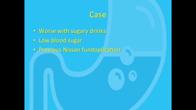
Dumping syndrome is a condition that can develop after surgery to remove all or part of your stomach or after surgery to bypass your stomach to help you lose weight. Also called rapid gastric emptying, dumping syndrome occurs when food, especially sugar, moves from your stomach into your small bowel too quickly.Diet: Eating too much sugar can cause sugars to pass into the colon, making the bacteria there get all excited and cause diarrhea. Other things like sorbitol, a sweetener in some sugarless candy, can also cause diarrhea through osmosis. Malabsorption: Some people don't digest sugars or fats properly.
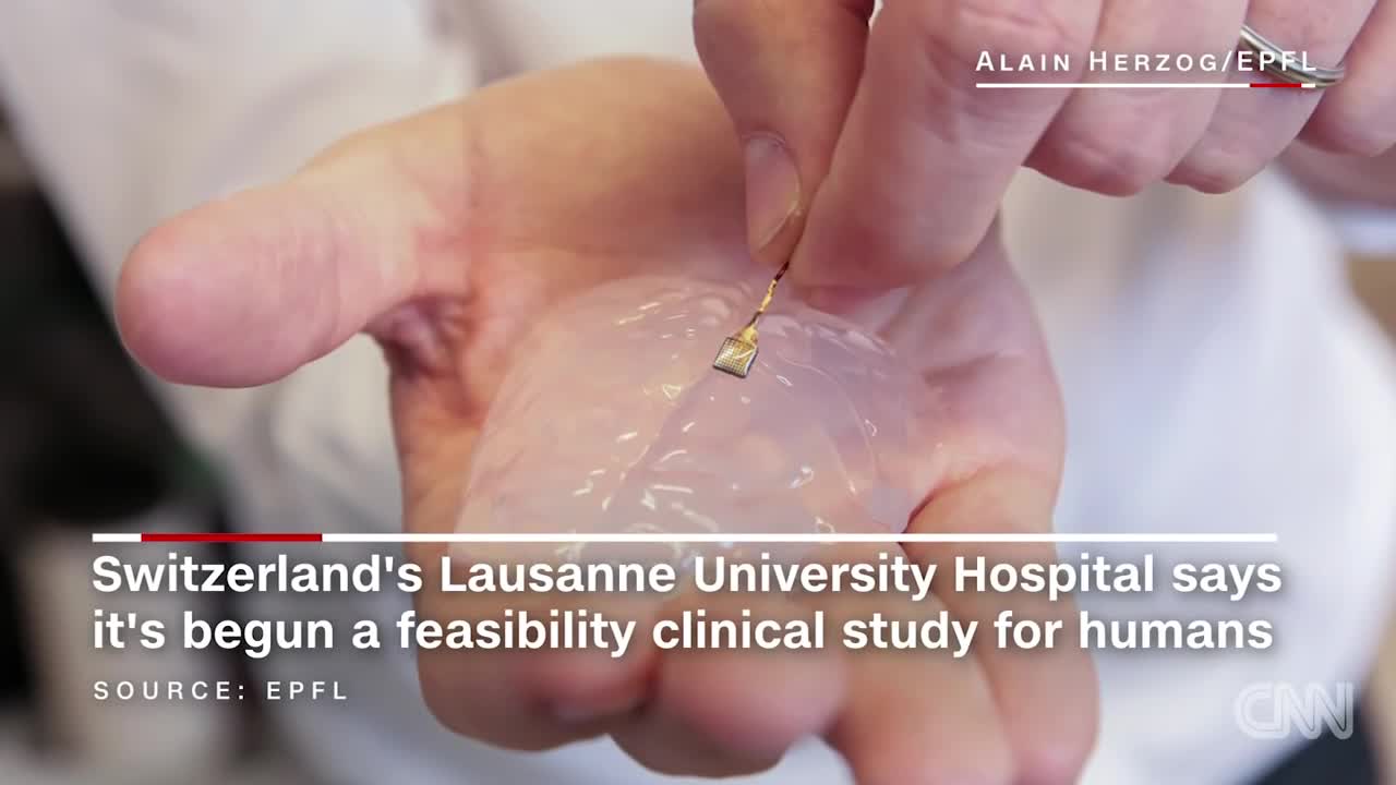
Scientists have developed a wireless brain implant that enabled a paralyzed monkey to walk again.
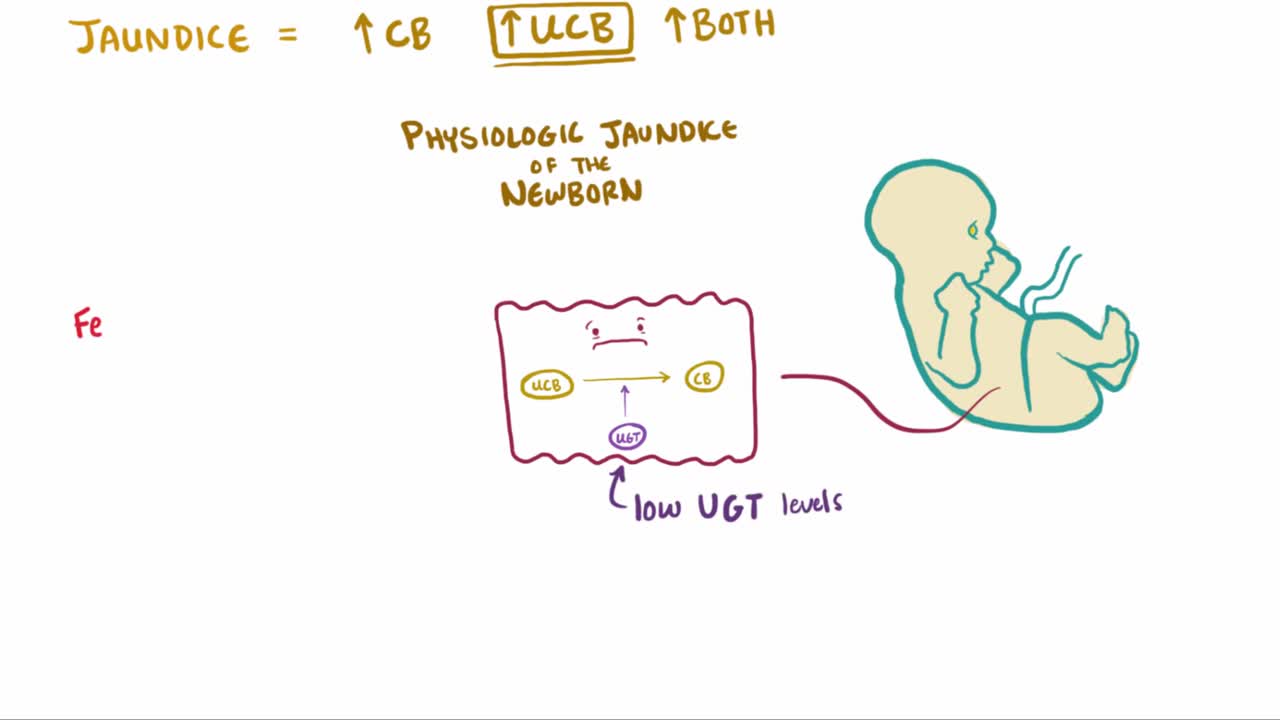
What is jaundice? Well, jaundice is a condition where the skin and eyes take on a yellowish color due to increased levels of bilirubin in the bloodstream
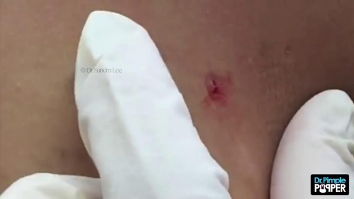
LASER SURGERY Pilonidal Cyst removal
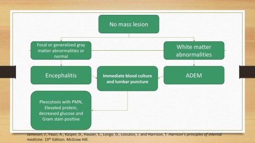
A detailed discussion of the causes, diagnosis and management of the causes of Meningitis and Encephalitis. Includes bacterial, viral, fungal and autoimmune conditions as well as treatment of these conditions. Includes antivirals such as Aciclovir and Ganciclovir as well as IVIG and plasma exchange for autoimmune encephalitis.
The spleen is one of the most overlooked organs. Rarely does it get attention unless there is some kind of accident or trauma. However, I find spleen dysfunction to be very prevalent. This video talks about some of the symptoms.
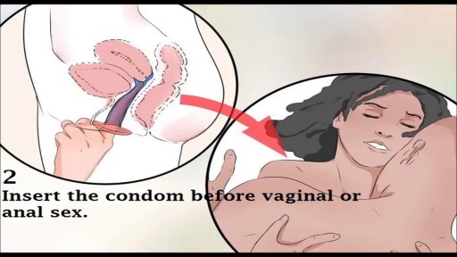
Watch that video to know How to Use Female Condom
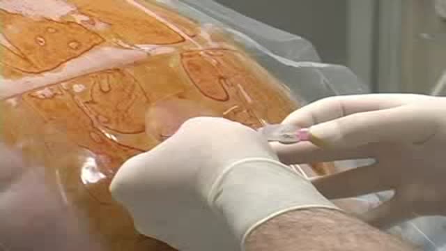
Thoracic Epidural Placement Paramedian Approach
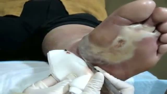
See http://nursing-resource.com for more on debridement.

Note: This video contains graphic surgical footage so viewer discretion is advised.
Director of the Penn Orthopaedics Robotics and Navigation Program, Dr. Christopher Travers, discusses robotic joint replacement surgery, which is one of the multiple options that Penn Orthopaedics offers for joint replacement surgery. He walks through a robotic knee replacement surgery, discussing what the procedure is, how it differs from traditional joint replacement surgery, and the benefits.
Refer a patient (physicians only):
https://www.pennmedicine.org/refer-your-patient
Learn more about the Penn Joint Replacement Program:
https://www.pennmedicine.org/f....or-patients-and-visi
Learn more about Dr. Travers:
https://www.pennmedicine.org/providers/profile/christopher-travers?fadf=pennmedicine&keyword=travers
#RoboticSurgery #JointReplacementSurgery #KneeReplacement #SurgicalFootage
