- Physical Examination
- Surgical Examination
- Ophthalmology
- Clinical Skills
- Orthopedics
- Surgery Videos
- Laparoscopy
- Pediatrics
- Funny Videos
- Cardiothoracic Surgery
- Nursing Videos
- Plastic Surgery
- Otorhinolaryngology
- Histology and Histopathology
- Neurosurgery
- Dermatology
- Pediatric Surgery
- Urology
- Dentistry
- Oncology and Cancers
- Anatomy Videos
- Health and Fitness
- Radiology
- Anaesthesia
- Physical Therapy
- Pharmacology
- Interventional Radiology
- Cardiology
- Endocrinology
- Gynecology
- Emergency Medicine
- Psychiatry and Psychology
- Childbirth Videos
- General Medical Videos
- Nephrology
- Physiology
- Diet and Food Health
- Diabetes Mellitus
- Neurology
- Women Health
- Osteoporosis
- Gastroenterology
- Pulmonology
- Hematology
- Rheumatology
- Toxicology
- Nuclear Medicine
- Infectious Diseases
- Vascular Disease
- Reproductive Health
- Burns and Wound Healing
- Other
Top videos
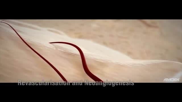
Median Sternotomy performed before open heart surgery !
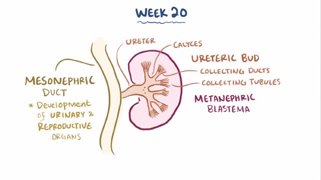
Renal agenesis is a condition in which a newborn is missing one or both kidneys. Unilateral renal agenesis (URA) is the absence of one kidney. Bilateral renal agenesis (BRA) is the absence of both kidneys. Both types of renal agenesis occur in fewer than 1 percent of births annually, according to the March of Dimes. Fewer than 1 in every 1,000 newborns has URA. BRA is much rarer, occurring in about 1 in every 3,000 births.
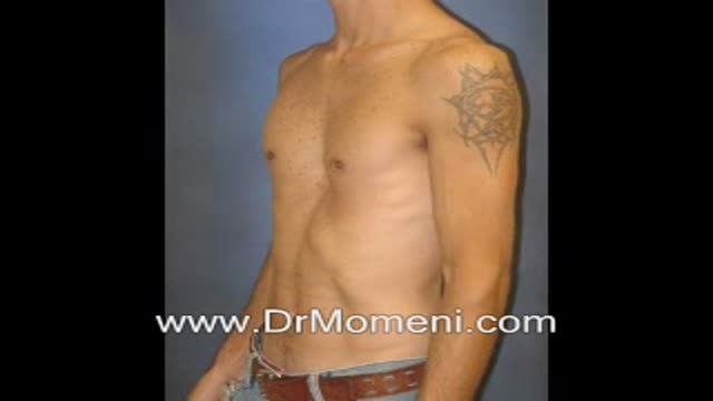
Pectus excavatum (hollow chest) deformity is not uncommon (sometimes mild and other times severe in its form). The chest deformity is often the source of self-consciousness for the patients while growing up. Several surgical techniques (Nuss procedure, Ravitch procedure, etc) are available.
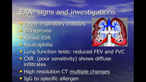
Occupational respiratory disease is any lung condition you get at work. Certain workplaces lend themselves to disease. The most common are coalmines and factories or areas with high amounts of toxins. These include asbestos and silica dust, as well as smoke, fumes, gases, and other particles. Types of occupational respiratory disease include: coal workers’ pneumoconiosis, also known as Black Lung Disease asbestosis silicosis farmers’ lung, also known as allergic alveolitis. It also includes forms of asthma, bronchitis, or emphysema.
A rare view into fertilization, embryo development, and laboratory procedures performed during an IVF cycle. Take an exclusive look inside one of the most advanced, state-of-the-art in vitro fertilization (IVF) laboratories to see how RMA of New York performs IVF and other advanced reproductive technologies using strict identification standards.
Medical and laboratory video footage documents egg retrieval, insemination, embryo development from cleavage stage (day 2-3) to blastocyst stage (day 5-6), intracytoplasmic sperm injection (ICSI), assisted hatching, embryo transfer and embryo cryopreservation.
Reproductive Medicine Associates of New York
www.rmany.com
635 Madison Avenue, 10th floor
New York, New York 10022
Telephone: (212) 756-5777
Facsimile: (212) 756-5770
15 North Broadway, Garden Level - Suite G
White Plains, New York 10601
Telephone: (914) 997-6200
Facsimile: (914) 997-8111
Reproductive Medicine Associates of New York, Long Island
400 Garden City Plaza, Suite 107
Garden City, NY 11530
Telephone: (516) 746-3633
Facsimile: (516) 746-3622
Reproductive Medicine Associates International Mexico, S.C.
Prolongacion Paseo de la Reforma 1232, Oficina 1213
Colonia Lomas de Bezares
Delegacion Miguel Hidalgo
Mexico, Distrito Federal 11910
Telephone: 011-52-55-2167-2515
Fax: 011-52-55-2167-6434
40 years old patient, Parity 3, wanted to have a sterilization. The surgery was perfomed laparoscopically with coagulation technique. This video is not edited and presented in full length.
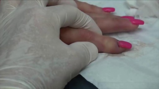
Worst Nail Infection: Paronychia

Surgery to treat men with prostate cancer is often followed by months of difficulty controlling urine flow, a condition known as urinary incontinence. But new research suggests that this problem may go away more quickly if the men perform certain exercises to strengthen their pelvic floor muscles.
Researchers from the Kaiser Permanente Medical Center in Los Angeles, California, found that men who were taught how to perform pelvic floor exercises before and after surgery were more likely to have regained continence three months later.
Men Doing Pelvic Exercises Recover Earlier
In the current study, the researchers randomly assigned 38 men scheduled for radical prostatectomy to either a treatment group or a control group. The men in the treatment group were referred to a physical therapist. They were instructed how to do Pelvic Floor Exercises both before and after surgery, using biofeedback to ensure they were using the proper muscles. The control group did not receive any formal instruction. All of the men completed questionnaires regarding bladder function at regular intervals over the next year.
Overall, 82% of the patients had regained continence (defined as not needing to use any absorbent pads) by the end of the year, including about equal numbers in both groups. But on average the men who had been educated about Pelvic exercises regained continence about one month earlier than those in the control group (at 12 weeks vs. 16 weeks).
Most of the men who did not regain continence within a year were still using at least three absorbent pads a day, indicating continued severe incontinence. The study authors explained that these men probably had extensive damage to the bladder sphincter or severe dysfunction of the bladder after surgery, and the exercises alone were unable to compensate for this.
But the exercises seemed to be effective. Pelvic floor exercise and education initiated prior to surgery is an effective noninvasive intervention useful for improving early return of urinary continence, the authors concluded. It would certainly have a positive impact on our patients undergoing radical prostatectomy in an effort to improve quality of life after major urological surgery.
The results of the study were published in the Journal of Urology (Vol. 170, No. 1: 130-133)
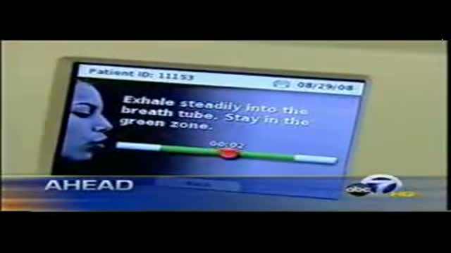
The video is a clip from ABC 7 News, KGO-TV. The video details the new FDA approved device Insight eNO system which uses exhaled nitric oxide for effective asthma management, in both adults and children.
Insight eNO has revolutionized asthma treatment. Apieron’s asthma products help in managing asthma for patients suffering from acute asthma attacks by detecting exhaled nitric oxide (eNO) present in the human breath.

Ventricular fibrillation is a heart rhythm problem that occurs when the heart beats with rapid, erratic electrical impulses. This causes pumping chambers in your heart (the ventricles) to quiver uselessly, instead of pumping blood. Sometimes triggered by a heart attack, ventricular fibrillation causes your blood pressure to plummet, cutting off blood supply to your vital organs. Ventricular fibrillation, an emergency that requires immediate medical attention, causes the person to collapse within seconds. It's the most frequent cause of sudden cardiac death. Emergency treatment includes cardiopulmonary resuscitation (CPR) and shocks to the heart with a device called a defibrillator. Treatments for those at risk of ventricular fibrillation include medications and implantable devices that can restore a normal heart rhythm.
How Does a Bone Heal? All broken bones go through the same healing process. This is true whether a bone has been cut as part of a surgical procedure or fractured through an injury. The bone healing process has three overlapping stages: inflammation, bone production and bone remodeling. Inflammation starts immediately after the bone is fractured and lasts for several days. When the bone is fractured, there is bleeding into the area, leading to inflammation and clotting of blood at the fracture site. This provides the initial structural stability and framework for producing new bone. Diagram of inflammation in a fractured bone Bone production begins when the clotted blood formed by inflammation is replaced with fibrous tissue and cartilage (known as soft callus). As healing progresses, the soft callus is replaced with hard bone (known as hard callus), which is visible on x-rays several weeks after the fracture. Bone remodeling, the final phase of bone healing, goes on for several months. In remodeling, bone continues to form and becomes compact, returning to its original shape. In addition, blood circulation in the area improves. Once adequate bone healing has occurred, weightbearing (such as standing or walking) encourages bone remodeling.

All Solution of Male Disorder Male Infertility Diagnostic and Treatment Re-Slim Care Latest Technology in Pakistan Dr. Aslam Naveed is a well known sexologist in Pakistan. He has treated more than 1 Lac patients since last 30 years of clinical Practice in sexology, he knows how to help the people facing sexual disorders. Contact: 02134965050, 03432821919, 0345-8314663 http://www.sexologistpakistan.com/ https://www.facebook.com/menssexcareclinic/ https://www.youtube.com/channel/UCagSSgdEgQJWl_xfFM12BwA https://twitter.com/bettersexcare https://www.instagram.com/dr.aslamnaveed/ ADDRESS: Men’s Care Modern Hospital, Opposite, Safari Park, University Road, Karachi, Pakistan.
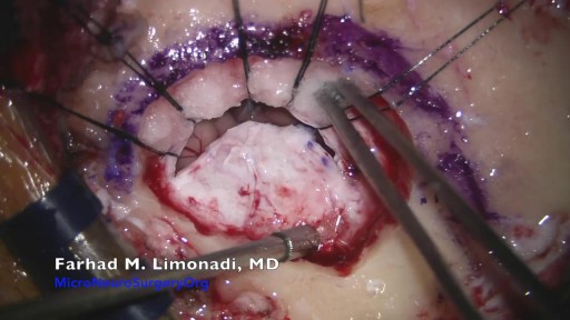
Brain Surgery: Microvascular Decompression of facial nerve for hemifacial spasm
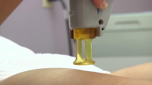
Laser Hair Removal for Dark Skin with YAG Laser
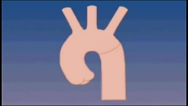
Although the exact cause of abdominal aortic aneurysms is unknown, a number of factors may play a role, including: Tobacco use. ... Hardening of the arteries (atherosclerosis). ... High blood pressure. ... Blood vessel diseases in the aorta. ... Infection in the aorta. ... Trauma. ... Heredity.

DMC Specialists use minimally invasive surgery to remove an extremely large uterine fibroid from a patient. ~ Detroit Medical Center
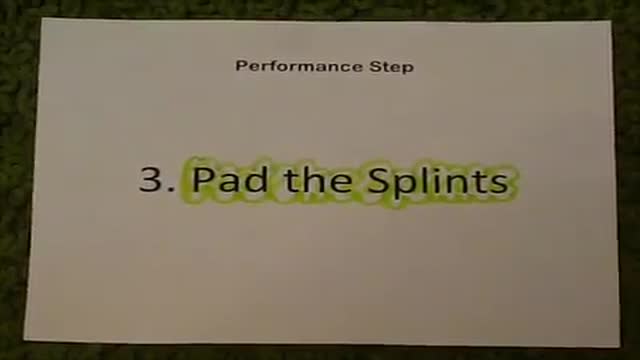
First Aid for a suspected Fracture

A central venous catheter, also called a central line, is a long, thin, flexible tube used to give medicines, fluids, nutrients, or blood products over a long period of time, usually several weeks or more. A catheter is often inserted in the arm or chest through the skin into a large vein.
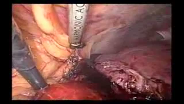
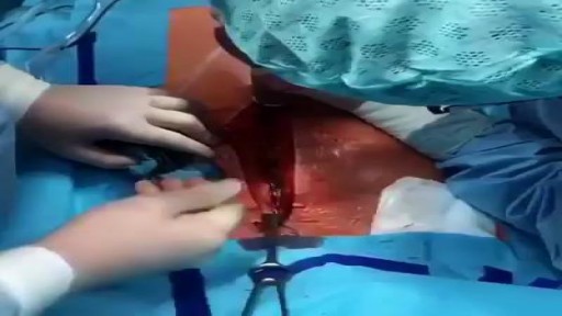
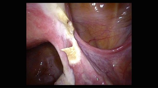
Laparoscopic resection of the right hepatic lobe for a 5 cm hepatoma
