I migliori video

This one goes out to all the student, resident and fellows trying to clarify what their bosses are trying to say to the patient
Ways to Help Pregnant Women Dilate HD

Mothers can do everything for her baby
Pediatric Urine Samples Collection
Migraine treatments can help stop symptoms and prevent future attacks. Many medications have been designed to treat migraines. Some drugs often used to treat other conditions also may help relieve or prevent migraines. Medications used to combat migraines fall into two broad categories: Pain-relieving medications. Also known as acute or abortive treatment, these types of drugs are taken during migraine attacks and are designed to stop symptoms. Preventive medications. These types of drugs are taken regularly, often on a daily basis, to reduce the severity or frequency of migraines. Your treatment strategy depends on the frequency and severity of your headaches, the degree of disability your headaches cause, and your other medical conditions. Some medications aren't recommended if you're pregnant or breast-feeding. Some medications aren't given to children. Your doctor can help find the right medication for you
An intra-aortic balloon pump (IABP) is a mechanical device that helps the heart pump blood. This device is inserted into the aorta, the body's largest artery. It is a long, thin tube called a catheter with a balloon on the end of it. If you are hospitalized, your doctor may insert an IABP.
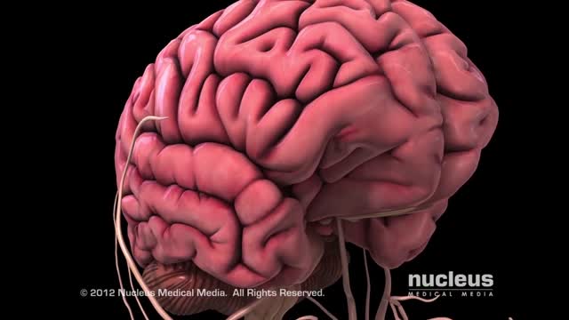
What is an Aneurysm? A cerebral or intracranial aneurysm is an abnormal focal dilation of an artery in the brain that results from a weakening of the inner muscular layer (the intima) of a blood vessel wall. The vessel develops a "blister-like" dilation that can become thin and rupture without warning. The resultant bleeding into the space around the brain is called a subarachnoid hemorrhage (SAH). This kind of hemorrhage can lead to a stroke, coma, and/or death. Aneurysms are usually found at the base of the brain just inside the skull, in an area called the subarachnoid space. In fact, 90 percent of SAHs are attributed to ruptured cerebral aneurysms and the two terms are often used synonymously.
Watch that video of a v
One of a series of films we produced to help patients, their families and carers learn more about some of the most common tests and procedures used to diagnose and treat blood diseases. Patients who have previously undergone these tests helped us to design the videos. Each film clearly explains what the procedure involves and addresses common issues and concerns including: Why your doctor recommended this procedure What you need to do to prepare What you can expect during the procedure What you need to do afterwards Not every patient will be referred for all of these tests and practice may differ slightly depending on where you are treated.
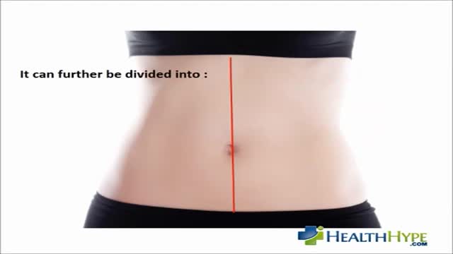
Dehydration can also be a cause of kidney stones. A common symptom is having a lower left abdominal pain, fever, nausea, groin pain and vomiting. Lower left abdominal pain can also be caused by an infection of the kidneys. It usually begins with the bladder and then reaches out to the kidneys.

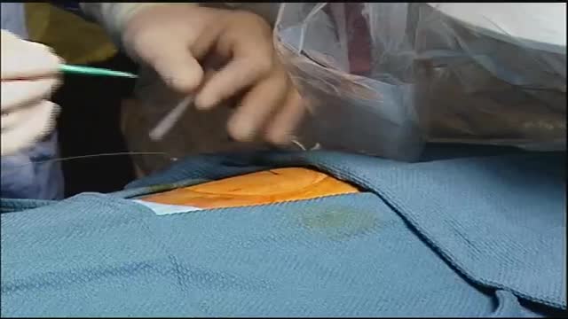
Nursing skills lab procedure for wound care dressing change with irrigation and packing.
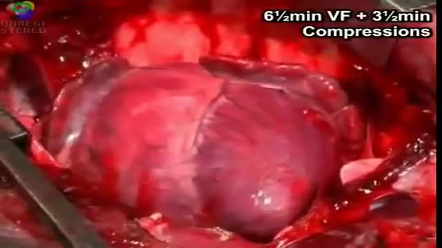
cardiac massage intermittent compression of the heart by pressure applied either over the sternum (closed cardiac massage) or directly to the heart through an opening in the chest wall (open cardiac massage). simple massage in the nursing interventions classification, a nursing intervention defined as stimulation of the skin and underlying tissues with varying degrees of hand pressure to decrease pain, produce relaxation, and/or improve circulation.
Watch that video to know the Serious Side Effects of STEROIDS on Human Body
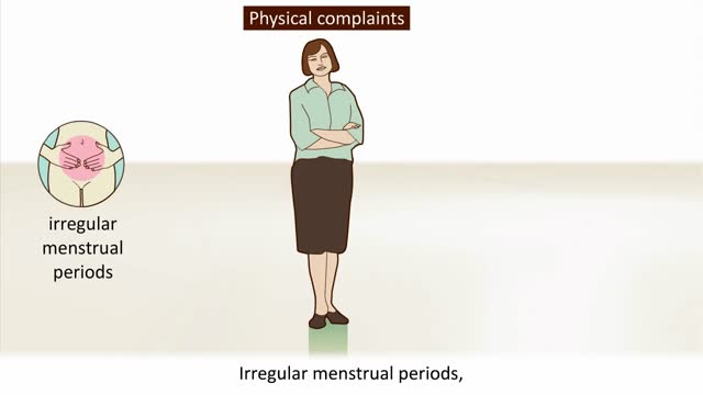
Menopause is defined as occurring 12 months after your last menstrual period and marks the end of menstrual cycles. Menopause can happen in your 40s or 50s, but the average age is 51 in the United States. Menopause is a natural biological process. Although it also ends fertility, you can stay healthy, vital and sexual. Some women feel relieved because they no longer need to worry about pregnancy. Even so, the physical symptoms, such as hot flashes, and emotional symptoms of menopause may disrupt your sleep, lower your energy or — for some women — trigger anxiety or feelings of sadness and loss. Don't hesitate to seek treatment for symptoms that bother you. Many effective treatments are available, from lifestyle adjustments to hormone therapy.
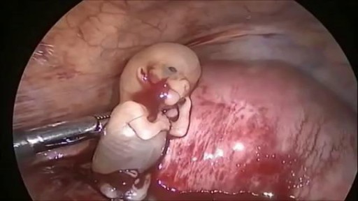
Watch that Ectopic Pregnancy Baby Abortion Surgery
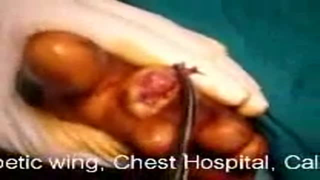
Diabetic Foot Surgical Debridement
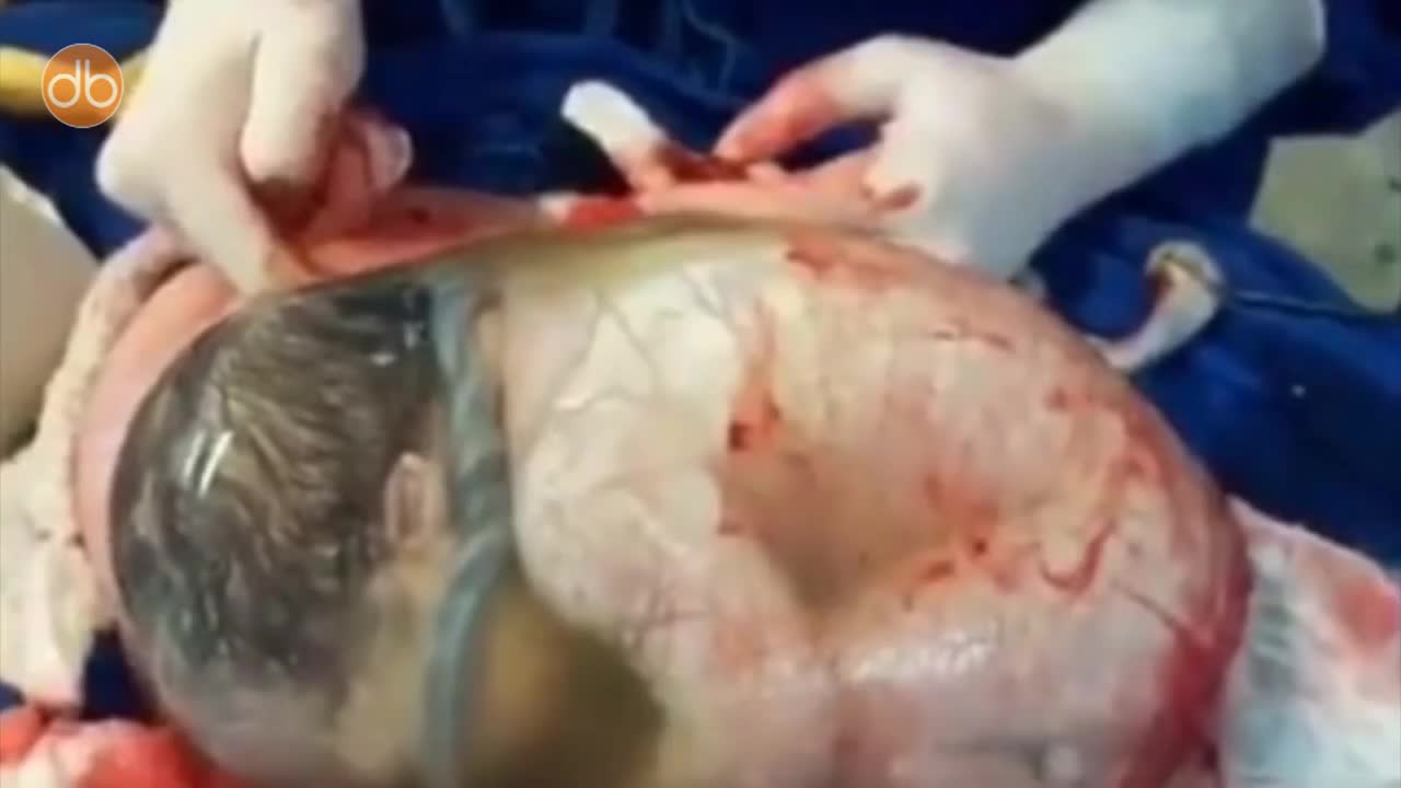
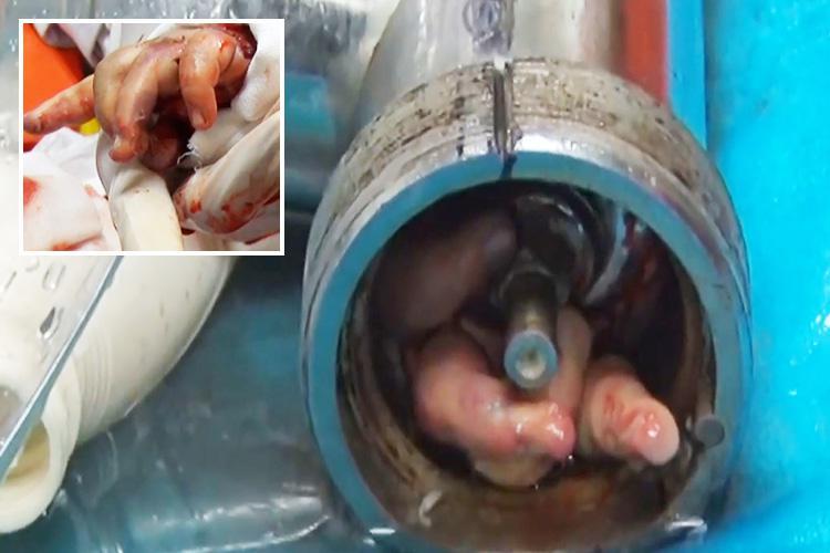
Baby Born Still Inside The Amniotic Sac
A carotid endarterectomy is performed in a sterile surgical suite or standard operating room. You may go home the same day or stay 1–2 nights after the procedure depending on your medical condition. You receive a local anesthetic or general anesthesia. Your vascular surgeon makes an incision at the front of your neck. After removing the plaque from the artery your vascular surgeon repairs the artery by stitching in a natural graft (formed from a piece of vein from elsewhere in your body) or a woven patch. The incision is closed
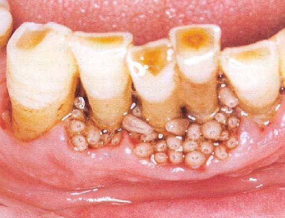
Watch that video of Removing Worms and Parasites From Girl's Mouth

Gastric Lavage Video
