


Below Knee Amputation

Below Knee Amputation
Video Transcript
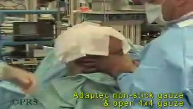
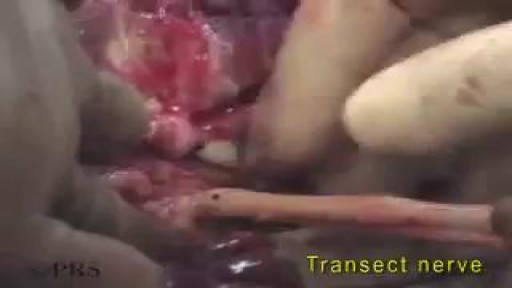
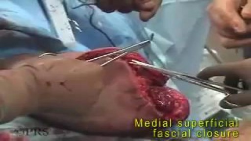
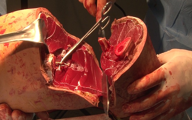
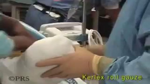
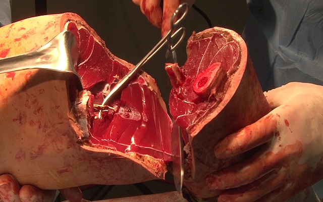
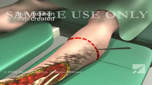
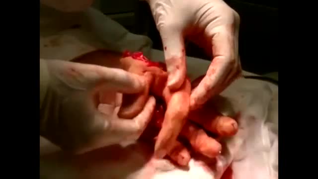
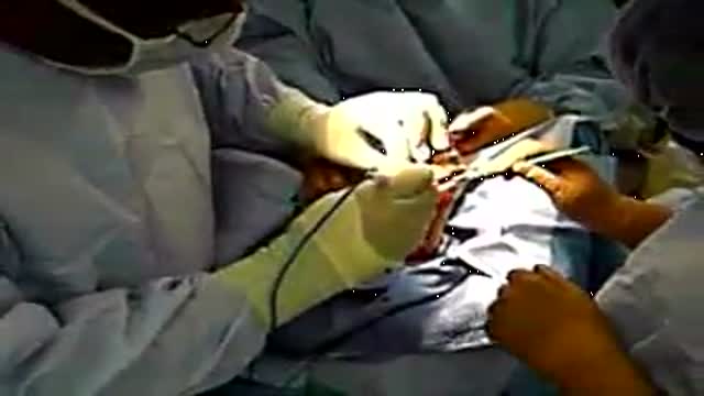
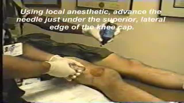
A narrated below-knee amputation. The surgeon describes measuring and cutting the extended posterior flap, carrying the incision down the tibia and fibula, freeing and securing the flap over the bone end, and shows how the flap settles at two, four and ten weeks after the operation.
Read the full transcript
Ammeter the limb at the level of the bone cut, which was 12 centimeters. A normal flap would be 13 centimeters and an extended flap 18. The anterior incision is carried through skin and subcutaneous. Soft section with the amputation knife carries down the back of the tibia and the fibula. At the distal end of the extended posterior flap, the flap is dissected free. The superficial pair irrigation solution is used to remove any debris. Extraceus, the flap is brought up over the bone bridge and secured in place by bringing the medial fascia to the soleus fascia.
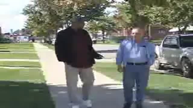
The extreme bulbous nature of the extended flap resolves rapidly. Enter op, two weeks, four weeks, and 10 week photos show the resolution of the flap and how it contours nicely to a very...
Transcript auto-generated from the video’s audio for accessibility and study. Educational content only — not medical advice.
Video Overview
From our Surgery Videos collection: Surgery videos: watch real operations step by step — general, laparoscopic and specialist surgery performed in the OR, explained for students and surgeons. Browse all Surgery Videos videos →
SORT BY-
תגובות ראשיות
-
תגובות אחרונות