
Carpal Tunnel Release Open

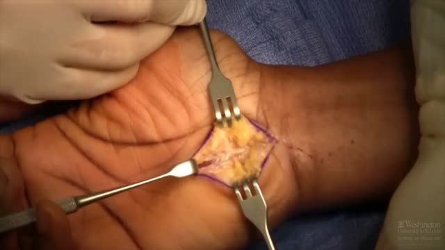
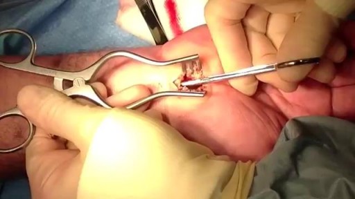
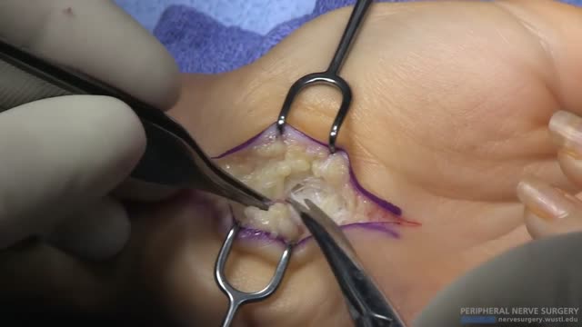
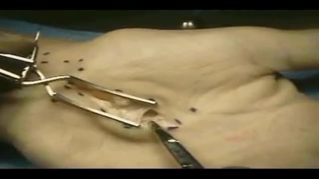
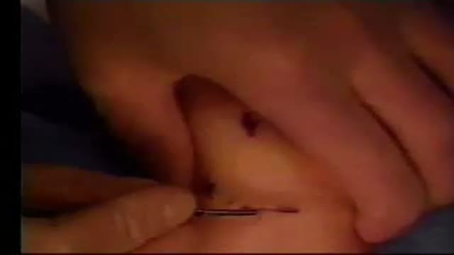
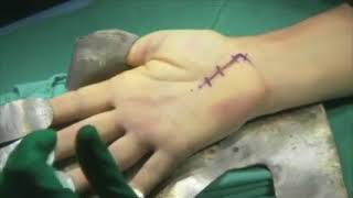
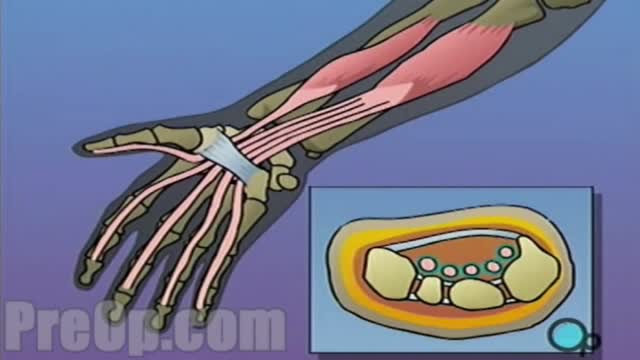
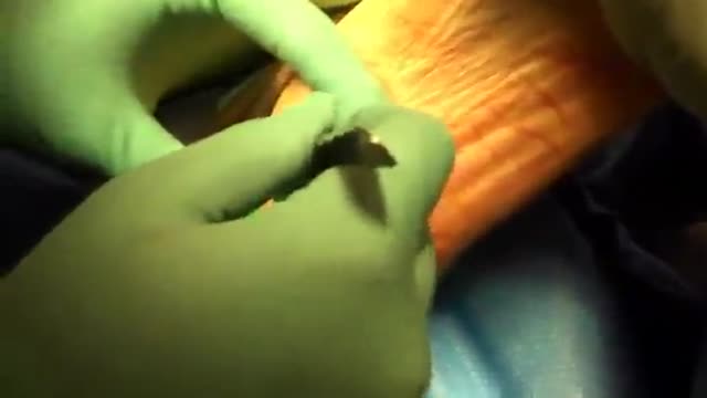
During open carpal tunnel release surgery, the transverse carpal ligament is cut, which releases pressure on the median nerve and relieves the symptoms of carpal tunnel syndrome. An incision is made at the base of the palm of the hand. This allows the doctor to see the transverse carpal ligament.
Video Transcript
A narrated open carpal tunnel release. The surgeon describes the local anesthetic mixture and landmarks, the longitudinal incision in the thenar crease, division of the flexor retinaculum and transverse carpal ligament until the fat pad is reached, inspection of the median nerve and its thenar branch, and closure with a soft dressing, with therapy starting within a few days.
Read the full transcript
As Richard and Dacus today will be discussing open carbotone release. We perform this procedure under MAC with local anesthesia. Here we're cleaning the area. MAC anesthesia has been applied and now an injection is made approximately. We then insert and again we'll inject distally, superficially along the area where we believe our incision will be made. We use a mixture of half of quarter percent marcane and half of one percent lidocaine with a total of 10 ccs. We then place a portion of the fluid in the carbotone as well.
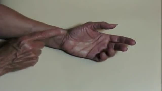
Now the anatomical landmarks are marked. Here the hook of the hammock, Kaplan's cardinal line and the radial portion of the ring finger are all used for identification. I like to use the thing our crease if possible. The incision may be extended as needed distally. Here the skin incision is made longitudinally. The person performing the procedure I've found does better if they're moving from distally to proximally. Sharp skin retractors are placed and the adipose tissue is released on top of the flexor retinaculum. The flexor retinaculum is now identified and incised longitudinally as well.
Again any excessive adipose is removed. Now the transverse carboligament is visualized as you can see here and this is entered again using sharp section. Once the compartment is entered you should feel a pop and flexor tendons should be identified at that point in time. We then use a scissor to then elevate the soft tissues superficial to the transverse carboligament and also to sound the carpal tone. Then we'll release the transverse carboligament proximally with care taken to spread and to palpate prior to cutting. Once we feel that we've had adequate release proximally we'll further inspect and evaluate
the nerve at this point in time. Occasionally the flexor retinaculum will be palpable and if possible this is released as well. We attempt to pass the scissor without cutting if possible or cut under direct visualization. Now distally the remaining portion of the transverse carboligament is released. It's released until the fat pad is identified. Once complete release is obtained it is easy to illustrate the free limbs of the transverse carboligament and now the median nerve is identified as well as its thin-art branch. The wound is copaciergated. The skin is closed using an interrupted mattress type fashion.
Upon closure additional local anesthesia is used if necessary. A soft dressing is placed and a 3 inch ace is applied. We typically will ask the patient to go to therapy within 3 to 5 days to begin range of motion to assess for any possible hangups post-operatively and to allow the patient to begin their therapeutic regimen. Typically therapy only requires 1 to 2 visits. By placing the incision in the thin-art crease the wound is barely evident at healing particularly at 3 months.
Transcript auto-generated from the video’s audio for accessibility and study. Educational content only — not medical advice.
Video Overview
From our Orthopedics collection: Orthopedic surgery videos: fracture fixation, joint replacement, arthroscopy and trauma procedures performed by orthopedic surgeons. Browse all Orthopedics videos →
SORT BY-
Topkommentarer
-
Seneste kommentarer