





Cesarean Section Childbirth

A video showing the cesarean section birth
Video Transcript
A hospital-produced film (MemorialCare / Miller Children's Hospital) of an elective repeat cesarean section. After a promotional introduction to the hospital, an obstetrician narrates the operation on a 39-week patient with a prior cesarean performed in Mexico — the abdominal layers, delivery of the baby, cord clamping, and the baby's first checks before transfer to the well-baby nursery.
Read the full transcript
At Memorial Care, we want you to know the real story. State-of-the-art technology and best practices are standard operating procedure at Miller Children's Hospital and Women's Pavilion, hailed by Fit Pregnancy Magazine as one of the top 18 hospitals in the entire country to have a baby. Memorial Care offers a comprehensive array of services all under the same roof, including one of the premier neonatal and birthing centers in the nation. As Executive Director of the Memorial Medical Center and Miller Children's Hospital, Dr. Michael Pinajot combines nearly three decades of OB-GYN surgical experience
with a well-earned reputation as a leading expert in the treatment of high-risk pregnancies. This patient is a term-pregnant patient who has been followed for prenatal care and has a history of a prior cesarean section having been performed in Mexico. The incision was thought to be a low-transverse incision, but we were not able to get her records and she was not interested in a trial of labor. She is 39 weeks gestation with a mature baby and she is consented to go to surgery for an elective repeat cesarean section.
Patients are also offered the opportunity to attempt a trial of labor or a vaginal birth after cesarean section, and the success rate with such an endeavor approaches 85% in certain populations, but it does require the ability to document the kind of surgery a patient has had in the past as well as an interest on the part of the patient. Since this patient was really not interested in a trial of labor and we were unable to get her prior surgical records, we offered her an elective repeat cesarean section at term.
People involved in the cesarean section include the surgeon and the assistant surgeon, the anesthesiologist, the scrub nurse. There's also a nurse for the baby that takes the baby to the incubator or vasinet and cleans the baby up. Those are the main people that are in the operating room. If you have multiple births, the team is doubled with respect to the nursery personnel for the babies and you have two vasinets where the babies are placed. This is the abdomen that has been prepped and draped now. The light handles are being placed as well as other necessary assistance for surgery.
There is a blue cord there that is the bovi cannula, which basically allows us to coagulate any bleeding vessels. There is also a suction cord that you will see, a suction device. We have something to control bleeding with an electrical signal going through it as well as a suction cord. The surgeon's hands are now on the lower abdomen and a different incision is going to be placed prior to the patient had had a vertical incision, but we're going to do what's called a Thanan steel or a low transverse skin incision,
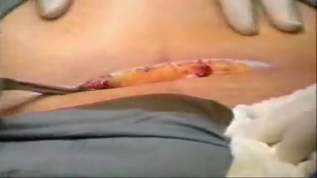
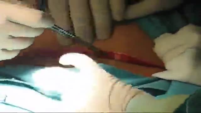
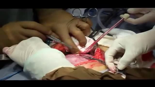
which you're about to see in a moment. We cut right through the plastic drape. At this point, the incision has begun through the skin and subcutaneous tissue. This is a transverse incision from the patient's left side to her right side. The assistant surgeon is on the right side of the image retracting the skin. You can see there are some bleeding vessels in the subcutaneous tissue. The effort here is to control any brisk bleeding as well as to stay in the same plane as one approaches the fascia, which is the tissue that surrounds the muscle
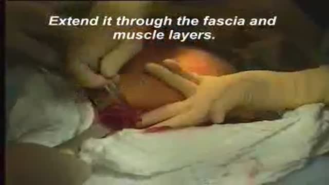
or the rectus abdominis muscle in the lower abdomen. This is a bleeder that is at the right lateral margin of the patient's skin incision, controlled with a clamp and then a bove coagulation. This is further incision with the sharp instrument of the scalpel. One is able to readily see the subcutaneous tissue as we are approaching the fascia. At this point, the fascia is being incised sharply with a scissors, and the tissue is being retracted with these retractors that are called goulay retractors. The scissors is a male scissors. It's curved,
and the sharp pickups are being used to lift the fascia under direct visualization and incising it, and the rectus muscle is what is beneath it. At this point, there is a scalpel being used to incise the scarred fascia over the rectus muscle from her prior surgery, so that this is done again under direct visualization, pushing the muscles down, which is the tissue that's pink below, and the white tissue on top is the fascia itself. We're trying to create a plane between the fascia and the muscle. The next procedure is to divide the midline of the rectus muscles
and to approach the peritoneal cavity under direct visualization. So the scissors are being used to incise in the midline in an avascular area where there are few, if no, blood vessels, and trying to sharply enter the peritoneal cavity. We try to enter the peritoneal cavity as superiorly as we can to avoid the bladder, so that the effort will be made here to lift the patients abdominal wall and enter the peritoneal cavity towards the umbilicus away from her bladder, which is indicated by the index finger right there.
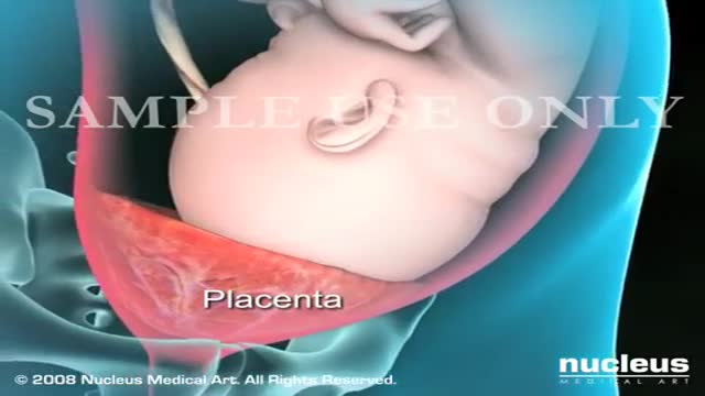
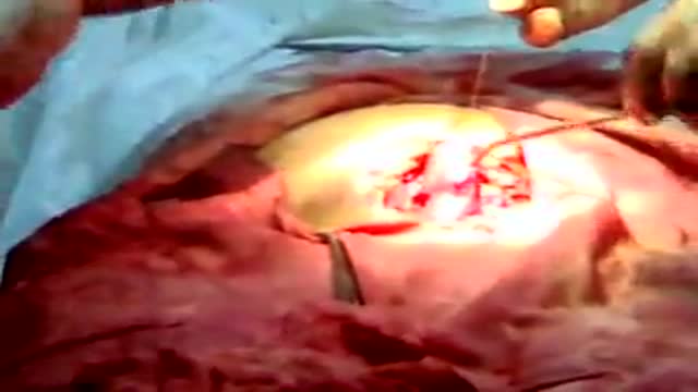
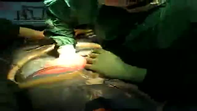
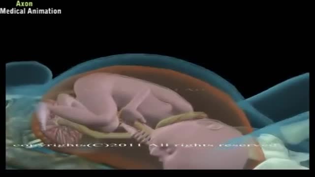
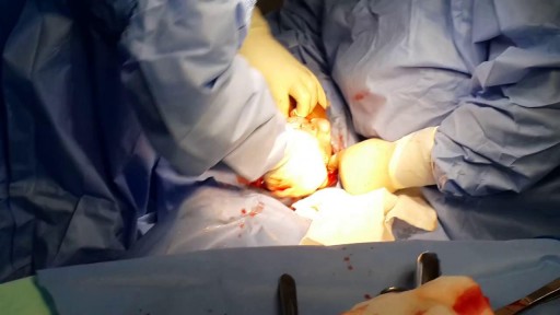
This is now the peritoneal cavity being entered, and you can see the uterus is right below there, but this is now bluntly and sharply being incised in a midline fashion. We are now within the peritoneal cavity, and you're looking right at the uterus itself that has got the fetus with inside. The surgeon's hand is introducing the peritoneal cavity to feel for the position of the uterus, specifically the round ligaments, to make sure that the uterus is not rotated unduly in one direction or the other because there's going to be, again, a low transverse incision now being made in the uterus,
much like in the skin, through the muscle of the uterus itself. And this is being done to free up the bladder below and to gently and carefully enter the uterine cavity using caution to not go into quickly and cut the baby. And you can see right there those are the membranes with the amniotic fluid within and the vernex floating around. This is now rupturing of the membranes and there's clear fluid. The surgeon's hand is now within the uterus itself feeling for the baby's presenting part, which is most likely a vertex, as can be seen at the top of the baby's head there.
It has got hair on it. Now the effort is to free up the head with fundal pressure. This is a nice, gentle delivery. The baby's head is facing to the screen left. The umbilical cord is reduced over the top of the baby. It was probably around the baby's neck, which is a very common finding with term births. The baby's nose and mouth are being suctioned before the baby's first cry. So the majority of the baby is still within the uterus at this point. Efforts are made to just really clear out that airway as much as possible.
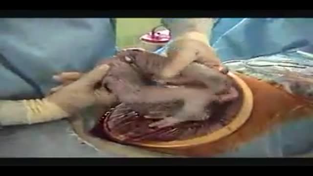
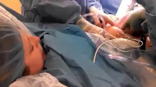
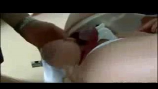
Now this is the posterior shoulder. An anterior shoulder of the baby is being freed up. So that just gently, you don't want to put too much traction on the baby's neck. And just assisting getting our fingers underneath the shoulders, the best the baby is delivered. One can see it's a good-sized baby. The baby's got good tone and movement and is crying. The baby is just lying on the mom's legs now. And we've clamped the cord and are cutting the cord. That's going to go to the baby's doctor, the pediatrician,
after we show it to the mom. The baby is taken to the incubator. The baby is cleaned off. The heart and lungs are listened to. There is a further shortening of the umbilical cord being done. The baby has footprints taken. And depending upon where the baby is born, there may be a scale brought in for weighing. In the operating room, we don't weigh the baby there, but we do in the delivery room. But in this case, the operating room was not where the baby was to be weighed.
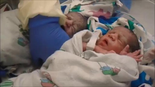
The baby was taken to the nursery to be weighed. Depending upon the size and how the baby is performing in the first several minutes after delivery, determines whether the baby goes to the well-baby nursery or to an intensive care setting for further assessment. This baby went to the well-baby nursery.
Transcript auto-generated from the video’s audio for accessibility and study. Educational content only — not medical advice.
Video Overview
From our Reproductive Health collection: Fertility, contraception, IVF and sexual health explained clearly by specialists. Browse all Reproductive Health videos →
SORT BY-
سرفہرست تبصرے
-
تازہ ترین تبصرے