- Physical Examination
- Surgical Examination
- Ophthalmology
- Clinical Skills
- Orthopedics
- Surgery Videos
- Laparoscopy
- Pediatrics
- Funny Videos
- Cardiothoracic Surgery
- Nursing Videos
- Plastic Surgery
- Otorhinolaryngology
- Histology and Histopathology
- Neurosurgery
- Dermatology
- Pediatric Surgery
- Urology
- Dentistry
- Oncology and Cancers
- Anatomy Videos
- Health and Fitness
- Radiology
- Anaesthesia
- Physical Therapy
- Pharmacology
- Interventional Radiology
- Cardiology
- Endocrinology
- Gynecology
- Emergency Medicine
- Psychiatry and Psychology
- Childbirth Videos
- General Medical Videos
- Nephrology
- Physiology
- Diet and Food Health
- Diabetes Mellitus
- Neurology
- Women Health
- Osteoporosis
- Gastroenterology
- Pulmonology
- Hematology
- Rheumatology
- Toxicology
- Nuclear Medicine
- Infectious Diseases
- Vascular Disease
- Reproductive Health
- Burns and Wound Healing
- Other
Latest videos
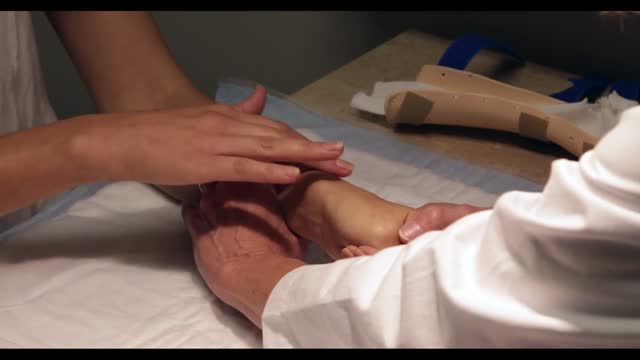
De Quervain's tenosynovitis (dih-kwer-VAINS ten-oh-sine-oh-VIE-tis) is a painful condition affecting the tendons on the thumb side of your wrist. If you have de Quervain's tenosynovitis, it will probably hurt when you turn your wrist, grasp anything or make a fist. Although the exact cause of de Quervain's tenosynovitis isn't known, any activity that relies on repetitive hand or wrist movement — such as working in the garden, playing golf or racket sports, or lifting your baby — can make it worse. Symptoms ShareTweet June 13, 2015 References Products and Services Mayo Clinic Sports Medicine Newsletter: Mayo Clinic Health Letter See also Prednisone risks, benefits Prednisone withdrawal: Why taper down slowly? Integrative approaches to treating pain Lifestyle strategies for pain management Nutrition and pain Pain rehabilitation Self-care approaches to treating pain Show more Advertisement Mayo Clinic does not endorse companies or products. Advertising revenue supports our not-for-profit mission. Advertising & Sponsorship PolicyOpportunitiesAd Choices Mayo Clinic Store Check out these best-sellers and special offers on books and newsletters from Mayo Clinic. NEW! – The Mayo Clinic Diet, Second Edition Healthy Heart for Life! Mayo Clinic on Better Hearing and Balance Treatment Strategies for Arthritis The Mayo Clinic Diet Online
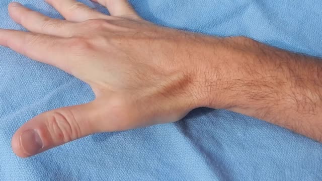
Tenosynovitis is inflammation of the lining of the sheath that surrounds a tendon (the cord that joins muscle to bone).
How to increase breast milk supply How to Naturally Boost & Increase your breast milk supply

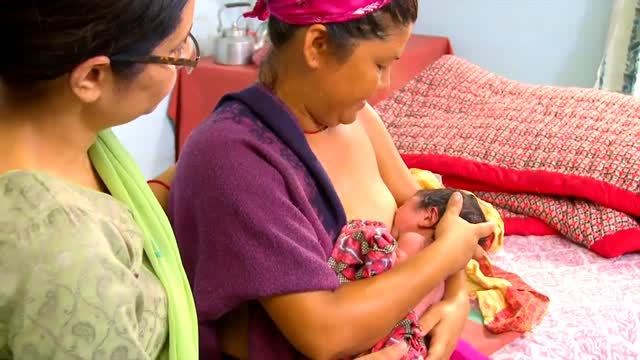
Breastfeeding attachment Attaching your baby at the breasT
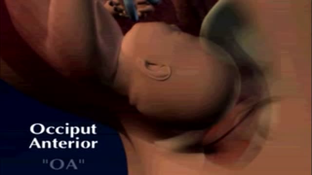
fetal position in womb at 34 weeks fetal position in womb week by week fetal position in womb at 19 weeksUnborn babies toss and turn and hold many different positions within the womb during the gestation period; pregnant women everywhere will attest to the fact that their children always start up the gymnastics at bedtime.

Exercise For Positioning Baby in Womb
Successful External Cephalic Version (ECV) - Turning a breech baby in just 2 minutes!
This animation describes tools and tests used to diagnose inflammatory bowel disease (IBD), determine IBD type, and predict its probable course and outcome.
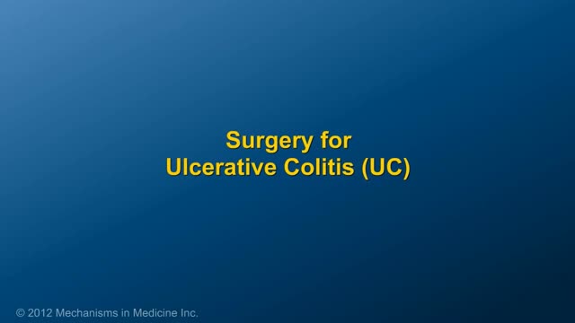
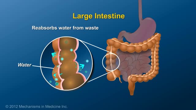
This animation describes surgery for patients with inflammatory bowel disease (IBD) -- IPAA, removal of colon, intestinal resection, & stricturoplasty.
An ileostomy is an opening in the belly (abdominal wall) that’s made during surgery. The end of the ileum (the lowest part of the small intestine) is brought through this opening to form a stoma, usually on the lower right side of the abdomen. A Wound Ostomy Continence nurse (WOCN or WOC nurse) or the surgeon will figure out the best location for your stoma. (A WOC nurse is a specially trained registered nurse who takes care of and teaches ostomy patients. This nurse may also be called an ostomy nurse.)
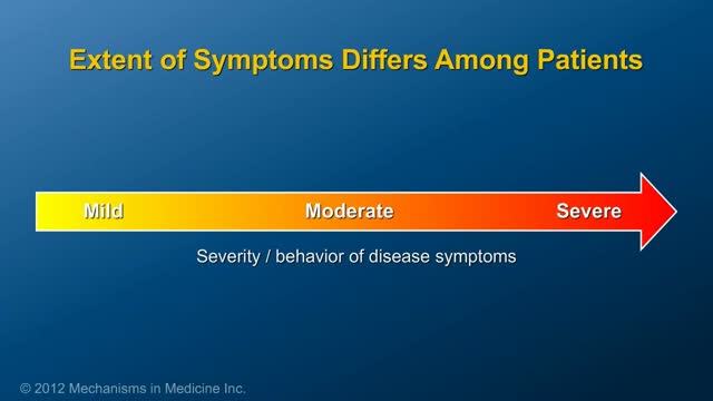
This animation describes the goals of inflammatory bowel disease (IBD) management and how patients can take an active role in managing their disease.
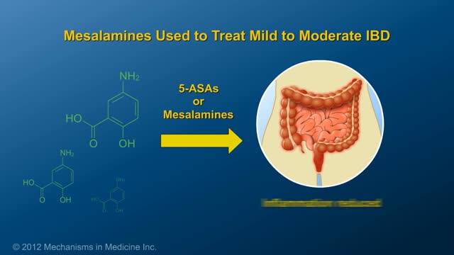
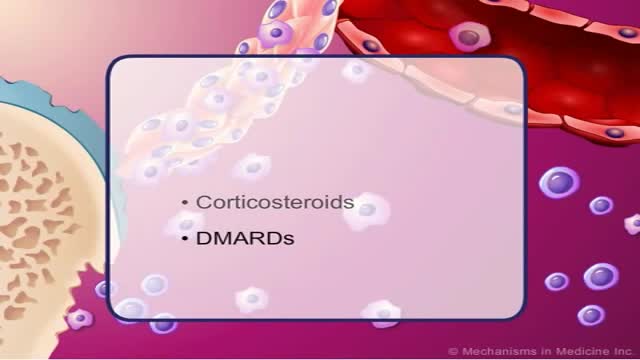
This animation describes risks of inflammatory bowel disease (IBD) and risks/benefits of medication (5-ASAs, steroids, immunomodulators, & biologics).
Rheumatoid arthritis is a chronic inflammatory disorder that can affect more than just your joints. In some people, the condition also can damage a wide variety of body systems, including the skin, eyes, lungs, heart and blood vessels. An autoimmune disorder, rheumatoid arthritis occurs when your immune system mistakenly attacks your own body's tissues. Unlike the wear-and-tear damage of osteoarthritis, rheumatoid arthritis affects the lining of your joints, causing a painful swelling that can eventually result in bone erosion and joint deformity. The inflammation associated with rheumatoid arthritis is what can damage other parts of the body as well. While new types of medications have improved treatment options dramatically, severe rheumatoid arthritis can still cause physical disabilities.
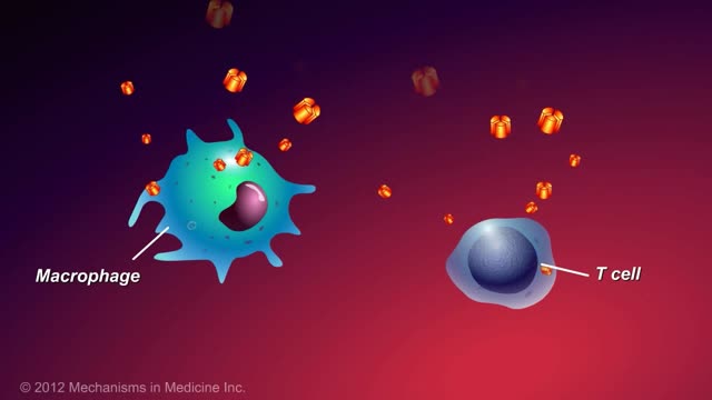
This animation describes what anti-TNF-alpha therapies are, how they work, and how patients with inflammatory bowel disease (IBD) can benefit.

This video explains briefly the use of anti-tnf alpha in therapy.
Impaired awareness of illness (anosognosia) is a major problem because it is the single largest reason why individuals with schizophrenia and bipolar disorder do not take their medications. It is caused by damage to specific parts of the brain, especially the right hemisphere.
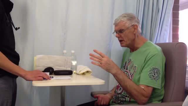
Testing for the four features of Gerstmann Syndrome in this patient with two separate left sided strokes (left frontoparietal ischaemic stroke followed by left posterior parietal haemorrhagic stroke). He exhibits (i) acalculia, (ii) agraphia, (iii) left-right disorientation, and (iv) finger agnosia. Complicating the issue is his obvious nonfluent aphasia (expressive dysphasia) with paraphasic errors (replacing words with associated words (e.g. says 'fork' instead of 'spoon')) and some comprehension issues.

When a stroke affects an extensive portion of the front and back regions of the left hemisphere, the result may be global aphasia. Survivors with global aphasia: May have great difficulty in understanding words and sentences. May have great difficulty in forming words and sentences. May understand some words. Get out a few words at a time. Have severe difficulties that prevent them from effectively communicating.

People with serious comprehension difficulties have what is called Wernicke’s aphasia and: Often say many words that don’t make sense. May fail to realize they are saying the wrong words; for instance, they might call a fork a “gleeble.” May string together a series of meaningless words that sound like a sentence but don’t make sense. Have challenges because our dictionary of words is shelved in a similar region of the left hemisphere, near the area used for understanding words.

Broca's Aphasia (expressive) When a stroke injures the frontal regions of the left hemisphere, different kinds of language problems can occur. This part of the brain is important for putting words together to form complete sentences. Injury to the left frontal area can lead to what is called Broca's aphasia.
