- Physical Examination
- Surgical Examination
- Ophthalmology
- Clinical Skills
- Orthopedics
- Surgery Videos
- Laparoscopy
- Pediatrics
- Funny Videos
- Cardiothoracic Surgery
- Nursing Videos
- Plastic Surgery
- Otorhinolaryngology
- Histology and Histopathology
- Neurosurgery
- Dermatology
- Pediatric Surgery
- Urology
- Dentistry
- Oncology and Cancers
- Anatomy Videos
- Health and Fitness
- Radiology
- Anaesthesia
- Physical Therapy
- Pharmacology
- Interventional Radiology
- Cardiology
- Endocrinology
- Gynecology
- Emergency Medicine
- Psychiatry and Psychology
- Childbirth Videos
- General Medical Videos
- Nephrology
- Physiology
- Diet and Food Health
- Diabetes Mellitus
- Neurology
- Women Health
- Osteoporosis
- Gastroenterology
- Pulmonology
- Hematology
- Rheumatology
- Toxicology
- Nuclear Medicine
- Infectious Diseases
- Vascular Disease
- Reproductive Health
- Burns and Wound Healing
- Other
Latest videos

#STOP VIOLENCE AGAINST DOCTORS#SAVE THE DOCTOR

STOP VIOLENCE AGAINST DOCTORs

today we talk about Amniotic fluid during your pregnancy! Looking forward to your comments.

How to Get Rid of Vaginal Discharge - Treating Normal Discharge.

Pregnancy is one of the beautiful phases of a woman’s life after their marriage. The feeling of the baby growing inside the womb is exceptional and very special.

A lot of women want to know what type of vaginal discharge is normal during pregnancy, and when you're not pregnant. So let's start out by talking about what's normal when you're not pregnant. It's normal to have about 1/2 teaspoon to 1 teaspoon of whitish, creamy, tannish discharge on most days of your cycle in between periods, with the exception of the time of ovulation. Actually, around the time of ovulation, it's normal to notice the discharge becoming more slippery and clear, almost like egg whites. And this is actually a sign that you can watch for to know when you're ovulating. And if you're seeing this type of discharge and you're trying to have a baby, then you should start to time intercourse with ovulation to increase your chances of conceiving.

It sounds like you're questioning whether or not your water may have broken, and this can actually be a hard thing for a lot of women to tell. Usually if your water breaks, it's just a trickle of fluid, and you're afraid to admit it to anyone because you think you peed your pants. And it is normal to pee your pants when you're pregnant because the bladder is right below the uterus, and if the baby moves just right, it might kick out a little bit of urine. So if you feel a trickle or a little tiny gush of fluid, what you want to do is put a pad or a pantie-liner on after going to the bathroom and emptying your bladder, and wait an hour and see if fluid continues to come out. And if it does, then you're not having bladder leakage issues - your water is probably broken.

Women are routinely invited to have cervical screening tests (also called smear tests). The tests are done to prevent cervical cancer, not to diagnose cancer. During each test some cells are removed from the neck of the womb (cervix), with a plastic brush. The cells are examined under a microscope to look for early changes that, if ignored and not treated, could develop into cancer of the cervix. You are very unlikely to develop cervical cancer if you have regular cervical screening tests at the times advised by your doctor. If the test shows any abnormality, you will have treatment to stop you ever getting cancer of the cervix. So, an abnormal test does not mean you have cancer. It means you should have some treatment to stop you getting cancer.

Cases of some sexually transmitted diseases have reached an all-time high, according to a new report from the Centers for Disease Control and Prevention. From 2014 to 2015, there was a 6% increase in diagnosed cases of chlamydia and a 13% increase in gonorrhea.

Colposcopy (kol-POS-kuh-pee) is a procedure to closely examine your cervix, vagina and vulva for signs of disease. During colposcopy, your doctor uses a special instrument called a colposcope. Your doctor may recommend colposcopy if your Pap test has shown abnormal results.
Remembering Medications & The Body Systems Affected

How to memorize more in pharma: Drug names, dental implications, numbers
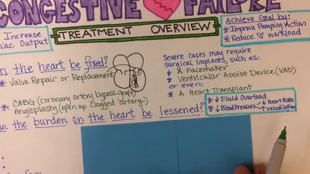
Congestive Heart Failure
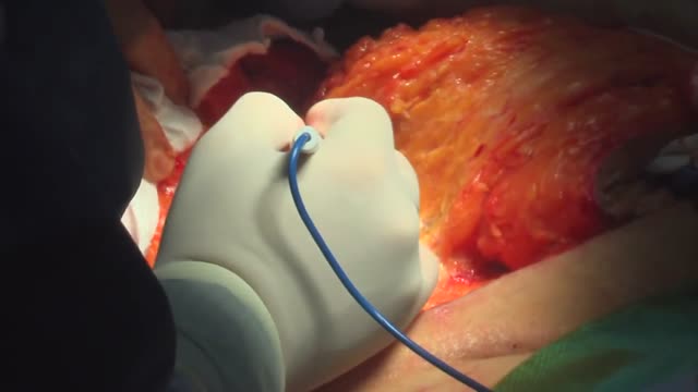
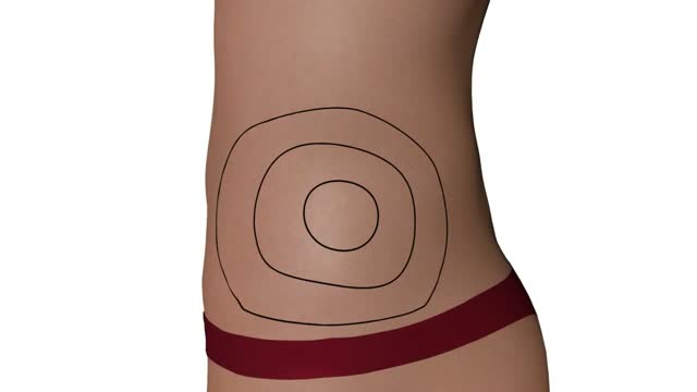
surgical procedure used to remove excess skin and fat from the abdomen and to tighten the muscles of the abdominal wall. Most tummy tuck patients are dealing with the effects of pregnancies and weight loss and find themselves with loose skin in spite of exercise and weight control. Each year, thousands of Americans undergo a tummy tuck to tone, firm and define the abdominal area.
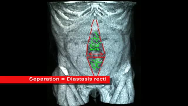
plastic surgeon demonstrates the results of a muscle separation(rectus diastasis) repair using 3 dimesional CAT scan and photographic images
Questo Video 3D illustra la tecnica della Microlipocavitazione: sistema chirurgico ad ultrasuoni per ottenere l'emulsione del grasso in eccesso da eliminare. La Microlipocavitazione è una tecnica di chirurgia ambulatoriale, che richiede una modesta anestesia locale con un recupero delle proprie attività pressoché immediato.

The hips are one of the hardest places to loose fat. Liposuction can be done on this area to dramatically help loose inches. This area is also one of the most successful areas to show visible improvement after liposuction is done. Liposuction of the hips can help patients to reduce dress and pant sizes. Disclaimer. The photographs on these pages illustrate typical results of some liposuction surgery procedures and may contain some nudity. Viewer discretion is advised. In providing the photos and statements on this web site, Liposuction.com does not state or imply any guarantee.
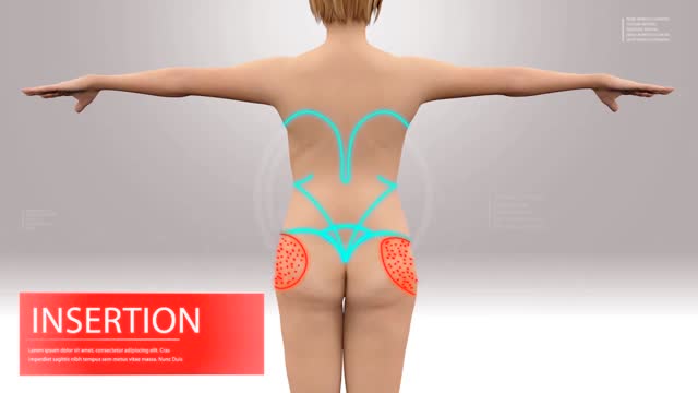
Liposuction & Facelift

Bacterial vaginosis is a type of vaginal inflammation caused by the overgrowth of bacteria naturally found in the vagina, which upsets the natural balance. Women in their reproductive years are most likely to get bacterial vaginosis, but it can affect women of any age. The cause isn't completely understood, but certain activities, such as unprotected sex or frequent douching, increase your risk.

Lichen sclerosus is a skin condition that mainly affects the genital skin (vulva) in women and the penis in men. It most commonly occurs in middle-aged women. Symptoms may include itch, soreness, and changes in the appearance of affected skin.
