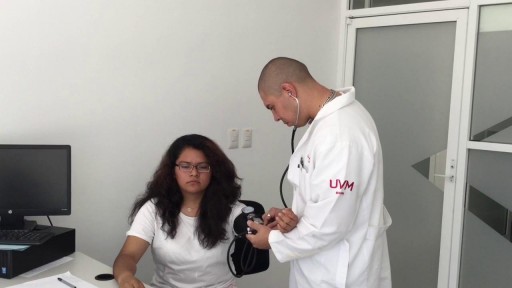
- Physical Examination
- Surgical Examination
- Ophthalmology
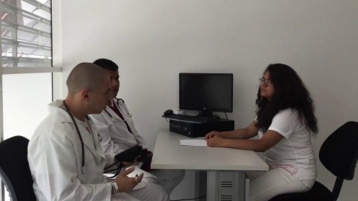
- Clinical Skills
- Orthopedics
- Surgery Videos
- Laparoscopy
- Pediatrics
- Funny Videos
- Cardiothoracic Surgery
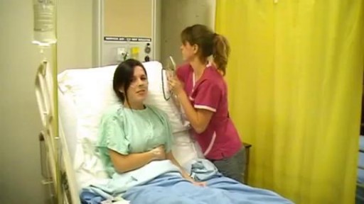
- Nursing Videos
- Plastic Surgery
- Otorhinolaryngology
- Histology and Histopathology
- Neurosurgery
- Dermatology
- Pediatric Surgery
- Urology
- Dentistry
- Oncology and Cancers
- Anatomy Videos
- Health and Fitness
- Radiology
- Anaesthesia
- Physical Therapy
- Pharmacology
- Interventional Radiology
- Cardiology
- Endocrinology
- Gynecology
- Emergency Medicine
- Psychiatry and Psychology
- Childbirth Videos
- General Medical Videos
- Nephrology
- Physiology
- Diet and Food Health
- Diabetes Mellitus
- Neurology
- Women Health
- Osteoporosis
- Gastroenterology
- Pulmonology
- Hematology
- Rheumatology
- Toxicology
- Nuclear Medicine
- Infectious Diseases
- Vascular Disease
- Reproductive Health
- Burns and Wound Healing
- Other
Latest videos
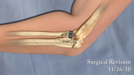
Tennis elbow is caused by doing the same forceful arm movements over and over. It creates small, painful tears in the tendons in your elbow. This injury can be caused by tennis, other racquet sports, and activities such as turning a wrench, prolonged typing, or chopping with a knife. The outside (lateral) elbow tendon is most commonly injured. The inside (medial) and backside (posterior) tendons can also be affected. This article discusses surgery to repair tennis elbow

UCLA Hand Transplant Procedure

A digital rectal examination (DRE) is a simple procedure doctors use to examine the lower rectum and other internal organs. A DRE is done for a number of reasons. It's a quick, easy way to check the health of a man's prostate gland. It can detect conditions like an enlarged prostate

Hemorrhoids repair: Disposable hemorrhoidal stapler
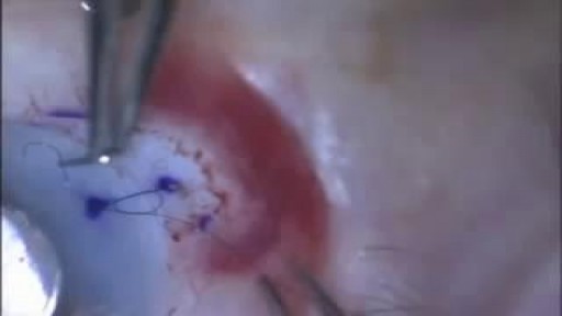
Video demonstrates the fundamental components of placing your first suture.
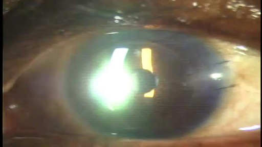
Stephen Slade shows a 1 week post op patient after DSAEK. DSAEK is an excellent option for many patients with corneal disease. In DSAEK, only the thin, inner layer is replaced, so the healing is typically much faster than a full thickness cornea graft.
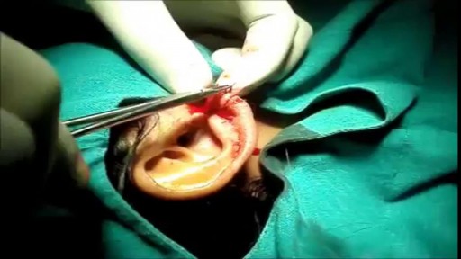
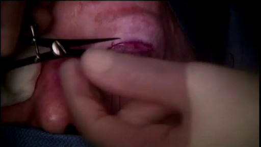
This 25 year young female wanted her split earlobe hole to be repaired.Ear lobe ring hole usually elongated due to continuous use of fancy heavy ear rings.most young ladies suffer from this problem, subsequently this get converted to complete split ear lobe.This needs surgical repair only.This is a cosmetic repair .watch the video , how this repair is done.Usually the split is completely closed with suture.After healing new hole to be done little distance from the repair site.
Surgery is performed by Kami Parsa M.D. The patient is a 55 year old with a history of previous upper eyelid blepharoplasty with excessive skin removed from both upper eyelids which resulted in bilateral lagophthalmos. Patient could not close her eyes and had problems with severe dry eyes.
This is a video of a Gender Reassignment Surgery, watch as surgeons change a male to a female its an extremely interesting procedure
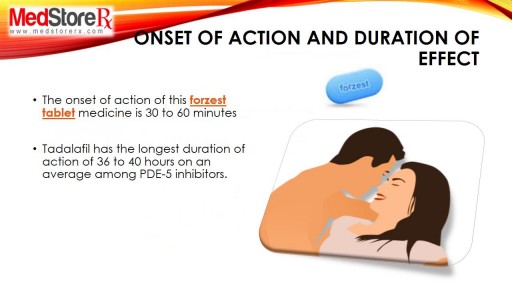
Forzest is FDA approved medicine, it is used bt men to improve erectile dysfunction dusring intercourse session with partner. for more information related side effects, dosage, etc kindly visit to http://www.medstorerx.com/forzest.aspx
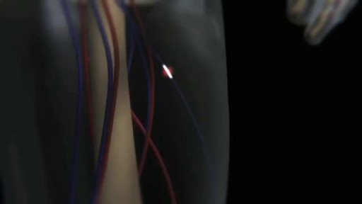
How a Clot Can Become a Pulmonary Embolism

Prompt treatment to break up the clot greatly reduces the risk of death. This can be done with blood thinners and drugs or procedures. Compression stockings and physical activity can help prevent clots from forming in the first place.
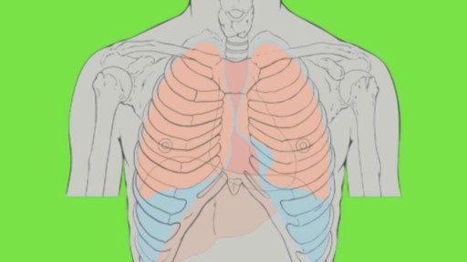
Most times, a pulmonary embolism is caused by blood clots that travel from the legs or, rarely, other parts of the body (deep vein thrombosis, or DVT). Symptoms include shortness of breath, chest pain, and cough. Prompt treatment to break up the clot greatly reduces the risk of death. This can be done with blood thinners and drugs or procedures. Compression stockings and physical activity can help prevent clots from forming in the first place.
Pulmonary edema is usually caused by a heart condition. Other causes include pneumonia, exposure to certain toxins and drugs, and being at high elevations. Depending on the cause, pulmonary edema symptoms may appear suddenly or develop over time. Mild to extreme breathing difficulty can occur. Cough, chest pain, and fatigue are other symptoms. Treatment generally includes supplemental oxygen and medications.
Swelling is a typical symptom of lymphedema and commonly affects legs and arms. Compression stockings work to encourage the movement of lymph out of an affected limb. Lymphedema is incurable. However, treatment can help reduce the swelling and pain

Heart failure can occur if the heart cannot pump (systolic) or fill (diastolic) adequately. Symptoms include shortness of bronicreath, fatigue, swollen legs, and rapid heartbeat. Treatments can include eating less salt, limiting fluid intake, and taking prescription medications. In some cases a defibrillator or pacemaker may be implanted.
Pulmonary edema is usually caused by a heart condition. Other causes include pneumonia, exposure to certain toxins and drugs, and being at high elevations. Depending on the cause, pulmonary edema symptoms may appear suddenly or develop over time. Mild to extreme breathing difficulty can occur. Cough, chest pain, and fatigue are other symptoms. Treatment generally includes supplemental oxygen and medications.
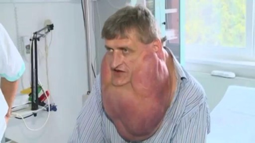
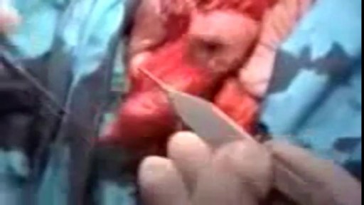
Massive Tumor Removed from Man's Face
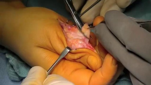
Recurrent Giant Cell Tumor of Tendon Sheath
