סרטונים מובילים

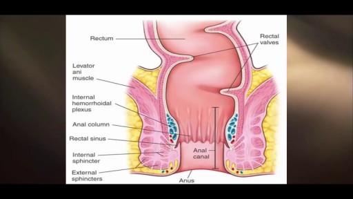
A flail chest occurs when a segment of the thoracic cage is separated from the rest of the chest wall. This is usually defined as at least two fractures per rib (producing a free segment), in at least two ribs. A segment of the chest wall that is flail is unable to contribute to lung expansion. Large flail segments will involve a much greater proportion of the chest wall and may extend bilaterally or involve the sternum. In these cases the disruption of normal pulmonary mechanics may be large enough to require mechanical ventilation.
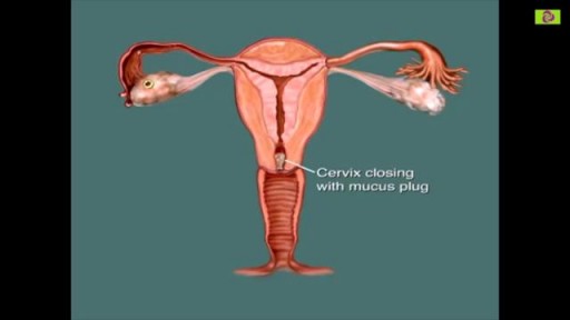
You are most fertile at the time of ovulation, (when an egg is released from your ovaries) which usually occurs 12-14 days before your next period starts. This is the time of the month when you are most likely to get pregnant. It is unlikely that you will get pregnant just after your period, although it can happen.
An intrauterine device (IUD), also known as intrauterine contraceptive device (IUCD or ICD) or coil, is a small, often T-shaped birth control device that is inserted into a woman's uterus to prevent pregnancy. IUDs are one form of long-acting reversible birth control (LARC).

A Texas baby, born with part of her heart outside her body ( Ectopia Cordis) , defies the odds and leaves hospital following a successful surgery.
Masturbating is totally healthy, and totally normal. There are tons of myths out there meant to scare you into thinking masturbation is wrong or bad. But the truth is masturbation is perfectly safe. Masturbating won't make you blind, crazy, or stupid. It won’t damage your genitals, cause pimples, or stunt your growth. It doesn’t use up all your orgasms or ruin other kinds of sex. In fact, masturbation can actually be good for you. Here are some benefits of masturbation: Masturbation is safer than any other type of sex. You can’t get pregnant or get any sexually transmitted infections from masturbating. Masturbation can help you learn what you like and don’t like sexually. And if you decide to have sex with someone, you can know what you do/don’t want to do. BONUS: getting comfortable talking about sex and your body with your partner makes it easier to talk about protecting yourself against STDs and pregnancy, too. Exploring your body and learning how to give yourself sexual pleasure can be empowering and help improve your body image. Masturbation can lower stress and help you relax. It even helps some people fall asleep. Having an orgasm releases endorphins — feel good chemicals in your brain. Orgasms can be a natural painkiller and can even help with period cramps. Mutual masturbation (masturbating with a partner) is a really safe way to have sex and let the other person know what feels good to you. If you share a sex toy, use condoms on the toy and clean it before swapping. And if you touch each other’s genitals, wash your hands before touching your own. Can I get an STD from masturbating? Nope. Masturbating is the safest sexual activity out there. There is virtually NO chance of getting an STD or any other infection from touching your own genitals (and there’s also no chance of pregnancy). STDs have to be passed from one person to another, so you can’t give yourself an STD. The one exception to this is herpes - so if you have any cold sores on your mouth and touch them, make sure to wash your hands before masturbating. But it IS possible to get an STD if you’re masturbating with another person and touching each other’s genitals. Anytime semen (cum) or vaginal fluids are spread to someone else’s body, or your genitals rub against each other, there’s a risk of STDs. So if you touch each other’s genitals, wash your hands before touching your own. STDs can also be spread by sharing sex toys with another person. You can help protect yourself by using condoms on any toys that you share (even if they’re not shaped like a penis). Put a new condom on anytime a different person uses it. If you’re the only one using your sex toys, you don’t have to worry about STDs. But if you use them with other people, protect those sex toys just like you’d protect your own genitals — put a condom on ‘em! It’s possible for masturbation to cause irritation or infections if your body is sensitive to the way you masturbate or the things you masturbate with — but this isn’t the same thing as an STD. Lotions, Vaseline, oils, and scented or flavored stuff may irritate your vulva and vagina. Masturbating roughly and not using lubrication can also lead to irritation because of friction. And germs from the anus can cause vaginal infections — so never put something in your vagina that’s been in your butt without washing it or covering it with a condom. If you’re worried that you have an STD because of pain, itching, or discomfort in your genitals, go to your doctor or your local Planned Parenthood health center.
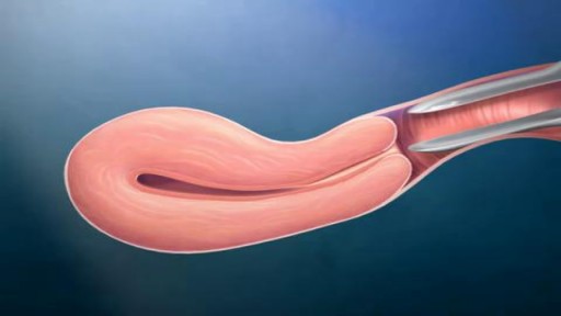
Penile implants are devices placed inside the penis to allow men with erectile dysfunction (ED) to get an erection. Penile implants are typically recommended after other treatments for ED fail. There are two main types of penile implants, semirigid and inflatable.

Ectopia cordis is a rare genetic defect. During a baby’s development in utero, their chest wall doesn’t form correctly. It also doesn’t fuse together as it normally would. This prevents the heart from developing where it should, leaving it defenseless and exposed outside of the protection of the chest wall. The defect affects about one in 126,000 births. In partial ectopia cordis, the heart is located outside the chest wall, but just under the skin. The heart can be seen beating through the skin.

The menstrual cycle is the regular natural change that occurs in the female reproductive system that makes pregnancy possible. The cycle is required for the production of oocytes, and for the preparation of the uterus for pregnancy.

Schizophreniform disorder is a mental disorder diagnosed when symptoms of schizophrenia are present for a significant portion of the time within a one-month period, but signs of disruption are not present for the full six months required for the diagnosis of schizophrenia.
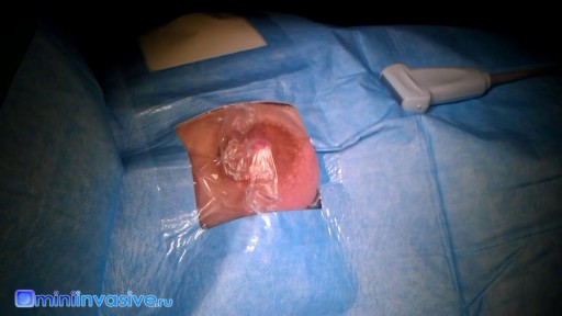
Breast implants do not last forever, and during its lifetime, it may rupture. Dr. Linder, Beverly Hills breast surgeon specialist, breaks down how removing breast implants works. To learn more about Dr. Stuart Linder and his expertise, Visit: www.drlinder.com

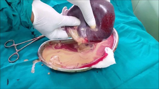
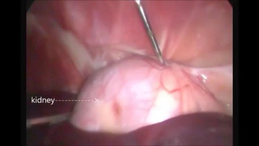
There are several reasons that your doctor may recommend that you have your spleen removed. These include having: a spleen that’s damaged from injury an enlarged spleen or ruptured spleen, which can occur from trauma certain rare blood disorders cancer or large cysts of the spleen infection
Once the diagnosis of a splenic abscess has been made, the patient must be admitted to the hospital and treated. Treatment depends on the patient's overall condition, comorbidities, and primary disorder (if any), as well as the size and topography of the abscess
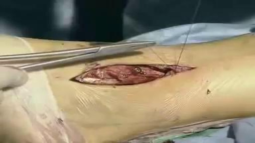
Unstable ankle joints after internal fixation of type B malleolar fractures exist. Residual instability most often occurs after trimalleolar fractures with initial joint dislocation. Treatment with an additional positioning screw generally produced a satisfactory result.
The only way to completely avoid anal sex risks is to abstain from anal sex. If you engage in anal sex, it is always important to use a condom to protect against the spread of infections and diseases.
Possible causes are a blocked milk duct or bacteria entering the breast. It usually occurs within the first three months of breast-feeding. Symptoms include breast pain, swelling, warmth, fever, and chills. Antibiotics are required. Mild pain relievers can help with discomfort.
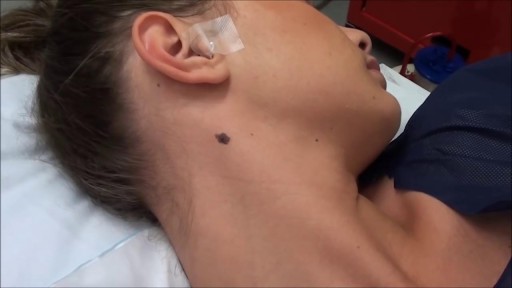
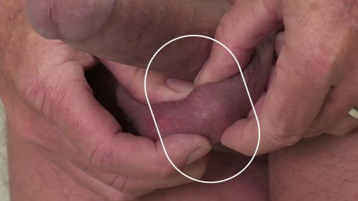
Most people develop several moles (nevi) throughout adulthood. Moles can be found anywhere on the body, usually in sun-exposed areas, and are usually brown, smooth, and slightly raised. In most cases, a nevus is benign and doesn't require treatment. Rarely, they turn into melanoma or other skin cancers. A nevus that changes shape, grows bigger, or darkens should be evaluated for removal.

This animation demonstrates how a unilateral complete cleft lip repair is performed. This video is meant for educational purposes for patients and families. There are many ways to fix a complete cleft lip, but the technique shown here is the most common known as the Millard Rotation Advancement Repair.
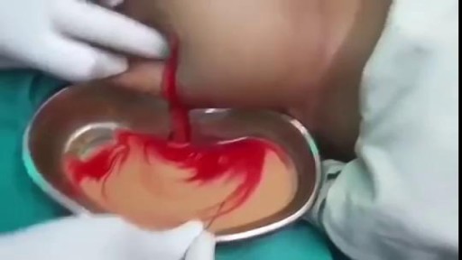
Draining HUGE back abscess
Nasal polyps are soft, painless, noncancerous growths on the lining of your nasal passages or sinuses. They hang down like teardrops or grapes. They result from chronic inflammation due to asthma, recurring infection, allergies, drug sensitivity or certain immune disorders. Small nasal polyps may not cause symptoms. Larger growths or groups of nasal polyps can block your nasal passages or lead to breathing problems, a lost sense of smell and frequent infections. Nasal polyps can affect anyone, but they're more common in adults. Medications can often shrink or eliminate nasal polyps, but surgery is sometimes needed to remove them. Even after successful treatment, nasal polyps often return.
