- Physical Examination
- Surgical Examination
- Ophthalmology
- Clinical Skills
- Orthopedics
- Surgery Videos
- Laparoscopy
- Pediatrics
- Funny Videos
- Cardiothoracic Surgery
- Nursing Videos
- Plastic Surgery
- Otorhinolaryngology
- Histology and Histopathology
- Neurosurgery
- Dermatology
- Pediatric Surgery
- Urology
- Dentistry
- Oncology and Cancers
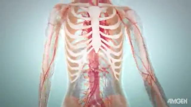
- Anatomy Videos
- Health and Fitness
- Radiology
- Anaesthesia
- Physical Therapy
- Pharmacology
- Interventional Radiology
- Cardiology
- Endocrinology
- Gynecology
- Emergency Medicine
- Psychiatry and Psychology
- Childbirth Videos
- General Medical Videos
- Nephrology
- Physiology
- Diet and Food Health
- Diabetes Mellitus
- Neurology
- Women Health
- Osteoporosis
- Gastroenterology
- Pulmonology
- Hematology
- Rheumatology
- Toxicology
- Nuclear Medicine
- Infectious Diseases
- Vascular Disease
- Reproductive Health
- Burns and Wound Healing
- Other
Top videos

laparoscopic cornuotomy using a temporary tourniquet suture and diluted vasopressin injection in interstitial pregnancy video
Thoracic Epidural Placement Paramedian Approach
Femoral Hernia Repair with Prosthetic PHS repair placed on anterior way
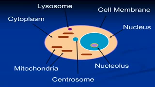
The video will describe aerobic and anaerobic metabolim in mitochondria. Please visit my website for disclaimer.
LCHI - Hernia repair done by medical students with guidance and assistance of Professor Luiz Eduardo C. Miranda. Description of surgery is in portuguese.

Anterior vaginal wall relaxation (cystocele) is one of the most commonly diagnosed forms of pelvic organ prolapse in women. More than 200,000 cystocele repairs are completed yearly, however to date the procedures that are completed do not provide very high cure rates and/or poor anatomic outcomes. Successful treatment of anterior vaginal wall prolapse remains one of the most challenging aspects of pelvic reconstructive surgery we face. We have developed very good procedures that provide excellent support for the posterior wall (ie rectoceles) and the apex of the vagina (ie vaginal vault prolapse) and reproduce normal anatomy. We were one of the first centers in the country to utilize grafts in rectocele repairs and have seen improved cure rates to over 90% with minimal complications. It has been known for many years that abdominal sacralcolpopexy with placement of a mesh graft at the top of the vagina for vaginal vault prolapse is the most successful procedure in the literature. We have made advancements with this procedure as well in being able to offer our patients a laparoscopic minimally invasive approach for sacralcolpopexy, with the same excellent cure rates (>92%) and with hospital stays typically less than 24 hours and reduced complications. However the anterior wall has been one of the most difficult compartments in the vagina to get good anatomic results and high cure rates with traditional repairs and at the same time not cause sexual dysfunction, pain with intercourse, voiding dysfunction (ie incontinence or urgency/frequency syndrome), or a shortened or scarred down vagina. The transobturator approach was developed as a less invasive way to place an anterior wall graft (see below) however this still involved blind needle passes and the graft did not support the apex of the vagina, therefore the search for improvements in these procedures is ongoing.
Recto-vaginal medical examination
Bone Remodeling and Modeling

Patient Greg Grindley communicates with host Bryant Gumbel and his wife for the first time while undergoing deep brain stimulation surgery at University Hospital's Case Medical Center in Cleveland, Ohio.
➡ Subscribe: http://bit.ly/NatGeoSubscribe
About National Geographic:
National Geographic is the world's premium destination for science, exploration, and adventure. Through their world-class scientists, photographers, journalists, and filmmakers, Nat Geo gets you closer to the stories that matter and past the edge of what's possible.
Get More National Geographic:
Official Site: http://bit.ly/NatGeoOfficialSite
Facebook: http://bit.ly/FBNatGeo
Twitter: http://bit.ly/NatGeoTwitter
Instagram: http://bit.ly/NatGeoInsta
Greg's First In-Surgery Conversation | Brain Surgery Live
https://youtu.be/zvqV_2zncNU
National Geographic
https://www.youtube.com/natgeo

http://www.landging.com/digestive-system-animation-colon.html
This digestive system animation demonstrates the procedure of colon cleansing.
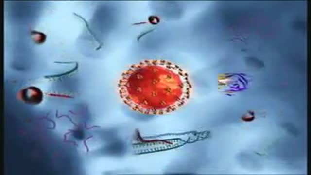
Binding and Fusion: HIV begins its life cycle
when it binds to a CD4 receptor and one of two
co-receptors on the surface of a CD4+
Tlymphocyte. The virus then fuses with the host
cell. After fusion, the virus releases RNA, its
genetic material, into the host cell.
Reverse Transcription: An HIV enzyme
called reverse transcriptase converts the singlestranded HIV RNA to double-stranded HIV DNA.
Integration: The newly formed HIV DNA
enters the host cell's nucleus, where an HIV
enzyme called integrase "hides" the HIV DNA
within the host cell's own DNA. The integrated
HIV DNA is called provirus. The provirus may
remain inactive for several years, producing few or
no new copies of HIV
Transcription: When the host cell receives a
signal to become active, the provirus uses a host
enzyme called RNA polymerase to create copies of
the HIV genomic material, as well as shorter
strands of RNA called messenger RNA (mRNA).
The mRNA is used as a blueprint to make long
chains of HIV proteins.
Assembly: An HIV enzyme called protease cuts
the long chains of HIV proteins into smaller
individual proteins. As the smaller HIV proteins
come together with copies of HIV's RNA genetic
material, a new virus particle is assembled.
Budding: The newly assembled virus pushes out
("buds") from the host cell. During budding, the new
virus steals part of the cell's outer envelope. This
envelope, which acts as a covering, is studded with
protein/sugar combinations called HIV
glycoproteins. These HIV glycoproteins are
necessary for the virus to bind CD4 and coreceptors. The new copies of HIV can now move
on to infect other cells.
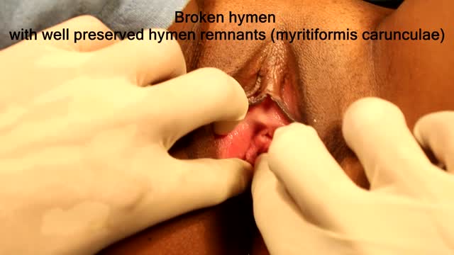
Best and 100% Successful Hymen Repair Surgery in Delhi with Latest Ultrafine Hymen repair Technology. 100% successful , Secure and Private. for more information visit: http://www.olmeccosmeticsurgery.com/best-hymenoplasty-surgery-india-delhi/
External cephalic version, or version, is a procedure used to turn a fetus from a breech position or side-lying (transverse) position into a head-down (vertex) position before labor begins. When successful, version makes it possible for you to try a vaginal birth.
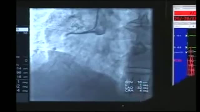
Cardiac catheterization (kath-uh-tur-ih-ZAY-shun) is a procedure used to diagnose and treat cardiovascular conditions. During cardiac catheterization, a long thin tube called a catheter is inserted in an artery or vein in your groin, neck or arm and threaded through your blood vessels to your heart. Using this catheter, doctors can then do diagnostic tests as part of a cardiac catheterization. Some heart disease treatments, such as coronary angioplasty, also are done using cardiac catheterization. Usually, you'll be awake during cardiac catheterization, but given medications to help you relax. Recovery time for a cardiac catheterization is quick, and there's a low risk of complications.
10 Animals Found Living Inside Humans
Respiratory syncytial virus (RSV) is a virus that causes infections of the lungs and respiratory tract. It's so common that most children have been infected with the virus by age 2. Respiratory syncytial (sin-SISH-ul) virus can also infect adults. In adults and older, healthy children, the symptoms of respiratory syncytial virus are mild and typically mimic the common cold. Self-care measures are usually all that's needed to relieve any discomfort. Infection with respiratory syncytial virus can be severe in some cases, especially in premature babies and infants with underlying health conditions. RSV can also become serious in older adults, adults with heart and lung diseases, or anyone with a very weak immune system (immunocompromised).
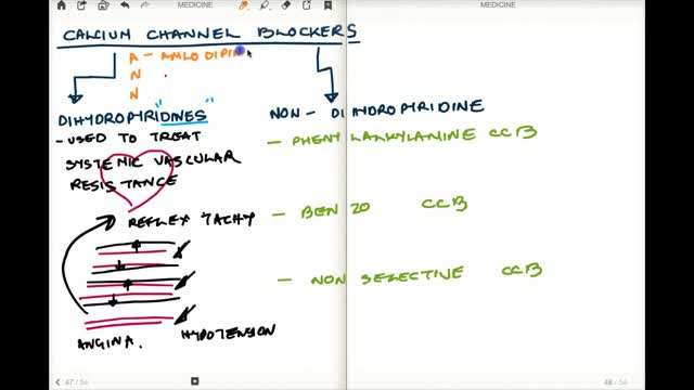
Calcium channel blockers prevent calcium from entering cells of the heart and blood vessel walls, resulting in lower blood pressure. Calcium channel blockers, also called calcium antagonists, relax and widen blood vessels by affecting the muscle cells in the arterial walls. Some calcium channel blockers have the added benefit of slowing your heart rate, which can further reduce blood pressure, relieve chest pain (angina) and control an irregular heartbeat. Examples of calcium channel blockers Some calcium channel blockers are available in short-acting and long-acting forms. Short-acting medications work quickly, but their effects last only a few hours. Long-acting medications are slowly released to provide a longer lasting effect. Several calcium channel blockers are available. Which one is best for you depends on your health and the condition being treated. Examples of calcium channel blockers include: Amlodipine (Norvasc) Diltiazem (Cardizem, Tiazac, others) Felodipine Isradipine Nicardipine Nifedipine (Adalat CC, Afeditab CR, Procardia) Nisoldipine (Sular) Verapamil (Calan, Verelan) In some cases, your doctor might prescribe a calcium channel blocker with other high blood pressure medications or with cholesterol-lowering drugs such as statins.
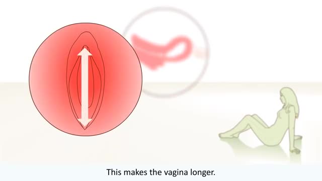
The big bang is the moment when the uterus, vagina, and anus contract simultaneously at 0.8-second intervals. A small orgasm may consist of three to five contractions; a biggie, 10 to 15. Many women report feeling different kinds of orgasms

10 Craziest Plastic Surgeries
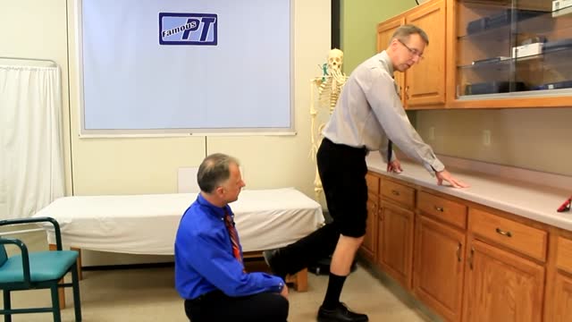
A torn meniscus is one of the most common knee injuries. Any activity that causes you to forcefully twist or rotate your knee, especially when putting the pressure of your full weight on it, can lead to a torn meniscus. Each of your knees has two menisci — C-shaped pieces of cartilage that act like a cushion between your shinbone and your thighbone. A torn meniscus causes pain, swelling and stiffness. You also might have trouble extending your knee fully. Conservative treatment — such as rest, ice and medication — is sometimes enough to relieve the pain of a torn meniscus and give the injury time to heal on its own. In other cases, however, a torn meniscus requires surgical repair.
