- Physical Examination
- Surgical Examination
- Ophthalmology
- Clinical Skills
- Orthopedics
- Surgery Videos
- Laparoscopy
- Pediatrics
- Funny Videos
- Cardiothoracic Surgery
- Nursing Videos
- Plastic Surgery
- Otorhinolaryngology
- Histology and Histopathology
- Neurosurgery
- Dermatology
- Pediatric Surgery
- Urology
- Dentistry
- Oncology and Cancers
- Anatomy Videos
- Health and Fitness
- Radiology
- Anaesthesia
- Physical Therapy
- Pharmacology
- Interventional Radiology
- Cardiology
- Endocrinology
- Gynecology
- Emergency Medicine
- Psychiatry and Psychology
- Childbirth Videos
- General Medical Videos
- Nephrology
- Physiology
- Diet and Food Health
- Diabetes Mellitus
- Neurology
- Women Health
- Osteoporosis
- Gastroenterology
- Pulmonology
- Hematology
- Rheumatology
- Toxicology
- Nuclear Medicine
- Infectious Diseases
- Vascular Disease
- Reproductive Health
- Burns and Wound Healing
- Other
Top videos

Full Tummy Tuck 3D Video - http://drlandsman.com
Look great... feel great
•Smart Liposuction + Liposculpture
•Abdominplasty (Tummy Tuck)
+ Full Mini Modified
•Brazilian Lift with Fat Transfer
•Vaginal Aesthetics & Rejuvenation
•Laser Hair Removal
•Full Body Lift
•Thigh lift
•Brachioplasty (Arm Lift) + Short Scar
Expertise in Body Contouring
Board Certified Plastic Surgeon
Expertise in body contouring combines skin excision techniques and advanced fat contouring technology
Weight control personalized training and smoking cessation results in a healthier lifestyle improved shape and longer lasting results
With over 2 decades of experience Dr Lloyd Landsman provides state of the art cosmetic and plastic surgery
Dr Landsman integrates the finest and safest products with the newest procedures
A customized treatment plan is created for each patient utilizing classic surgical and minimally invasive techniques for optimal results
Call for your complimentary consultation to learn how Dr Landsman can help you look your very best
Visit http://drlandsman.com Call 631 864 4111
Main Office 994 W Jericho Tpke Smithtown NY 11787
Affiliates East Islip • Westbury • Jackson Heights • Manhattan
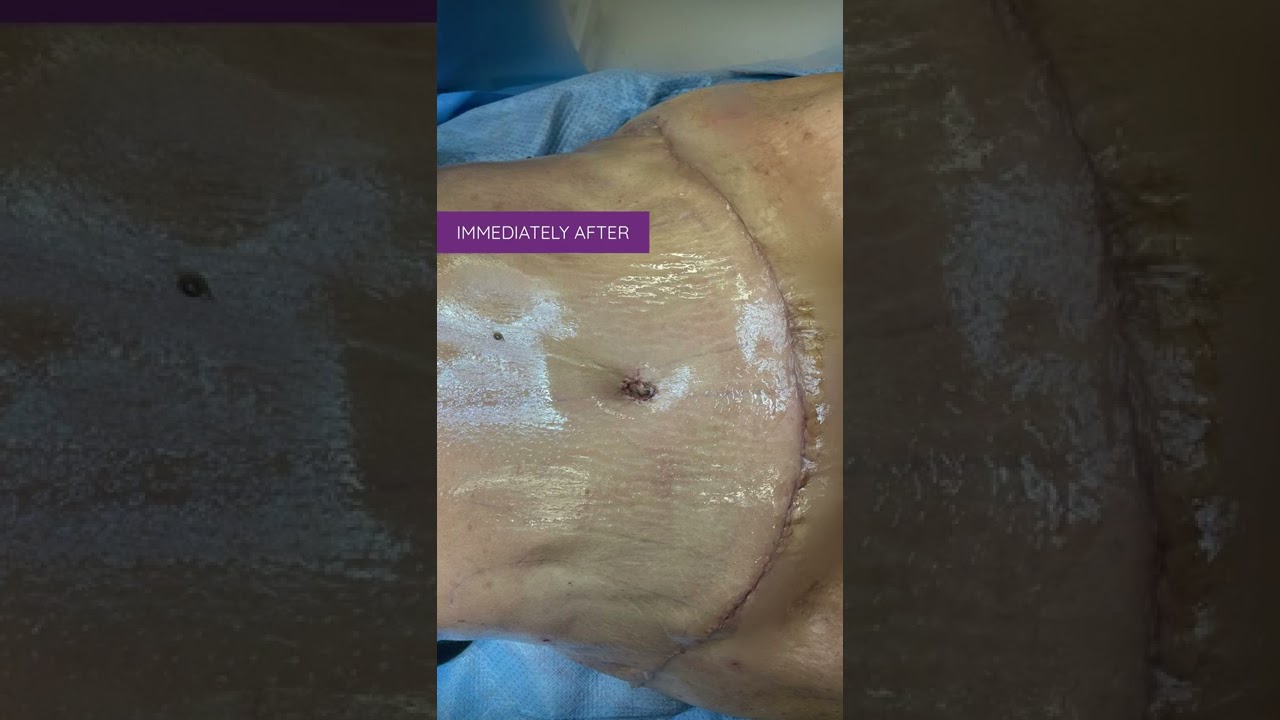
Liposuction in tummy tuck requires special planning and technique. I need to ensure that the blood circulation is well maintained for good healing. Yet proper liposuction is important to have a nice flat and contoured tummy.
#hdliposuction #tummytuck #lipoabdominoplasty #surgicalplanning #skinremovalsurgery #imeediatelyafter #plasticsurgeondubai #cocoonaclinic #drsanjayparashar #dubai
For more information visit www.drsanjayparashar.com
For more content, follow me on my social media
Instagram : https://www.instagram.com/drsanjayparashar/
Facebook : https://www.facebook.com/drsanjayparashar

#abdomenliposuction #laserskintightening #drprashantyadav #cosmeticsurgery #plasticsurgery #dezireclinicindia #weightloss #shorts #360degreeabdomenliposuction #lowerbackliposuction
Weight Loss After 360° Abdomen liposuction result, Abdomen Liposuction, lower back liposuction, 360 degree abdomen liposuction
☎️ For more info:
WhatsApp Your Details to know the Cost
Delhi - 8956880644, 9717470550, Pune - 9222122122, Bangalore- 8971224700, Gurugram - 9272007896, Ahmedabad - 9711162746
Why choose Dezire Clinic For Your Cosmetic and plastic surgery treatment ?
Dezire Clinic is a top searched clinic surgical and nonsurgical cosmetic procedure in India when comes to “Cosmetic, Skin ,Laser and Hair transplantation”.
Like and Share the video if you find it useful. Do not forget to Subscribe to our channel to get more updates.
Subscribe on YouTube https://youtube.com/dezireclin....ic?sub_confirmation=
https://youtube.com/dezireplas....ticsurgerycenter?sub
🎦 https://www.youtube.com/dezireclinic
🎦 https://www.youtube.com/DezirePlasticSurgeryCenter
👍🏻 https://www.facebook.com/drprashantmch/
👍🏻 https://www.facebook.com/dezireclinic
📸 https://www.instagram.com/drprashantdezireclinic/
📸 https://www.instagram.com/dezireclinics/
🐥 https://twitter.com/drprashantmch
👍🏻 https://www.linkedin.com/in/drprashantyadav/
🌐 Website: https://www.dezireclinic.in/
📧 dezireclinicindia@gmail.com
📧 info@dezireclinic.in
Dr. Prashant Yadav (M.S., M.Ch. Plastic Surgery ) & Founder of Dezire Clinic
Disclaimer: The content of this channel is for informational and educational purposes only. This content should not be considered a substitute for advice provided by a certified plastic or cosmetic surgeon. Patients must be properly diagnosed by a healthcare professional on an individual basis in order to achieve the desired results. There is no guarantee of getting the results and outcomes shown in videos, as the results can vary at the end. We will not be held liable for any harm caused by someone misusing our name.
#plasticsurgery #cosmeticsurgery #dezireclinic #drprashantyadav

Traditional Liposuction VS Vaser Liposuction
A side-by-side comparison of traditional liposuction and a #Vaser liposuction. Both of these were performed by our skilled surgeons at Divine Cosmetic Surgery.
#vaserliposuction #liposuction #liposuctionDelhi #liposuctionresults #shorts #vaserliposuctionDelhi
Know more about liposuction
https://www.divinecosmeticsurg....ery.com/liposuction-
Traditional Liposuction vs 360 High Def Vaser Liposuction - https://www.youtube.com/watch?v=r_bBI2p9fVI&t=14s
-------------------------------------------------------------------------------
Why Vaser Is Best For Thigh Liposuction - https://youtu.be/dlzpdDEZcS4
-------------------------------------------------------------------------------
Abdomen Vaser Liposuction - Live - https://www.youtube.com/watch?v=_Cvl2Txn8LQ
-------------------------------------------------------------------------------
Back Vaser Liposuction In Female - https://youtu.be/OC60UdgtIWU
-------------------------------------------------------------------------------
For more details about Liposuction Visit - https://www.divinecosmeticsurgery.com/
-------------------------------------------------------------------------------
Dr. Amit Gupta
MBBS, M.S., DNB (Plastic & Cosmetic Surgery)
Divine Cosmetic Surgery | +91 9811994417
info@divinecosmeticsurgery.com | 01141828787
Delhi | Mumbai | Gurgaon
𝗦𝗼𝗰𝗶𝗮𝗹 𝗠𝗲𝗱𝗶𝗮 𝗮𝗻𝗱 𝗬𝗼𝘂𝘁𝘂𝗯𝗲 𝘃𝗶𝗱𝗲𝗼 𝗰𝗵𝗮𝗻𝗻𝗲𝗹 : -
🎦 https://www.youtube.com/c/DrAm....itGuptaBestPlasticCo
👍🏻 https://www.facebook.com/dramitguptaplasticsurgeon
📷 https://www.instagram.com/divineaesthetics_delhi/
🐥 https://twitter.com/dramitguptajee
🖇️ https://www.linkedin.com/compa....ny/divinecosmeticsur
📌 https://pinterest.com/divinesurgery
#Liposuction #vaserliposuction #liposuctioncostinindia #liposuctiondelhi #liposuction #liposuctioncost #liposuctioncostfactors #liposuctioncostindelhi #DrAmitGuptaPlasticSurgeon #DivineCosmeticSurgery #dramitgupta
Disclaimer: The information on our videos & social media is provided for informational purposes only and is not meant for the advice provided by your surgeon.
We are not responsible for any harm if anyone misguides you from our name. Our all-social media official handles are linked up on our website. All images & content used on our videos & social media are for illustrative concerns only, original results and processes may vary.


In this episode of Crash Course Anatomy & Physiology, Hank gives you a brief history of histology and introduces you to the different types and functions of your body's tissues.
Pssst... we made flashcards to help you review the content in this episode! Find them on the free Crash Course App!
Download it here for Apple Devices: https://apple.co/3d4eyZo
Download it here for Android Devices: https://bit.ly/2SrDulJ
Chapters:
Introduction 00:00
Nervous, Muscle, Epithelial & Connective Tissues 1:23
History of Histology 2:07
Nervous Tissue Forms the Nervous System 5:17
Muscle Tissue Facilitates All Your Movements 7:00
Identifying Samples 9:03
Review 9:48
Credits 10:22
Crash Course is on Patreon! You can support us directly by signing up at http://www.patreon.com/crashcourse
Want to find Crash Course elsewhere on the internet?
Facebook - http://www.facebook.com/YouTubeCrashCourse
Twitter - http://www.twitter.com/TheCrashCourse
Instagram - https://www.instagram.com/thecrashcourse/
CC Kids: http://www.youtube.com/crashcoursekids

Give away to who ever that will correctly mention the sample specimen used to make this video? (Be specific)
DROP ON THE COMMENT BOX

► Sign up here and try our FREE content: http://lectur.io/freecontentyt
► If you’re an medical educator or faculty member, visit: http://lectur.io/medytb2u
This video “Connective Tissue” is part of the Lecturio course “Histology” ► WATCH the complete course on http://lectur.io/connectivetissue
► LEARN ABOUT:
- Cells and Basic Tissue
- Nerve Tissues
- Muscle Tissues
- Epithelial Tissues
- Connective Tissues
► THE PROF: Your lecturer is Professor Geoff Meyer. He is currently teaching at the School of Anatomy, Physiology and Human Biology at the University of Western Australia (UWA). As a leading anatomy and histology expert he is also coordinating the Federative International Program for Anatomical Terminologies (FIPAT) of the International Federation of Associations of Anatomists (IFAA). Besides medical research on the ovarian function, steroidogenesis, corpus luteum, angiogenesis, and microcirculation, Geoff Meyer’s research activities also focus on developing innovative, computer-aided learning and teaching tools. For his inventiveness, Geoff Meyer has received a number of awards, including the Australian University Teaching Award.
► LECTURIO is your single-point resource for medical school:
Study for your classes, USMLE Step 1, USMLE Step 2, MCAT or MBBS with video lectures by world-class professors, recall & USMLE-style questions and textbook articles. Create your free account now: http://lectur.io/connectivetissue
► INSTALL our free Lecturio app
iTunes Store: https://app.adjust.com/z21zrf
Play Store: https://app.adjust.com/b01fak
► READ TEXTBOOK ARTICLES related to this video:
Types of Tissue: Connective Tissue, Muscle Tissue, Epithelial Tissue, and Nervous Tissue
http://lectur.io/connectivetissuearticle
► SUBSCRIBE to our YouTube channel: http://lectur.io/subscribe
► WATCH MORE ON YOUTUBE: http://lectur.io/playlists
► LET’S CONNECT:
• Facebook: https://www.facebook.com/lectu....rio.medical.educatio
• Instagram: https://www.instagram.com/lecturio_medical_videos
• Pinterest: https://www.pinterest.de/lecturiomedical
• LinkedIn: https://www.linkedin.com/company/lecturio-medical/

| MBBS मतलब JOHARI MBBS I
Download Johari MBBS APP ( For Online LIVE Classes, Notes, Books PDFs, Test Series )
Johari MBBS ( iPhone IOS Users ) LINK { FOR Online LIVE Classes }
https://apps.apple.com/in/app/....johari-mbbs/id647466
JOHARI MBBS APP ( Android ) LINK { FOR Online LIVE Classes }
https://play.google.com/store/....apps/details?id=co.d
CRASH COURSE LINK ( Anatomy in 30Days with Biochemistry In 7Days Series )
https://zczob.on-app.in/app/oc/389813/zczob
IMPORTANT LINKS :-
1) ORDER Anatomy Next Edition Module , Biochemistry in 7Days & Physiology MODULE
https://joharimbbs.com/
2) Join INSTAGRAM ( For Notes, Revision REELs, Updates )
https://www.instagram.com/johari_mbbs_lectures/
3) INSTA Broadcast Channel ( FOR Daily VLOGS, Life Update )
https://ig.me/j/Abal9xRcXcUyrYpT/
4) Telegram ( For FREE BOOKS PDFs )
https://t.me/joharimbbsofficial
5) Whatsapp Channel ( Daily Update )
https://whatsapp.com/channel/0....029VaEeWKWHAdNb0xOzl
6) Follow On Twitter ( For Latest Updates )
https://twitter.com/JohariMbbs
CRASH COURSE LINK ( Anatomy in 30Days with Biochemistry In 7Days Series )
https://zczob.on-app.in/app/oc/389813/zczob
#mbbs #joharimbbs #anaatomy #biochemistry #physiology #medico #doctors
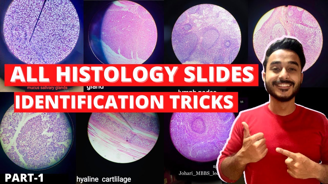
histology slide identification tricks
histology slides identification tricks
histology slide
histology slides
histology slides identification
histology slide preparation
histology slides identification epithelium
histology slides identification connective tissue
histology slides identification tricks
histology slides of epithelium
histology slide identification
#anatomy#clinicalanatomy #MBBS #neroanatomy #bdc #medsudent #medicalcollege
For notes IG - johari_mbbs_lectures
link- https://www.instagram.com/p/CNOwFgEJmJL/
Join Telegram Channel ( JOhari MBBS Public 20x )
https://t.me/JohariMBBS
For Tag :-
histology , histology slides , histology slide identification , histology slides identification , histology slides preparation , tongue histology slide , histology slides asked in exam , histology slide identification trick , histology slides of connective tissue , mbbs histology , histology slides identification epithelium , trachea histology slide , slides , duodenum histology slide , histology slides tricks , histology slides review , examine histology slides

An animated description of the composition of bones.
Visit www.orthofilms.com for more videos and info.
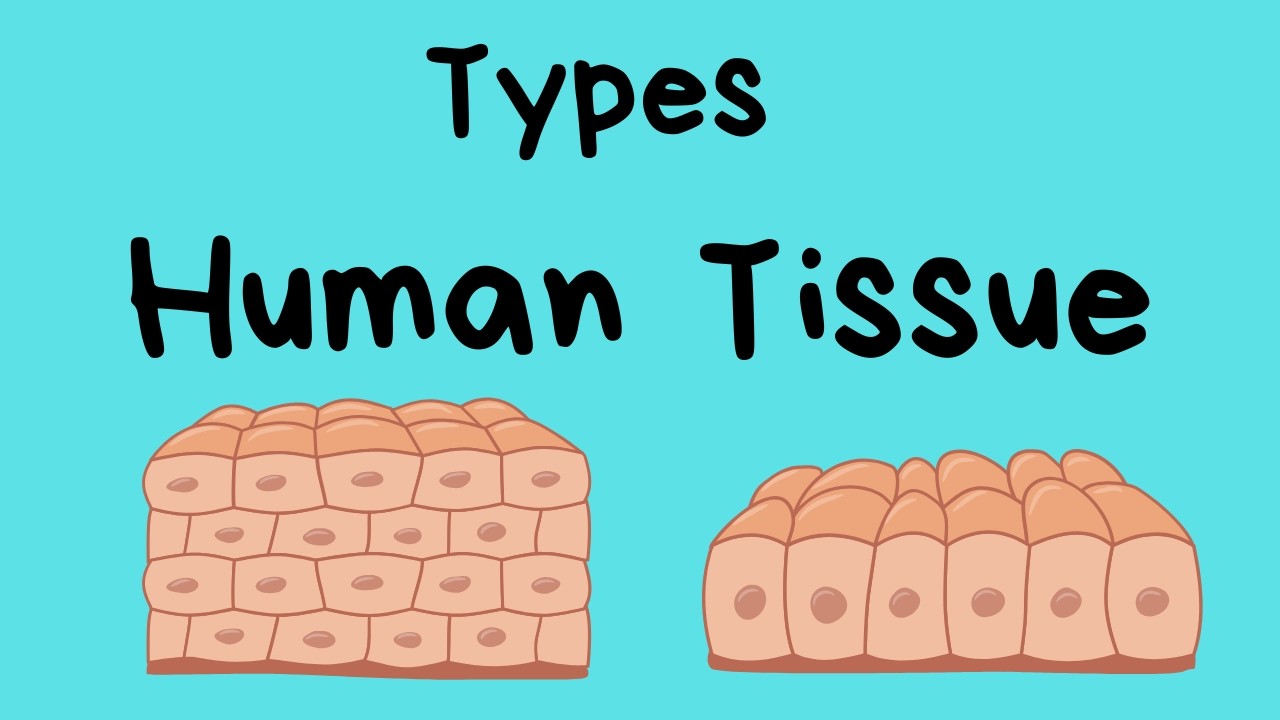
Types of Human Body Tissue
In this video, I review four types of tissue.
Connective tissue, epithelial tissue, muscle tissue, and nerve tissue.
Tissues are made up of cells working together.
*
*
For more Life Science videos and summaries see,
http://www.moomoomath.com/Midd....le-School-Science-an

Website : https://www.udemy.com/course/h....istology/?referralCo
Human Histology is one of the basic subject in a Medical Student career. By learning Histology in a proper way, this will help you to get a Visual memory of the Human body. Using this Visual memory, you can Learn any other subjects with little effort.
This Course is very well organized with lot of Histology images, Line diagrams, simple presentations and clear Explanations. This course has 33 videos, 19 chapters, 6 hours long covering all topics. Every topic is made Simple and Complete. Dr Ram has a great teaching style and has a good experience in teaching medical subjects to students.
After finishing this course, you will be better in your basics, with ability to visualize the human body and this will create an intense thirst to learn more. We give 100% guarantee that you will have a complete and in-depth understanding in short time, You will start to enjoy Learning Medicine because of the visualization of human body you get from this course and you will be ready to face any Medical exams in world.
Course features:
- Complete Histology lectures covering all chapters
- 19 chapters | 33 Videos | 6 Hours
- Clear Histology images
- Line diagrams for easy understanding
- Lot of memory tips
- High quality audio and Videos
- Can be viewed in Pc, or Phones or TV
Course content: ( 19 Chapters, 33 videos )
I The Cell - 3 Lessons
1. Nucleus
2. Cytoplasm
3. Cell Junctions
II Tissues - 11 Lessons
4. Epithelial tissues
5. Connective tissues
6. Muscular tissues
7. Nervous tissues
8. Bones
9. Cartilage
10. Lymphoid tissues
III Organ systems - 19 Lessons
11. Cardiovascular system
12. Respiratory system
13. Gastrointestinal system
14. Liver and Exocrine pancreas
15. Endocrine system
16. Urinary system
17. male reproductive system
18. Female reproductive system
19. The skin
Instructor : Dr Ram , Med Madness
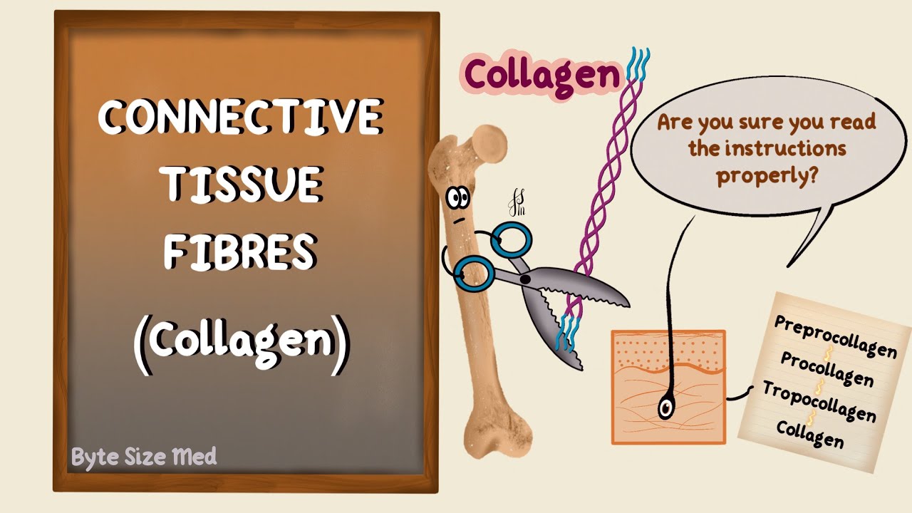
✨This video is on the protein fibres of connective tissue, the types, structure and synthesis of collagen and elastin. I hope it helps! ☀️
🌟What's in this video?
0:00 - Intro
0:07 - Connective Tissue Recap
0:39 - Connective Tissue Fibres
1:22 - Collagen
1:46 - Types of Collagen
3:40 - Structure of Collagen
4:40 - Collagen Synthesis
8:50 - Elastin
✨ Other videos you may need:
🔅 Connective Tissue : https://youtu.be/xw_ALdt5n-A
🔅 Cartilage : https://youtu.be/4inWF4H6pKE
🔅Epithelial Tissue: https://youtu.be/Gw5fC0zXaeU
🔅Structure of Blood Vessels: https://youtu.be/BAo2UqqyL3g
🔅Histology: https://www.youtube.com/playli....st?list=PL1rG930trF2
💫 For more videos like this, subscribe to my channel!
Byte Size Med: https://youtube.com/channel/UC....ZghvlgylH3r_CWfA18eF
📚Factual References & for Further Reading:
- DiFiore's Atlas of Histology
- Junqueira's Basic Histology
- Harper's Biochemistry
- Gartner's Concise Histology
- Openstax Anatomy and Physiology
https://openstax.org/details/b....ooks/anatomy-and-phy
- Openstax Biology
https://openstax.org/details/books/biology-2e
(The last two are links to open-source references. They are NOT affiliate links)
🌤 Note:
These are just a collection of my notes. So use them the way you would use borrowed notes from a friend. 📝
The images in this video are hand-drawn for illustration and explanation only.✍️ Hence, they may not be anatomically accurate. I am just one person making these videos. If there are any errors, that is unintentional. I try super hard to avoid them. Please let me know if you find any, so it gets clarified for other viewers. Science constantly evolves and changes. New discoveries are made everyday. So some of the information in these videos may become outdated. If you notice that, please let me know so I can update them.
⚡️Disclaimer:
These videos are NOT a substitute for a medical textbook. Textbooks are written by experts (which I do not claim to be), edited, proofread and referenced. Please use them.
The information has been sourced from multiple references as mentioned above. I draw all the pictures myself. But if I have inadvertently infringed on any copyright, that is completely unintentional. I only make these videos to impart education. If I have accidentally violated copyright in any way, do let me know so I can make the necessary changes or give credit to anyone who is owed the same.
These videos are NOT intended for patient education. They are NOT a substitute for diagnosis and treatment by a licensed medical professional. Always seek the advice of a qualified health care provider for any questions you may have regarding any medical condition, so that they can address your individual needs.
🔅They are ONLY meant to help students of medicine and health sciences with studying, and should be used for just that purpose and absolutely nothing else.
Byte Size Med. All Rights Reserved.

http://www.highimpact.com - This brain surgery animation was used to demonstrate a young girl's craniotomy, cranioplasty, and reconstructive skull surgery after her vehicle was struck by a tractor-trailer. The procedures included the evacuation of a large epidural hematoma, the draining of the epidural space, and the reassembly of bone fragments to repair the skull.
More Brain Surgery Animations: https://tinyurl.com/y6m4lkdf
WHAT HAPPENED
A teenage girl was riding home with her parents and boyfriend from a Wednesday night church service when a tractor-trailer struck the back driver’s side of their car as they were traveling through an intersection. The impact sent the car spinning into oncoming traffic where it struck another vehicle. When paramedics arrived, the 17-year-old was unresponsive with bleeding from her left ear and a laceration from behind her left ear.
She was rushed to the hospital where she underwent a series of CT scans that showed a severely comminuted open skull fracture with an underlying 1.1 cm subdural hematoma. She was taken to the operating room where an emergency craniotomy was performed to evacuate the hematoma and reassemble the skull fragments. The patient gradually began to wake up and was discharged six days later, after she showed she could maneuver up and down the hallway.
The biggest challenge in a traumatic brain injury case like this - where most of the damages are deeply underlying and undetectable on the surface - is that the only visual evidence is in the form of 2D black-and-white radiographic films. This can look ambiguous to the typical juror because it’s often difficult to discern where these snapshots are located inside the person’s skull. Tony Seaton, Esq., and Robert Bates, Esq., needed to reinforce this 2D radiographic evidence with maximum 3D context.
We equipped them with a custom Diagnostic Slice Chooser: an interactive presentation that presents radiographic slides within a three-dimensional model of the patient’s head. We also designed the model accurately to the patient’s likeness and colorized the films to highlight key areas of damage. The attorneys could show the complete depth and magnitude of his client’s injuries at every level both before and after the surgery. After establishing the full extent of damages, we also created an animation to walk viewers through the surgical experience the patient would undergo as a result of her injuries.
The visual presentation helped jurors understand the destructive impact this collision had on this young teenager’s life, and Mr. Seaton and Mr. Bates, Esq., were able to acquire a $4.5M settlement for his client.
Read the Full Case Study: https://tinyurl.com/yy4v2dyh

This 3D animation of brain surgery, shows how a ventriculostomy is performed, which is a neurosurgical procedure of creating a hole within a cerebral ventricle for drainage. It is most commonly performed on those with hydrocephalus, an abnormal buildup of fluid in the ventricles (cavities) deep within the brain. It's done by surgically penetrating the skull, dura mater, and brain such that the ventricular system ventricle of the brain is accessed.
When catheter drainage is temporary, it is commonly referred to as an external ventricular drain (EVD). When catheter drainage is permanent, it is usually referred to as a shunt.
There are many catheter-based ventricular shunts that are named for where they terminate, for example, a ventriculi-peritoneal shunt terminates in the peritoneal cavity, a ventriculoarterial shunt terminates within the atrium of the heart, etc. The most common entry point on the skull is called Kocher's point. An EVD ventriculostomy is done primarily to monitor the intracranial pressure as well as to drain cerebrospinal fluid (CSF), primarily, or blood to relieve pressure from the central nervous system (CNS).
For more information about custom medical animation, please visit https://www.amerra.com/.
Watch additional medical animations:
Craniectomy brain surgery - 3D animation: https://youtu.be/1RkseDeYS9g
Accessing an implantable port training - 3D animation: https://youtu.be/xSTpxjyv4O4
Open Suctioning with a Tracheostomy Tube - 3D animation: https://youtu.be/wamB7jpWCiQ
Suctioning the endotracheal tube - medical animation: https://youtu.be/pN6-EYoeh3g
Functional endoscopic sinus surgery (FESS) - 3D animation: https://youtu.be/qKTRyowwaLA
How to insert a nasogastric tube for NG intubation - 3d animation: https://youtu.be/Abf3Gd6AaZQ
Oral airway insertion - oropharyngeal airway technique - 3D animation: https://youtu.be/caxUdNwjt34
Nasotracheal suctioning (NTS) - 3D animation: https://youtu.be/979jWMsF62c
Learn about hemorrhoids with #3d #animation: https://youtu.be/R6NqlMpsiiY
LASIK eye surgery - 3D animation: https://youtu.be/Bb8bnjnEM00
CPR cardiopulmonary resuscitation - 3D animation: https://youtu.be/G87knTZnhks
What are warts (HPV)? - 3D animation: https://youtu.be/guJ1J7rRs1w
How Macular Degeneration Affects Your Vision - 3D animation: https://youtu.be/ozZQIZ_52YY
NeoGraft hair transplant procedure – animation: https://youtu.be/C-eTdH2UPXI

http://www.nucleushealth.com/ - This 3D medical animation depicts two operations, called craniotomy and craniectomy, in which the skull is opened to access the brain. The normal anatomy of the skull and tissues surrounding the brain are shown, including arteries and veins. The animation lists the common reasons for these procedures, and briefly introduces intracranial pressure.
Video ID: ANH13109
Transcript:
Your doctor may recommend a craniotomy or a craniectomy procedure to treat a number of different brain diseases, injuries, or conditions.
Your skull is made of bone and serves as a hard, protective covering for your brain. Just inside your skull, three layers of tissue, called meninges, surround your brain. The thick, outermost layer is the dura mater. The middle tissue layer is the arachnoid mater and the innermost layer is the pia mater. Between the arachnoid mater and the pia mater is the subarachnoid space, which contains blood vessels and a clear fluid called cerebrospinal fluid. Blood vessels, called bridging veins, connect the surface of your brain with the dura mater. Other blood vessels, called cerebral arteries, bring blood to your brain.
Inside your skull, normal brain function requires a delicate balance of pressure between the blood in your blood vessels, the cerebrospinal fluid that surrounds your brain, and your brain tissue. This is called normal intracranial pressure. Increased intracranial pressure may result from: brain tumors, head injuries, problems with your blood vessels, or infections in your brain or spinal cord. These conditions put pressure on your brain and may cause it to swell or change shape inside your skull, which can lead to serious brain injury.
Your doctor may recommend a craniotomy to remove: abnormal brain tissue, such as a brain tumor, a sample of tissue by biopsy, a blood clot, called a hematoma, excess cerebrospinal fluid, or pus from an infection, called an abscess.
A craniotomy may also be done to: relieve brain swelling,
stop bleeding, called a hemorrhage, repair abnormal blood vessels, repair skull fractures, or repair damaged meninges.
Finally, a craniotomy may also be done to: treat brain conditions, such as epilepsy, deliver medication to your brain, or implant a medical device, such as a deep brain stimulator.
The most common reason for a craniotomy is to remove a brain tumor.
#Craniotomy #Craniectomy #BrainSurgery
