
- Physical Examination
- Surgical Examination
- Ophthalmology
- Clinical Skills
- Orthopedics
- Surgery Videos
- Laparoscopy
- Pediatrics
- Funny Videos
- Cardiothoracic Surgery
- Nursing Videos
- Plastic Surgery
- Otorhinolaryngology
- Histology and Histopathology
- Neurosurgery
- Dermatology
- Pediatric Surgery
- Urology
- Dentistry
- Oncology and Cancers
- Anatomy Videos
- Health and Fitness
- Radiology
- Anaesthesia
- Physical Therapy
- Pharmacology
- Interventional Radiology
- Cardiology
- Endocrinology
- Gynecology
- Emergency Medicine
- Psychiatry and Psychology
- Childbirth Videos
- General Medical Videos
- Nephrology
- Physiology
- Diet and Food Health
- Diabetes Mellitus
- Neurology
- Women Health
- Osteoporosis
- Gastroenterology
- Pulmonology
- Hematology
- Rheumatology
- Toxicology
- Nuclear Medicine
- Infectious Diseases
- Vascular Disease
- Reproductive Health
- Burns and Wound Healing
- Other
Top videos


Watch that video of an Ingrown Hair Causes Huge Tumor in a Man's Stomach
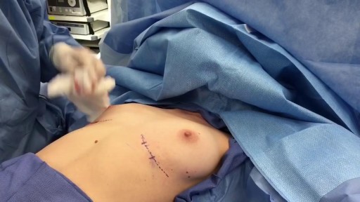
Breast Augmentation Short Scar Technique Slicone Implants
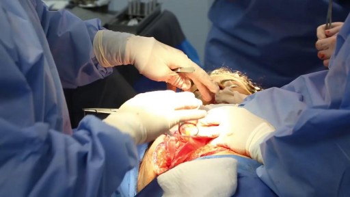
Combination Plastic Surgery Procedure (Face and Body) with Premium Care
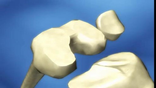
Knee replacement, also known as knee arthroplasty, is a surgical procedure to replace the weight-bearing surfaces of the knee joint to relieve pain and disability.
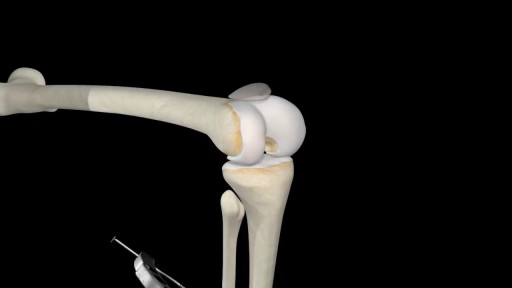
Anterior cruciate ligament reconstruction is a surgical tissue graft replacement of the anterior cruciate ligament, located in the knee, to restore its function after an injury.
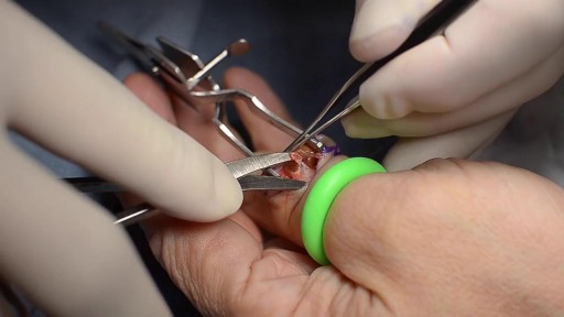
Giant cell tumors of the tendon sheath are common lesions and are the second most frequent tumors in the hand, after synovial cysts. They are diagnosed by means of clinical examination and complementary examinations (simple radiography and magnetic resonance). Erosion and invasion of the phalangeal bone affected may be seen on radiological examination. Magnetic resonance may show a “fluorescent or radiant effect” may be observed, caused by the high quantity of hemosiderin inside the tumor. Surgical treatment is the commonest practice, and complete excision is important for avoiding recurrence of the tumor, especially when bone invasion is observed on imaging examinations, which is generally related to greater tumor recurrence. In this paper, a case of a giant cell tumor of the tendon sheath in the middle phalanx of the third finger of a 45-year-old female patient is presented. This was successfully treated by means of surgery using a double access approach (dorsal and volar)
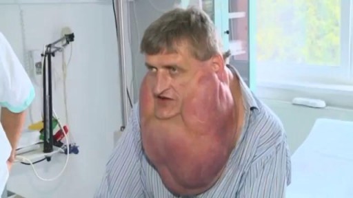
Massive Tumor Removed from Man's Face

Forzest is FDA approved medicine, it is used bt men to improve erectile dysfunction dusring intercourse session with partner. for more information related side effects, dosage, etc kindly visit to http://www.medstorerx.com/forzest.aspx
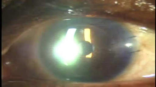
Stephen Slade shows a 1 week post op patient after DSAEK. DSAEK is an excellent option for many patients with corneal disease. In DSAEK, only the thin, inner layer is replaced, so the healing is typically much faster than a full thickness cornea graft.

UCLA Hand Transplant Procedure
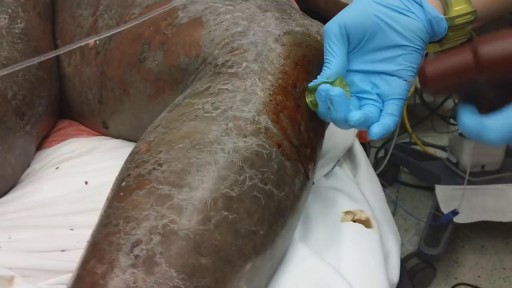
For patients in extremis from respiratory failure or shock, securing vascular access is crucial, along with establishing an airway and ensuring adequacy of breathing and ventilation. Peripheral intravenous catheter insertion is often difficult, if not impossible, in infants and young children with circulatory collapse. Intraosseous (IO) needle placement, shown in the images below, provides a route for administering fluid, blood, and medication. An IO line is as efficient as an intravenous route and can be inserted quickly, even in the most poorly perfused patients.
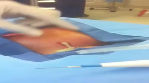
Huge pus in the neck after a bad cold and sore throat!
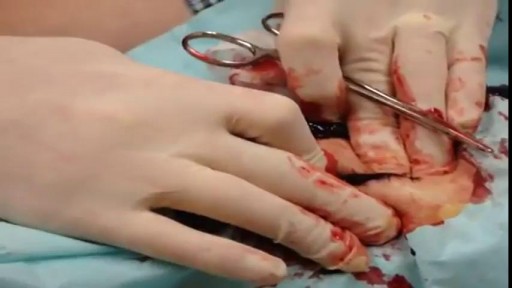
Draining a Blood Clot from the left thigh after knife stab wound sutured by some ER!

» A kidney stone, also known as a renal calculus (from the Latin rēnēs, “kidneys,” and calculus, “pebble”), is a solid concretion or crystal aggregation formed in the kidneys from dietary minerals in the urine. » Urolithiasis (UL) is one of the most common diseases, with approximately 7.6% incidence in Western India. Although most patients have only one stone episode, 25% of patients experience recurrent stone formation. UL therefore has a significant impact on quality of life and socioeconomic factors.
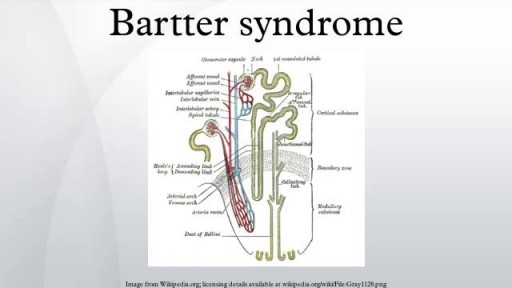
Bartter syndrome is a rare inherited defect in the thick ascending limb of the loop of Henle. It is characterized by low potassium levels (hypokalemia), increased blood pH (alkalosis), and normal to low blood pressure. There are two types of Bartter syndrome: neonatal and classic

Gitelman and Bartter Presentation and Magnesium Supplementation
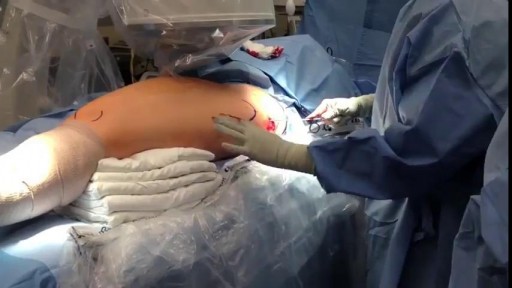
This is an example of a surgery to fix a femur (thigh bone) fracture utilizing an intramedullary nail. This is a minimally invasive way of fixing this surgical problem and allows for immediate range of motion and full weight-bearing.
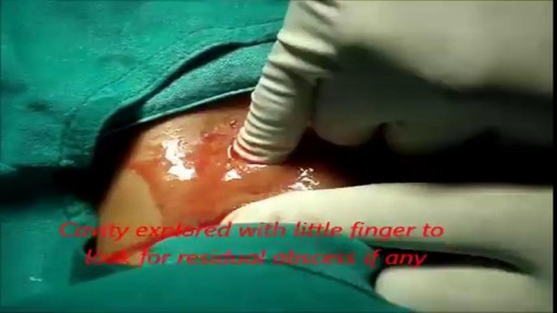
She is a twenty years young female presented with large cystic swelling in anterior aspect of neck. The swelling was of size 6cmx 6cm x5 cm ,tense tender, cystic just above sternal nutch.This was diagnosed as large neck abscess ./nRepeated aspiration done but the swelling reappeared. So Incision & Drainage planned under local anaesthesia./nPatient in supine position. Surgery part painted and draped. Local anaesthesia 2% xylocaine with adrenaline used for field block.After giving local anaesthesia, I used a no 11 blade for stab incision at the most prominent part of the swelling, where skin was thin and fluctuation present./nPus drained form that opening. Little dilatation of opening to be done with artery forceps or sinus forceps. Complete pus drainage to be ensured.Little finger can be introduced inside the pus cavity to ensure proper drainage of pus. The cavity I use to clean with a gauge piece. If necessary curette biopsy can be taken from the wall of the cavity.These wounds usually need daily proper dressing for faster healing.
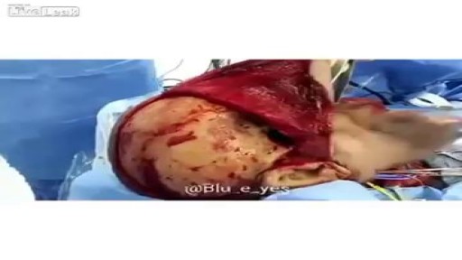
After trauma, surgery or illness, the face can be affected both in appearance and in function. Our physicians have the experience and expertise to reconstruct facial structures for a return to full functionality and comfort with one’s outside appearance. Our facial plastic surgeons are board certified in both otolaryngology and facial plastic surgery. This dual training makes their expertise absolutely critical for rebuilding major structures in the face and reshaping them for a return to one’s original appearance.
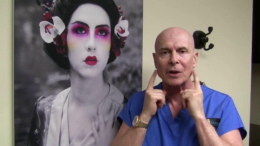
Mandibular Angle Augmentation with Injectable Filler
