- Physical Examination
- Surgical Examination
- Ophthalmology
- Clinical Skills
- Orthopedics
- Surgery Videos
- Laparoscopy
- Pediatrics
- Funny Videos
- Cardiothoracic Surgery
- Nursing Videos
- Plastic Surgery
- Otorhinolaryngology
- Histology and Histopathology
- Neurosurgery
- Dermatology
- Pediatric Surgery
- Urology
- Dentistry
- Oncology and Cancers
- Anatomy Videos
- Health and Fitness
- Radiology
- Anaesthesia
- Physical Therapy
- Pharmacology
- Interventional Radiology
- Cardiology
- Endocrinology
- Gynecology
- Emergency Medicine
- Psychiatry and Psychology
- Childbirth Videos
- General Medical Videos
- Nephrology
- Physiology
- Diet and Food Health
- Diabetes Mellitus
- Neurology
- Women Health
- Osteoporosis
- Gastroenterology
- Pulmonology
- Hematology
- Rheumatology
- Toxicology
- Nuclear Medicine
- Infectious Diseases
- Vascular Disease
- Reproductive Health
- Burns and Wound Healing
- Other
Top videos
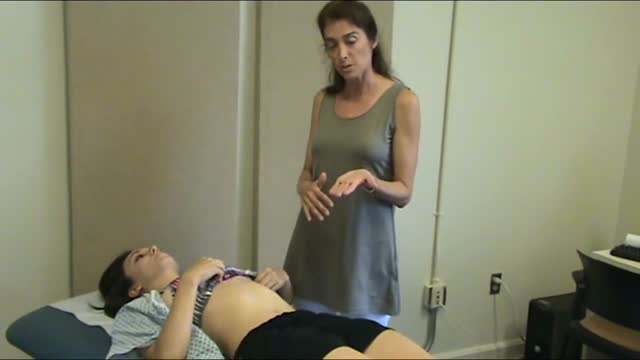

The best way to prepare yourself for your small group exercises is to first watch the cardiac exam video from beginning to end, then proceed through all the explanations of cardiac anatomy and physiology that follow. To go through the material in the recommended sequence, just click the "Next" button in the upper right corner of the screen. But you may also jump to any section using the menu to the left.

Ganglion Cyst Drained with BIG Needle
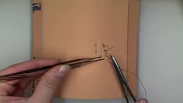
Demonstration of vertical mattress suturing technique for laceration repair or wound closure in the operating room.
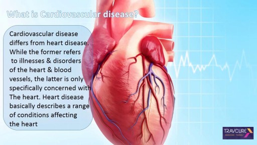
Cardiovascular surgery basically treats a number of diseases and medical disorders that affect your heart and the network of arteries and veins connecting it to every part of the body. https://goo.gl/iphEi9

Hip Examination
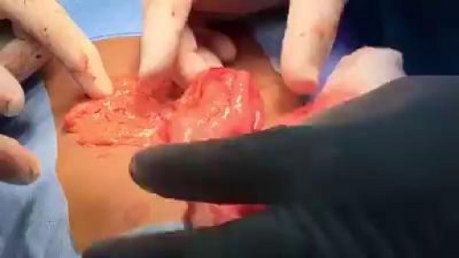
Hairs in Stomach.

Duke Sports Medicine Specialists Jocelyn Wittstein, MD, Janna Fonseca, ATC, and Michael Messer ,PT, present on Soccer Injury Prevention including Concussion Management and the 11+ program that significantly reduces ACL tear rates in soccer.

Linen Changes (with Patient in Bed)- Nursing Skills
FREE Nursing School Cheat Sheets at: http://www.NURSING.com
Get the full lesson on Patient Linen Changes here:
https://nursing.com/lesson/ski....lls-01-02-linen-chan
Get the full lesson on Bed Baths here:
https://nursing.com/lesson/skills-01-01-bed-bath/
Check out our new Nurse Care Plan Lessons here:
https://bit.ly/3BPRfPL
Get Access to Thousands of Lessons here:
https://nursing.com/courses/
Welcome to the NURSING Family, we call it the most supportive nursing cohort on the planet.
At NURSING.com, we want to help you remove the stress and overwhelm of nursing school so that you can focus on becoming an amazing nurse.
Check out our freebies and learn more at: (http://www.nursing.com)
Linen Changes (with Patient in Bed)- Nursing Skills
In this video, we’re going to show you how to change the linens with a patient in the bed. This might be after a bed bath or during incontinence care. So check out the bed bath video to see what got us up to this point. We love you guys! Go out and be your best selves today! And, as always, happy nursing!
Bookmarks:
0.05 Linen change introduction
0.16 Linen change supplies
0.30 Adjusting the patient/ sheet removal
1.00 Secure new fitted sheet
1.12 Pro tip
1.40 Roll patient back over
1.50 Repeat linen removal
2.02 Linen disposal
2.20 Wrinkle check
2.31 Reposition the patient for comfort
2.40 Covering the patient/ tuck-in
2.48 Pillowcase change (trick)
3.30 Making the patient comfortable
3.40 Linen change outro
Visit us at https://nursing.com/medical-disclaimer/ for disclaimer information.
NCLEX®, NCLEX-RN® are registered trademarks of the National Council of State Boards of Nursing, INC. and hold no affiliation with NURSING.com.

This video contains five segments with best practices on how to prevent infection in patients with catheters, fistulas or grafts. It also includes segments on hand hygiene and glove use and dialysis station disinfection. The video is intended to be used by outpatient hemodialysis facilities as an educational tool to help remind their frontline staff, including technicians and nurses, about infection prevention measures. It can be used as an orientation video for new staff and as an annual in-service training tool to remind staff of proper protocols.
See the Spanish captioned version at: http://youtu.be/L5ypnOvOFMQ
Comments on this video are allowed in accordance with our comment policy: http://www.cdc.gov/SocialMedia..../Tools/CommentPolicy
This video can also be viewed at http://streaming.cdc.gov/vod.p....hp?id=dc66d96228817d
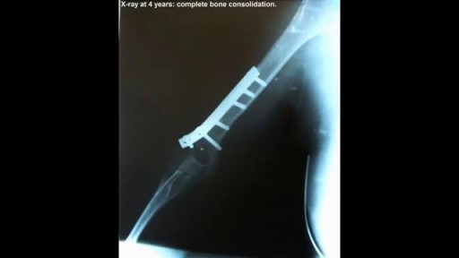
A case of replantation of a completely amputated arm
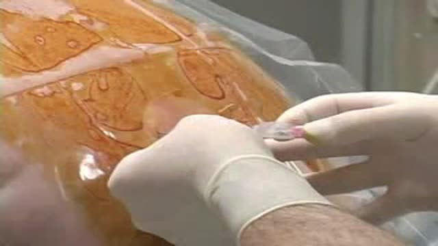
Paramedian Thoracic Epidural Anaesthesia
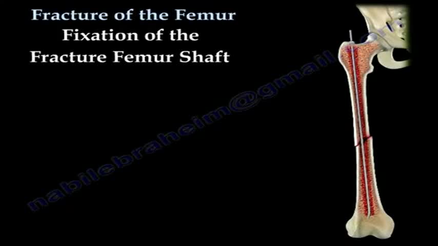
Open reduction and internal fixation (ORIF) is surgery used to stabilize and heal a broken bone. You might need this procedure to treat your broken thighbone (femur). The femur is the large bone in the upper part of your leg. Different kinds of trauma can damage this bone, causing it to fracture into 2 or more pieces. This might happen to the part of the femur near your knee, near the middle of the femur, or in the part of the femur that forms part of your hip joint. In certain types of femur fractures, your femur has broken, but its pieces still line up correctly. In other types of fractures (displaced fractures), the trauma moves the bone fragments out of alignment. If you fracture your femur, you usually need ORIF to bring your bones back into place and help them heal. During an open reduction, orthopedic surgeons reposition your bone pieces during surgery, so that they are back in their proper alignment. This contrasts with a closed reduction, in which a healthcare provider physically moves your bones back into place without surgically exposing your bone.
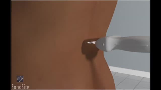
Lumbar puncture is a common emergency department procedure used to obtain information about the cerebrospinal fluid (CSF) for diagnostic and, less commonly, therapeutic reasons. Please refer to the full article on Lumbar Puncture for more details on the lumbar puncture procedure. Lumbar puncture is typically performed via “blind” surface landmark guidance. The surface landmark technique is reported to be successful in a high percentage of attempted lumbar punctures; however, surface landmark identification of underlying structures has been shown to be accurate only 30% of the time. [1] Unsuccessful identification of proper landmarks often leads to increased difficulty in obtaining CSF, if the procedure is performed, and a higher rate of complications. Few alternatives are available in these cases. If available, fluoroscopic-guided lumbar puncture may be performed. If not, treatment is sometimes initiated empirically without obtaining CSF. Disadvantages of using fluoroscopy include limited availability or necessary transport of the patient outside of the emergency department, inability to directly visualize the spinal canal, and inherent radiation exposure
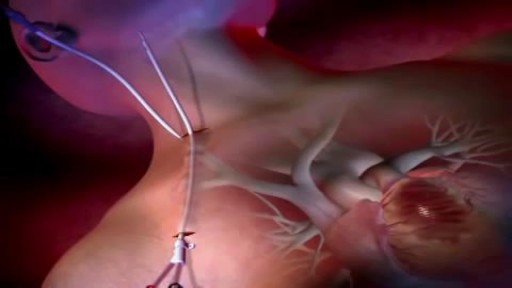
Learn more about Merit Medical's ProGuide™ Chronic Dialysis

Is that knee pain just a sprain or a more serious ACL injury? Orthopedic surgeon Paul Fadale, M.D., offers tips on how to tell the difference. http://www.orthopedicsri.org/
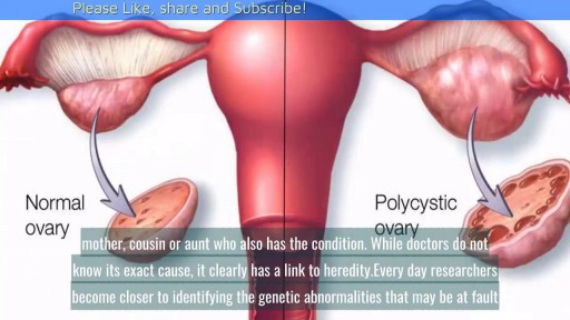
Causes of Polycystic Ovarian Syndrome|| Common gynaecological problems in women Polycystic ovarian syndrome, or PCOS, is a condition where a woman's ovaries and adrenal glands produce more androgens than normal, resulting in increased body hair, acne and irregular periods. While researchers are not certain of the exact cause of PCOS, it is known that an imbalance of the endocrine system is responsible for many of the changes associated with it. However, it is still not known exactly what causes those changes.

Elizabeth Stephens, MD joined the Department of Cardiovascular Surgery at Mayo Clinic Rochester, Minnesota in 2019. To learn more about Dr. Stephens’ practice: https://www.mayoclinic.org/bio....graphies/stephens-el
Elizabeth H. Stephens, M.D., Ph.D., is an Assistant Professor of Surgery in Cardiovascular Surgery specializing in congenital cardiac surgery. She received her medical degree from Baylor College of Medicine and Ph.D in Bioengineering from Rice University focusing on tissue engineering heart valves. Her adult cardiothoracic training was completed at Columbia University and congenital training at Lurie Children's Hospital in Chicago. Her clinical areas of expertise include the treatment of:
• Neonates, infants, and children with complex congenital heart disease
• Adult patients with congenital heart disease, including patients previously repaired
• Valve disease, including Ebstein's anomaly
• Pediatric patients with heart failure, including mechanical circulatory support and heart transplantation
• Patients with vascular rings and tracheal stenosis
In addition to her clinical areas of expertise, Dr. Stephens is active in outcomes research relative to congenital heart disease and is extensively published on various cardiac surgery conditions. She has a particular interest in education, including serving on national committees and mentoring trainees of all levels.
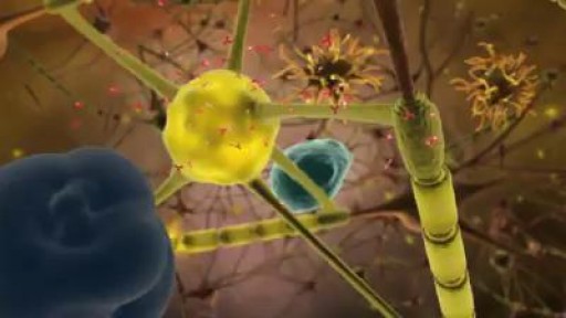
Multiple sclerosis (MS) affects the brain and spinal cord. Early MS symptoms include weakness, tingling, numbness, and blurred vision. Other signs are muscle stiffness, thinking problems, and urinary problems. Treatment can relieve MS symptoms and delay disease progression.